


Education Journalist | Study Abroad Lead
X-rays or x-radiations are a form of electromagnetic radiation consisting of powerful waves of electromagnetic energy. Most of the X-Rays are of wavelength ranging from 0.01 to 10 nanometers, corresponding to frequencies in the range from 30 petahertz to 30 exahertz and energies in the range of 100 eV to 100 KeV.
| Table of Content |
Keyterms: X-rays, X-radiations, Electromagnetic radiation, Waves, Electromagnetic energy, Wavelength, Nanometers, Energies, Frequencies, Electromagnetic Spectrum
History of X-Rays
[Click Here for Sample Questions]
The discovery of X-Rays is credited to the German physicist Wilhelm Röntgen who discovered them in 1895. The credit goes to him because he was the first to comprehensively study them, but he was not the first to have seen and perceived these effects.
X-Rays
X-Rays were found emanating from Crookes Tube, experimental discharge tubes which were invented around 1875 by the scientists looking into cathode rays, which are energetic electron beams that were first formed in tubes.
Also check:
| Related Concepts | ||
|---|---|---|
| Maxwell Equations | Impedance of free space | Scintillation Counter |
| Electromagnetic Waves MCQ | Types of Radiation | Wavelength Formula |
How do X-Rays Work?
[Click Here for Sample Questions]
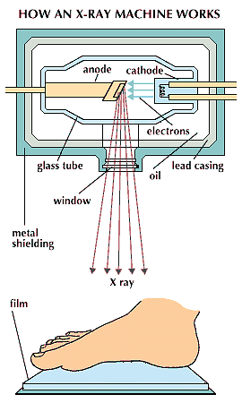
X-rays are formed through the collision of high-velocity electrons with metal sheets. The energy is released in the form of x-rays, which are themselves absorbed by the metal plates.
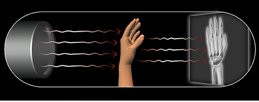
- X-Ray beam travels through the air and comes in contact with the body tissue, and then produces an image on the metal film.
- The soft tissue including skins and organs are unable to absorb these high energy rays and the beams pass through them.
- Dense material inside our body, for example, bones absorbs these radiations.
X-Rays Work
This works quite like the normal camera, when you see on the metal film you will find some white areas and some are black. The white area indicates to the bones, as the bones absorb the x-rays, whereas the black area indicates the soft tissue as it is non-absorbable to the X-Rays.
Types of X-Rays
[Click Here for Sample Questions]
Different types of X rays are identified by medical science. A few of these types which can be important are:
Computed Tomography Scan- This is also popularly known as CT scan is a medical imaging technique that is used in radiology to get a detailed image of the body for diagnostic purposes. Rotating x-ray tubes and a row of detectors are framed within the scanner to measure x-ray attenuation by different tissue inside the body.

Computed Tomography Scan
Teeth and Bones X-ray- They are also referred to as dental radiographs. Dentists use radiographs for many reasons that are to find hidden dental structures, malignant or cavities. A controlled burst of x-ray radiation results in the formation of a radiographic image that penetrates oral structure at a different level, depending on different densities, before striking the film or sensor.

Teeth and Bones X-ray
Chest X-ray- Chest x-ray is also known as chest radiograph is a projection radiograph of the chest which is used in the diagnosis of the conditions affecting the chest. Chest radiography uses ionizing radiation (in X-ray form) to generate images.

Chest X-ray
Chest radiographs are used to diagnose many conditions some of which are chest walls, structures within the thoracic cavity including lungs, heart. Pneumonia and Congestive Heart Failure can also be diagnosed through chest radiographs.
Abdomen X-ray- An abdominal x-ray is the x-ray of the abdomen. The standard abdominal x-ray is usually a single anteroposterior projection. Coverage on the x-ray must be included from the diaphragm (or the top of the liver) to the public symphysis.

Kidney, Ureter and Bladder X Ray

Properties of X-Rays
[Click Here for Sample Questions]
- They are not easily refracted.
- X-rays are not affected by the electromagnetic field.
- They can propagate independently which means that they don't require any medium for their transfer.
- X rays are diverging which means they can never be focused on one single point.
- The intensity of X rays completely relies on the number of electrons that are hitting the target.
X-Ray: Uses
[Click Here for Sample Questions]
Since the discovery of X rays, they have been extremely useful due to their uses in various fields and for various purposes. Some key uses of X ray are as follows:
- Medical Science- An x-ray is an image created on a photographic film or digitally to diagnose illness and injuries. The x-ray machine during this process is used to take pictures of the inside of the body. The x-ray machine passes through the body to produce images of tissues, organs and bones.

X-Ray Medical Uses
- Astronomy- X-ray astronomy is the observational branch which deals with the study of x-ray observation and detections from various astronomical objects. As the x-ray is absorbed by the ground, the detectors are taken to high altitude with the help of balloons, rockets, and satellites. X-ray astronomy takes help of a special space telescope that can be used to see x-ray radiations which a normal optical telescope cannot.
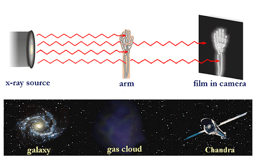
X-Ray Astronomy Uses
- Restoration- X-ray can also be used for the restoration purposes. It is most popularly known for finding defects (if any) in the walls. X-ray beams are projected on the walls and after that they provide a digital image of the walls.
- Security- X-rays are also used for security purposes at airports, railway stations, bus stands, etc. They can be used to find whether any illegal item is being carried within the luggage, or within the body. They are chiefly used to catch smugglers carrying any sort of drugs, guns or other contraband.

X-Ray Security Uses
- Industry- Industrial use of x-ray which is also termed as industrial radiography is used to inspect materials or objects with the objective of locating defects in material which could lead to the failure of engineering structures.
Also check:
| Chapter Related Links | ||
|---|---|---|
| Gamma Radiation | Faraday's Law | Huygens Principle |
| Displacement Current | Electrical Insulators | Radio Waves |
| Wave Speed | Transverse and Longitudinal Waves | Maxwell's Equations |
Things to Remember
- X-rays are a form of electromagnetic radiation consisting of powerful waves of electromagnetic energy.
- They were discovered in 1895 by German physicist Wilhelm Röntgen.
- X-rays are formed through the collision of high-velocity electrons with metal sheets.
- Different types of X-rays include CT scans, Dental X-rays, Chest Radiographs, Abdominal X-rays etc.
- X-Rays are used in a number of fields, mainly medicine, astronomy, security, industry, etc.
Sample Questions
Ques. How can X-rays be produced in the normal x-ray machine? (1 mark)
Ans. In a normal x-ray machine, x-rays are produced by the bombarding of cathode rays on a radioactive material. Then, there is an emission of electrons and energy. This energy is being used up in the x-ray machines.
Ques. Name the term that is used to describe the dental x-ray. (1 mark)
Ans. Orthopantomography is the term that is being used for the description of dental x-ray.
Ques. What is the minimum distance at which x-ray is being taken? (1 mark)
Ans. The minimum distance at which x-ray is being taken is 50 m.
Ques. Where can X rays be recorded? (1 mark)
Ans. x-rays can be recorded on a silver halide coated plate. Silver halide when exposed to heat starts turning black which results in the x-ray image.
Ques. Which organ needs the longest exposure to x-ray? (1 mark)
Ans. Spine needs the longest exposure to x-ray. Spine is exposed for 0.2 seconds resulting in a proper image.
For Latest Updates on Upcoming Board Exams, Click Here: https://t.me/class_10_12_board_updates
Check-Out:


Comments