Jasmine Grover Content Strategy Manager
Content Strategy Manager
Muscle tissue is a type of connective tissue that aids in muscle contraction. Muscle tissue allows muscles to contract by exerting forces on other regions of the body. They have several qualities, one of which is movement. It is found in the space between two bones. Its to contract aids in the smooth movement of bones. Myocytes are another name for muscle cells. Myofibrils, which are made up of actin and myosin myofilaments, are found in them. Muscle tissues are mainly divided into three categories: Smooth muscle, Skeletal muscle, Cardiac muscle There are no striations or stripes on smooth muscle cells. Stripes or striations can be found on skeletal muscles. The only place where cardiac muscle may be found is in the heart. In this article, we will have a look at the functions of muscular tissue and their different types.
| Table of Contents |
Key Takeaway: Skeletal muscle, involuntary, striated, Contractibility, Extensibility, Excitability, smooth muscle
Muscle Tissue
Muscle tissue is a type of specialized tissue found in animals that works by contracting and applying forces to various parts of the body. Muscle tissue is made up of sheets and fibres made up of muscle cell fibres.They control an organism's movement as well as numerous other contractile functions. Animals have three different types of muscle, depending on how they are used. Although these muscles differ slightly in appearance, they act in the same way.

Muscle Tissue
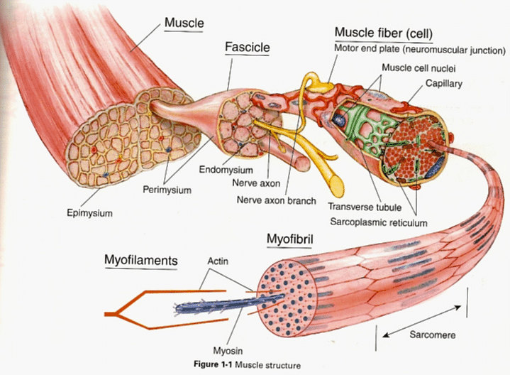
Myofibrils, which are made up of actin and myosin myofilaments, are found in the muscles. They glide past each other, creating stress and causing the myocyte's shape to change. Many myocytes can be found in a single gram of muscle tissue. Movement happens when muscle fibers contract and relax in response to stimuli.
Read More- Tissues
Function of Muscle Tissue
Muscle tissue is generally attached to the same nerve bundles and works as a single unit. The muscle contracts in response to a nerve impulse from the brain or another external stimulus which is sent to all of the nerve cells in the muscle tissue almost instantly, causing the entire muscle to contract.
Each muscle cell has a protein complex combining actin and myosin at the cellular level. When the signal to contract is received, these proteins move past one another. The filaments are attached to the cell's ends, and when they glide past each other, the cell shrinks in size. A single cell can contract up to 70% of its length, shortening the entire muscle when it does so. Muscle tissue is capable of moving bones, compressing chambers, and squeezing different organs.

Muscle Tissue
Properties
The properties of muscle tissues are:
- Contractibility: It refers to the ability to forcefully shorten.
- Extensibility: A muscle can be stretched.
- Excitability: The muscles respond to a stimulus that is delivered from a hormone or a motor neuron.
- Elasticity: Muscles can recoil to their original length after they are stretched.
Structure of Muscular Tissue
The structure of muscle tissue is as given below:
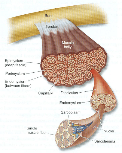
- The muscle tissues are bundled together and surrounded with epimysium, a stiff connective tissue that looks like cartilage.
- The epimysium surrounds a bundle of nerve cells that run in long fibers called fascicles.
- Perimysium is a kind of protective layer that surrounds the fascicles. It permits nerves and blood to pass freely to the individual fibers.
- The endomysium, another protective layer, surrounds the fibers.
- These layers and muscles aid in the contraction of various muscle components. As the bundle’s contract, they move past one another.
- The epimysium attaches to the tendons that run along the periosteum, the connective tissue that surrounds the bones. When the muscles contract, this aids in the movement of the bones.
- The epimysium joins with other connective tissues to exert pressure on the organs and regulate everything from circulation to food processing.

Muscle Structure
Read More- Difference between Cell and Tissue
Types of Muscle Tissue
Muscle tissue can be classified as either voluntary or involuntary muscles, & striated or non-striated muscles. Striation can be understood as the presence of visible banding within the myocytes that is caused as a result of the organization of myofibrils in order to produce a constant tension direction. Voluntary refers to whether the muscle is under conscious control;
Using the aforementioned classifications, three types of muscle tissue can be characterized, each of which performs a wide range of activities.
- Smooth Muscle
- Skeletal Muscle
- Cardiac Muscle
Smooth Muscle
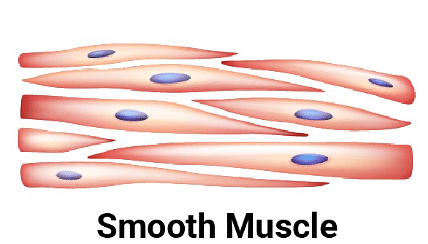
There are no striations or stripes on the smooth muscle cells. Involuntary muscles are another name for them. The cells have a single nucleus and are formed like a spindle. They can be found in the walls of hollow organs such as the stomach and the uterus.
- Their primary purpose is to transport substances through the body.
- The brain is in charge of controlling involuntary muscles.
- Smooth muscle fibers are not as well structured as other forms of muscle tissue in terms of myosin and actin fibers.
- Smooth muscle contractions are smooth and continuous rather than quick.
- Many organs, blood arteries, and other fluid-transporting vessels are surrounded by smooth muscles.
- Smooth muscle tissue can contract to exert force on an organ. This can be utilized to transport blood or food throughout the body.

Smooth Muscle
Skeletal Muscle
The most widespread and widely dispersed muscle tissue in the body is skeletal muscles. They account for roughly 40% of total body mass. These muscles are controlled by us and are voluntary.
- They are attached to the skeleton and mostly aid in our mobility.
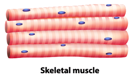
- The cells are cylindrical and lengthy, with many nuclei. Skeletal muscles can be found in the muscles of the limbs, face, neck, and other parts of the body.
- Skeletal muscle tissue is a striated muscle, which means it has definite bands visible under a microscope.
- The striated muscle may contract and release quickly because of these structured bundles.
- Tendons, which are highly elastic connective tissue parts, connect muscle tissue to the bones.
- Although many muscles appear to govern a single appendage, each one is only responsible for a small portion of the movement.
- The somatic nervous system can voluntarily control skeletal muscle tissue. The involuntary or autonomous nervous system is in charge of the other muscle kinds.

Skeletal Muscle
Cardiac Muscle
Cardiac muscle can only be found in the heart. The pumping of blood through the blood arteries to various regions of the body is aided by the rhythmic contractions of this muscle.
- This is an involuntary muscle that is controlled by the brain.
- Cardiac muscular tissue's cells are branching and cylindrical, with a single nucleus and striations.
- The cardiac muscle tissue's cells are shorter than those of skeletal muscle tissue.
- Between the heart muscle, cells are intercalated discs of the overlapping cell membranes. This aids in the tissue's tightening up.
- This location also provides for the rapid transmission of electrochemical impulses across cells.
- Myofibrils and mitochondria are abundant in heart muscle tissue. This offers the tissue a lot of strength and endurance when it comes to pumping blood.

Cardiac Muscle
Read More- Connective Tissues
Things To Remember
- Skeletal muscles make up 40% of our bodily weight.
- Actin and myosin filaments are extremely thin and organized in a random pattern.
- Cardiomyocytes are striated cells that make up the heart muscles.
- Smooth muscle cells have a single nucleus and are formed like a spindle.
- Myofibrils are found in every skeletal tissue.
- The most widespread and widely dispersed muscle tissue in the body is skeletal muscles.
- Sarcomeres, which are highly ordered structures of actin, myosin, and proteins, are the light and dark bands.
- Cardiac muscle can only be found in the heart and its rhythmic contractions aid the pumping of blood through the blood arteries to various regions of the body.
Sample Questions
Ques. Where is muscular tissue found? (2 marks)
Ans. These muscles are individual organs made up of skeletal muscle tissue, blood vessels, tendons, and nerves. The heart, digestive organs, and blood vessels all contain muscle tissue. Muscles in these organs help to transport chemicals throughout the body.
Ques. What cells are in muscle tissue? (2 marks)
Ans. Myocytes are the cells that make up muscular tissue and are also known as muscle cells. In the human body, there are three types of muscle cells: cardiac, skeletal, and smooth.
Due to their lengthy and fibrous nature, cardiac and skeletal myocytes are commonly referred to as muscle fibers.
Ques. What is the main function of muscular tissue? (3 marks)
Ans. Muscle tissue's primary purpose is movement. They can contract, and this is what causes body components to move. They also aid in the preservation of bodily posture and position. The muscular system is a complex network of muscles that are essential to the human body's survival. Muscles are involved in every action you take. They regulate your heart rate and respiration, aid digestion, and allow you to move around. When you exercise and eat well, your muscles, like the rest of your body, thrive.
Ques. What are the distinguishing features of the three types of muscular tissues? (3 marks)
Ans. In the human body, each type of muscle tissue has its structure and function. Skeletal muscle is a type of muscle that moves bones and other tissues. The heart's cardiac muscle contracts to pump blood. Slow oxidative (SO), rapid oxidative (FO), and fast glycolytic muscle fibers are the three types of muscle fiber. SO fibers are slow to fatigue and utilize aerobic metabolism to create low-power contractions over long periods. FO fibers make ATP through aerobic metabolism, however, they have stronger tension contractions than SO fibers.
Ques. How are slow-twitch muscle fibers different from fast-twitch muscle fibers? (3 marks)
Ans. Fast-twitch muscle fibers exhaust sooner and are used in intense bursts of action, whereas slow-twitch muscle fibers allow for long-endurance activities like distance running. Slow-twitch muscles conserve energy by releasing it slowly and evenly over time. This allows them to contract (work) for an extended period without running out of energy. Fast-twitch muscles expend a lot of energy quickly, then get exhausted (fatigued) and require rest.
Ques. How is muscle tissue formed? (2 marks)
Ans. Muscle tissue develops in the embryo's mesoderm layer in response to fibroblast growth factor, serum response factor, and calcium signals. Myoblasts fuse into multinucleated myotubes in the presence of fibroblast growth factors, which constitute the foundation of muscle tissue.
Ques. What tissues make up skeletal muscles? (2 marks)
Ans. Connective tissue, blood vessels, and nerves are all found in skeletal muscles. Epimysium, endomysium, and perimysium, are the 3 layers of connective tissue. Fascicules are structured groupings of skeletal muscle fibres. Blood vessels and nerves branch in the cell after entering the connective tissue.
Ques. What are the components of cardiac muscle? (3 marks)
Ans. The plasma membrane and transverse tubules in register with the Z lines, the longitudinal sarcoplasmic reticulum and terminal cisternae, and the mitochondria are all important components of each cardiac muscle cell involved in excitation and metabolic recovery processes. The muscle that surrounds the heart is made up of cardiac muscular tissue. Unlike skeletal muscles, the muscle's role is to cause the mechanical motion of pumping blood throughout the rest of the body. The movement is involuntary to maintain life.


Comments