




NEET PG 2026 Question Paper is available for download here. NBEMS conducted NEET PG 2026 exam on 30 August from 9 AM to 12:30 PM. NEET PG 2026 was conducted in online CBT mode for a duration of 3 hours 30 minutes. NEET PG question paper comprises 180 questions carrying a total weightage of 720 marks.
- NEET PG Question Paper is divided into 5 sections with 36 questions each. Candidates have to attempt each section in 42 minutes.
- Each correct answer carries 4 marks and there is negative marking of 1 for incorrect answers.
Candidates can download NEET PG 2026 Question Paper with Answer Key and Solutions PDF from the links provided below. According to initial student reaction, NEET PG 2026 Paper was of moderate to difficult level.
NEET PG 2026 Question Paper with Solution PDF
| NEET PG Question Paper 2026 | Download PDF | Check Solutions |
Atomic Energy Regulatory Board (AERB) clearance or registration is NOT required for which of the following diagnostic modalities?
View Solution
Concept:
The Atomic Energy Regulatory Board (AERB) is the statutory body in India responsible for framing safety codes, standards, and regulatory licensing for all ionizing radiation-emitting equipment and radioactive sources.
Diagnostic and therapeutic modalities are categorized based on whether they emit ionizing or non-ionizing forms of radiation.
All facilities housing ionizing radiation devices must obtain procurement permission, site layout approval, and an operational license from AERB via the e-LORA portal.
Explanation:
Step 1: Regulatory Scope of AERB:
The Atomic Energy Regulatory Board (AERB) enforces safety regulations exclusively for equipment and processes that emit ionizing radiation.
Diagnostic modalities such as conventional X-rays, Computed Tomography (CT), fluoroscopy, C-arms, mammography, and portable radiography emit ionizing photons, posing potential deterministic and stochastic radiation hazards.
Consequently, strict layout approval, quality assurance tests, and operating licenses from AERB are legally mandatory for these installations.
Step 2: Physics and Regulation of Non-Ionizing Modalities:
Ultrasonography (USG) relies entirely on high-frequency sound waves (mechanical acoustic pressure waves) to generate images.
Because USG emits zero ionizing radiation, it does not fall under the statutory purview of the AERB and does not require AERB clearance or registration.
Instead, ultrasound units and diagnostic centers in India are strictly regulated under the Pre-Conception and Pre-Natal Diagnostic Techniques (PC-PNDT) Act to prevent illegal sex determination.
Step 3: Comparative Assessment of Options:
$\bullet$ Option (A) X-ray: Emits ionizing radiation $\rightarrow$ Requires AERB clearance.
$\bullet$ Option (B) CT scan: High-dose ionizing radiation $\rightarrow$ Requires AERB clearance.
$\bullet$ Option (C) USG: Non-ionizing acoustic waves $\rightarrow$ Exempt from AERB clearance.
$\bullet$ Option (D) Portable radiography: Ionizing radiation source $\rightarrow$ Requires AERB clearance.
Final Answer:
Because Ultrasonography (USG) is a completely non-ionizing imaging modality, it does not require AERB clearance or permission.
USG and MRI utilize non-ionizing physics and do NOT need AERB registration.
USG centers in India must be registered under the PC-PNDT Act.
A 42-year-old female presents with symptomatic uterine fibroids and heavy menstrual bleeding. She is advised a hysterectomy but strongly wishes to preserve her uterus. Which of the following is the most appropriate first-line non-surgical management option?
View Solution
Concept:
Uterine leiomyomas (fibroids) are benign monoclonal smooth muscle tumors of the myometrium that frequently cause heavy menstrual bleeding (menorrhagia) and pelvic pain.
In patients who wish to avoid hysterectomy or definitive surgery, medical therapies aimed at inducing local or systemic endometrial suppression provide excellent symptomatic relief.
Local intrauterine progestin release provides maximal endometrial suppression while avoiding systemic steroidal adverse effects.
Explanation:
Step 1: Clinical Presentation and Goal of Therapy:
The patient is a perimenopausal woman with symptomatic uterine leiomyoma presenting with menorrhagia who desires uterine conservation.
The therapeutic goal is to suppress excessive menstrual blood loss, correct secondary anemia, and avoid invasive major surgery (hysterectomy).
Step 2: Mechanism and Efficacy of LNG-IUS:
The Levonorgestrel-releasing Intrauterine System (LNG-IUS, e.g., Mirena) delivers a steady local intrauterine dose of $20\ \mu\text{g}$ levonorgestrel per 24 hours.
This continuous local release causes extensive decidualization of the stroma and profound atrophy of endometrial glandular epithelium.
Clinical trials demonstrate a reduction in menstrual blood volume by $80\text{--}90\%$ within 3 to 6 months, making it the most effective medical alternative to hysterectomy for abnormal uterine bleeding in non-cavity distorting fibroids.
Step 3: Evaluation of Alternative Options:
$\bullet$ Danazol: Although it reduces bleeding, it causes significant androgenic side effects (weight gain, voice deepening, acne, hirsutism) and hepatic toxicity, limiting its long-term utility.
$\bullet$ Oral Estrogen: Contraindicated as estrogens promote leiomyoma cellular proliferation and exacerbate menorrhagia.
$\bullet$ Copper Intrauterine Device (Cu-T): Induces local sterile inflammatory changes that typically increase menstrual blood loss and dysmenorrhea.
Final Answer:
The LNG-IUS is the most effective and safest uterus-sparing medical intervention to control heavy menstrual bleeding in uterine fibroids.
Copper-containing IUDs are strictly contraindicated in menorrhagia as they increase menstrual blood loss.
A 48-year-old woman presents with progressive deepening of her voice and persistent hoarseness for several months. She has a heavy smoking history. Laryngoscopy reveals diffuse, symmetric, polypoid, bilateral gelatinous fluid accumulation in the superficial layer of the lamina propria of both true vocal cords. What is the most likely diagnosis?
View Solution
Concept:
Reinke's edema (polypoid corditis) is a chronic inflammatory disorder of the vocal folds characterized by the accumulation of gelatinous, mucoid fluid within the potential subepithelial space known as Reinke's space.
Reinke's space corresponds anatomically to the superficial layer of the lamina propria of the true vocal cords.
The accumulation of fluid increases the vibrating mass of the vocal folds, significantly lowering the fundamental frequency of phonation.
Explanation:
Step 1: Pathophysiological Basis:
Chronic exposure to cigarette smoke and vocal strain triggers increased vascular permeability and chronic subepithelial inflammation within Reinke's space.
Because Reinke's space has limited lymphatic drainage, thick mucoid, gelatinous fluid progressively accumulates in the superficial layer of the lamina propria.
Step 2: Clinical and Laryngoscopic Features:
The increased mass and decreased tension of the vocal folds cause a profound drop in the fundamental frequency of phonation.
Middle-aged female patients characteristically present with an abnormally low-pitched, coarse, mannish voice ("whiskey voice").
Laryngoscopy demonstrates bilateral, symmetric, diffuse, pale, translucent, bag-like polypoid swelling running the entire length of both membranous vocal cords.
Step 3: Ruling Out Alternative Diagnoses:
$\bullet$ Post-cricoid malignancy: Presents with progressive dysphagia, pooling of secretions in the hypopharynx, and stridor, not isolated voice deepening.
$\bullet$ Sulcus vocalis: Shows a longitudinal groove/furrow along the medial vocal cord edge causing a weak, breathy voice.
$\bullet$ Laryngeal papillomatosis: Presents with multiple exophytic, verrucous, raspberry-like lesions due to HPV 6 and 11 infection.
Final Answer:
The clinical presentation of progressive voice deepening in a female smoker with bilateral subepithelial fluid collection is characteristic of Reinke's edema.
Anatomical site: Superficial layer of lamina propria (Reinke's space).
Definitive management: Smoking cessation followed by vocal cord microflap decortication surgery.
A patient presents with unilateral high-pitched tinnitus and progressive sensorineural hearing loss (SNHL). A retrocochlear lesion involving the cerebellopontine angle / posterior aspect of the temporal bone is suspected. What is the gold standard investigation of choice?
View Solution
Concept:
Unilateral sensorineural hearing loss and asymmetric tinnitus are cardinal clinical indicators of a retrocochlear pathology involving the vestibulocochlear nerve ($8^{\text{th}}$ cranial nerve) in the internal auditory canal (IAC) or cerebellopontine angle (CPA).
The most common CPA mass is a vestibular schwannoma (acoustic neuroma), followed by meningioma and epidermoid cyst.
Accurate visualization requires high-resolution soft-tissue imaging with paramagnetic contrast enhancement.
Explanation:
Step 1: Clinical Suspicion of Retrocochlear Lesions:
A lesion at the CP angle or internal auditory canal compresses the eighth cranial nerve, leading to progressive unilateral high-frequency sensorineural hearing loss, disproportionately poor speech discrimination, and continuous high-pitched tinnitus.
Vestibular schwannoma accounts for approximately $80\text{--}90\%$ of all cerebellopontine angle tumors.
Step 2: Diagnostic Modality of Choice:
Gadolinium-enhanced Magnetic Resonance Imaging (MRI) with dedicated thin-slice ($1\text{--}2\text{ mm}$) T1-weighted and constructive interference in steady state (CISS/FIESTA) sequences is the gold standard investigation.
Gadolinium contrast avidly enhances vestibular schwannomas, allowing detection of micro-schwannomas confined strictly within the internal auditory meatus ($1\text{--}2\text{ mm}$ size) before extra-canalicular extension occurs.
Step 3: Limitations of Other Modalities:
$\bullet$ HRCT of the temporal bone: Excellent for cortical bone details and erosion of the internal acoustic meatus, but fails to identify small soft-tissue intracanalicular tumors.
$\bullet$ Carotid angiography: Used for evaluating vascular lesions (e.g., glomus jugulare tumors, aneurysms), not nerve sheath tumors.
$\bullet$ Echocardiography: Plays no role in auditory pathway assessment.
Final Answer:
Gadolinium-enhanced MRI of the cerebellopontine angle is the diagnostic investigation of choice for suspected retrocochlear and acoustic nerve lesions.
HRCT detects bone erosion; MRI detects soft-tissue tumor mass.
A 15-year-old male with Juvenile Nasopharyngeal Angiofibroma (JNA) presents with an expanding nasopharyngeal mass and severe, recurrent, profuse epistaxis. What is the primary pathophysiological basis for the severe, torrential bleeding characteristic of JNA?
View Solution
Concept:
Juvenile Nasopharyngeal Angiofibroma (JNA) is a histologically benign but locally destructive, highly vascular neoplasm that arises almost exclusively in adolescent males.
The tumor originates near the sphenopalatine foramen and consists of an irregular meshwork of fibrous stroma interspersed with variable-sized vascular spaces.
The unique ultrastructural composition of its blood vessels explains the severe bleeding tendencies of this tumor.
Explanation:
Step 1: Histopathological Architecture of JNA:
Histologically, JNA consists of two distinct components: an irregular, proliferating collagenous stroma and a dense network of thin-walled, irregular vascular spaces of varying calibers.
Step 2: Ultrastructural Defect and Impaired Hemostasis:
The vascular channels within JNA consist of an endothelial lining embedded directly in fibrous tissue, completely lacking a complete muscular layer (tunica media) and an internal elastic lamina.
Under normal physiological conditions, vascular injury induces instant vasoconstriction mediated by the smooth muscle of the tunica media to achieve initial hemostasis.
Because JNA vessels lack smooth muscle contractile elements, they cannot constrict, retract, or spasm when injured or incised, leading to continuous, torrential, life-threatening hemorrhage.
Step 3: Clinical Consequence:
Because of the absence of contractile elements and the resulting inability to control bleeding mechanically, direct incisional biopsy of a suspected JNA in an outpatient setting is strictly contraindicated.
Preoperative transarterial embolization is routinely performed prior to surgical resection to minimize intraoperative blood loss.
Final Answer:
The absence of a contractile smooth muscle layer (tunica media) in the vascular walls prevents vasoconstriction, causing profuse hemorrhage.
$\bullet$ Classic patient: Adolescent male with recurrent painless profuse epistaxis.
$\bullet$ Histology: Blood spaces lacking tunica media (smooth muscle) in fibrous stroma.
$\bullet$ Cardinal Rule: Never perform an incisional biopsy due to risk of fatal hemorrhage!
A 25-year-old female presents with bilateral progressive hearing loss. Pure Tone Audiometry (PTA) demonstrates an air-bone gap along with a characteristic notch in bone conduction thresholds at 2000 Hz. What is the most likely diagnosis?
View Solution
Concept:
Otosclerosis (otospongiosis) is a primary metabolic bone disease of the otic capsule characterized by abnormal bone remodeling, leading to stapedial ankylosis at the oval window.
It typically presents in young adults, with a female-to-male preponderance ($2:1$), and frequently accelerates during pregnancy.
Pure Tone Audiometry (PTA) demonstrates characteristic conductive hearing loss with an artifactual dip in bone conduction.
Explanation:
Step 1: Pathophysiology of Bone Conduction Artifact:
In normal individuals, bone conduction hearing involves an inertial ossicular component mediated by the free vibration of the middle ear ossicles within the tympanic cavity.
In stapedial otosclerosis, fixation of the stapes footplate at the oval window abolishes this inertial component, leading to an artifactual elevation of bone conduction thresholds.
Step 2: Identification of Carhart's Notch:
This artifactual dip in the bone conduction curve is maximal at $2000\text{ Hz}$ ($2\text{ kHz}$) and is termed Carhart's notch.
The typical threshold elevations are $5\text{ dB}$ at $500\text{ Hz}$, $10\text{ dB}$ at $1000\text{ Hz}$, $15\text{ dB}$ at $2000\text{ Hz}$, and $5\text{ dB}$ at $4000\text{ Hz}$.
Carhart's notch does not represent true cochlear hair cell damage and characteristically overcorrects or disappears following successful stapedotomy.
Step 3: Differentiation from Other Audiological Notches:
$\bullet$ Noise-induced hearing loss causes a sensorineural notch at $4000\text{ Hz}$ (Boilermaker's notch).
$\bullet$ Acoustic trauma from blast injury typically shows a dip at $6000\text{ Hz}$.
$\bullet$ Presbyacusis manifests as a bilateral down-sloping high-frequency sensorineural deficit in elderly patients.
Final Answer:
A bone conduction dip at 2000 Hz in a young female with progressive hearing loss represents Carhart's notch seen in otosclerosis.
$\bullet$ $2000\text{ Hz}$ Bone Conduction Dip = Carhart's Notch $\rightarrow$ Otosclerosis (Reversible after stapedotomy).
$\bullet$ $4000\text{ Hz}$ Dip = Acoustic/Noise-induced notch $\rightarrow$ Acoustic trauma.
$\bullet$ $6000\text{ Hz}$ Dip = Firearm/Blast injury.
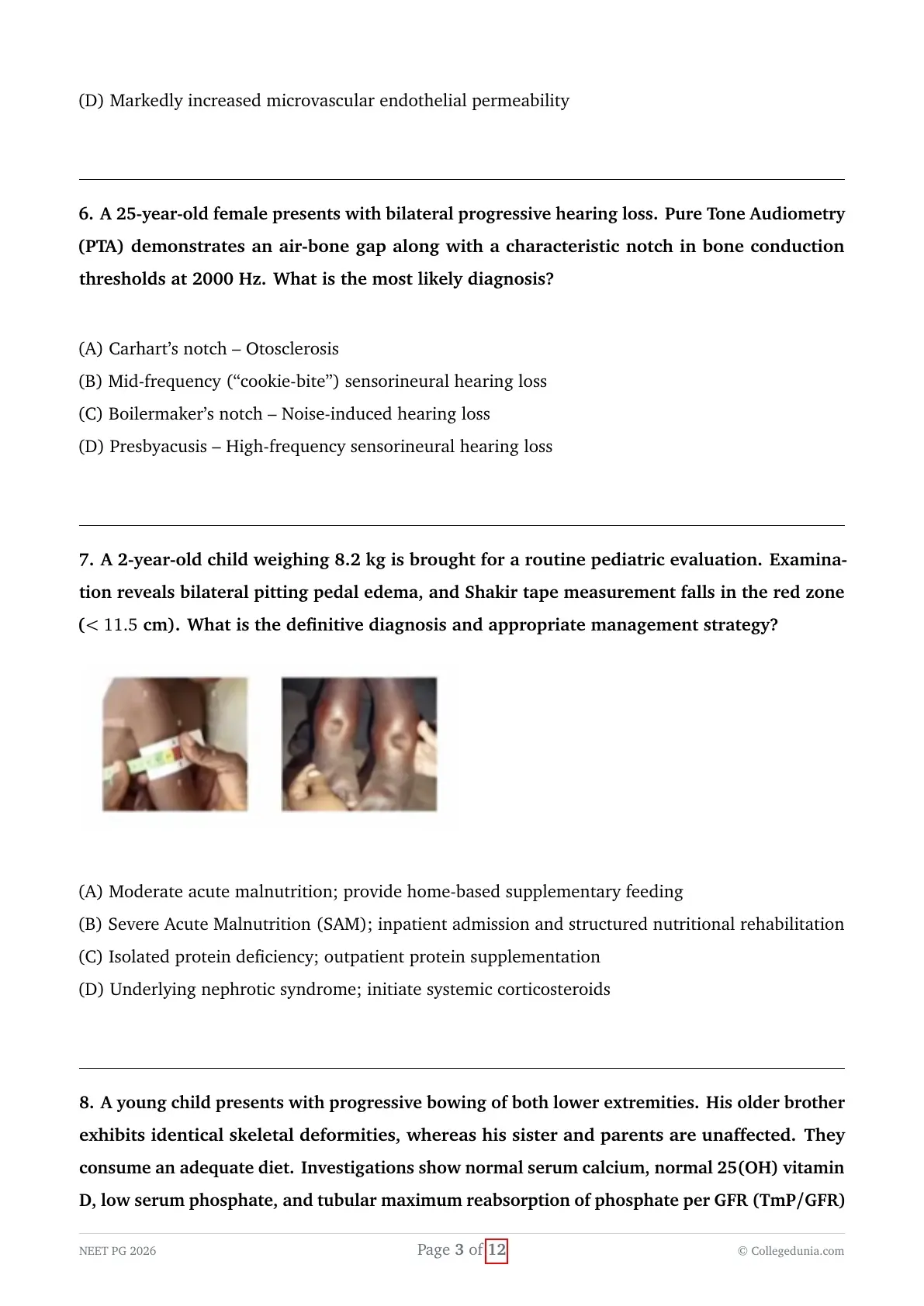
A 2-year-old child weighing 8.2 kg is brought for a routine pediatric evaluation. Examination reveals bilateral pitting pedal edema, and Shakir tape measurement falls in the red zone (<11.5 cm). What is the definitive diagnosis and appropriate management strategy?

View Solution
Concept:
Severe Acute Malnutrition (SAM) is a life-threatening form of undernutrition in children aged $6\text{--}59$ months defined by standardized anthropometric criteria and clinical signs established by the World Health Organization (WHO).
The presence of bilateral nutritional pitting edema or severe wasting signifies high mortality risk requiring immediate, protocol-based therapeutic intervention.
Explanation:
Step 1: WHO Diagnostic Criteria for SAM:
According to WHO and IAP guidelines, SAM is diagnosed in children aged $6\text{--}59$ months when any of the following are met:
1. Mid-Upper Arm Circumference (MUAC) $< 115\text{ mm}$ ($11.5\text{ cm}$), corresponding to the RED zone on Shakir tape.
2. Weight-for-height $Z\text{-score} < -3\ \text{SD}$.
3. Presence of nutritional bilateral pitting pedal edema (kwashiorkor).
Step 2: Clinical Severity and Triage:
The patient satisfies both the MUAC criterion ($<11.5\text{ cm}$) and presents with bilateral pedal edema.
Any child with edematous SAM is automatically categorized as complicated SAM and mandates urgent inpatient admission to a Nutrition Rehabilitation Centre (NRC).
Step 3: Staged Management Protocol:
$\bullet$ Stabilization Phase (Days 1--7): Treat and prevent hypoglycemia, hypothermia, dehydration, and infections; initiate starter low-protein, low-sodium formula feed F-75 ($75\text{ kcal/100 mL}$).
$\bullet$ Rehabilitation Phase (Weeks 2--6): Promote vigorous catch-up growth using high-calorie formula F-100 ($100\text{ kcal/100 mL}$) or Ready-to-Use Therapeutic Food (RUTF).
$\bullet$ Follow-up Phase: Transition to home-cooked energy-dense diet and developmental stimulation.
Final Answer:
The child has Severe Acute Malnutrition (SAM) and requires urgent hospital admission followed by structured inpatient nutritional rehabilitation.
$\bullet$ MUAC $< 11.5\text{ cm}$ (Red zone on Shakir strip).
$\bullet$ Weight-for-height $Z$-score $< -3\ \text{SD}$.
$\bullet$ Bilateral pitting pedal edema.
Formula feeds: F-75 for stabilization; F-100 for catch-up rehabilitation.
A young child presents with progressive bowing of both lower extremities. His older brother exhibits identical skeletal deformities, whereas his sister and parents are unaffected. They consume an adequate diet. Investigations show normal serum calcium, normal 25(OH) vitamin D, low serum phosphate, and tubular maximum reabsorption of phosphate per GFR (TmP/GFR) <80%. What is the most likely diagnosis?
View Solution
Concept:
Hypophosphatemic rickets (vitamin D-resistant rickets) is a genetic disorder of phosphate homeostasis characterized by impaired renal tubular phosphate reabsorption and abnormal vitamin D metabolism.
The X-linked dominant form (caused by PHEX gene mutations) is the most frequent hereditary cause of non-nutritional rickets.
Excess Fibroblast Growth Factor 23 (FGF-23) impairs renal phosphate conservation, causing chronic hypophosphatemia.
Explanation:
Step 1: Molecular Pathogenesis:
Loss-of-function mutations in the phosphate-regulating gene PHEX (on the X chromosome) lead to inappropriately elevated circulating concentrations of Fibroblast Growth Factor 23 (FGF-23).
FGF-23 downregulates sodium-phosphate co-transporters (NaPi-IIa and NaPi-IIc) in renal proximal tubule cells, leading to marked urinary phosphate wasting and reduced tubular maximum reabsorption of phosphate (TmP/GFR $<80\%$).
Step 2: Biochemical Profiling:
$\bullet$ Serum Calcium: Normal.
$\bullet$ Serum Phosphate: Markedly reduced.
$\bullet$ Serum Alkaline Phosphatase (ALP): Elevated (active rickets).
$\bullet$ Serum 25(OH) Vitamin D: Normal.
$\bullet$ FGF-23 also suppresses renal $1\alpha$-hydroxylase, causing inappropriately low/normal $1,25\text{-(OH)}_2\text{D}_3$ levels.
Step 3: Exclusion of Other Conditions:
$\bullet$ Vitamin D-dependent rickets: Presents with severe hypocalcemia, secondary hyperparathyroidism, and tetany.
$\bullet$ Fanconi syndrome: Presents with generalized proximal tubular dysfunction (aminoaciduria, renal glucosuria, and normal anion-gap metabolic acidosis), which are absent here.
Final Answer:
Isolated renal phosphate wasting with normal calcium and vitamin D levels confirms the diagnosis of X-linked hypophosphatemic rickets.
$\bullet$ Calcium = Normal, Phosphate = Low, ALP = High, Vit D = Normal.
$\bullet$ Cause: PHEX mutation $\rightarrow$ $\uparrow$ FGF-23 $\rightarrow$ Renal phosphate wasting.
$\bullet$ Treatment: Oral Phosphate + Calcitriol ($1,25\text{-(OH)}_2\text{D}_3$) or Burosumab (anti-FGF-23 monoclonal antibody).
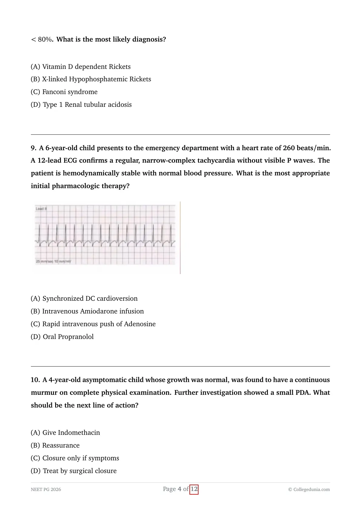
A 6-year-old child presents to the emergency department with a heart rate of 260 beats/min. A 12-lead ECG confirms a regular, narrow-complex tachycardia without visible P waves. The patient is hemodynamically stable with normal blood pressure. What is the most appropriate initial pharmacologic therapy?

View Solution
Concept:
Supraventricular Tachycardia (SVT) is the most common symptomatic pathologic tachyarrhythmia in pediatric patients.
It is characterized by rapid, regular rhythm with narrow QRS complexes ($<0.09\text{ seconds}$) and heart rates typically exceeding $220\text{ bpm}$ in infants and $>180\text{ bpm}$ in children.
Management algorithms are stratified by hemodynamic stability according to Pediatric Advanced Life Support (PALS) protocols.
Explanation:
Step 1: ECG and Hemodynamic Evaluation:
The ECG displays a regular rhythm, narrow QRS complexes, absent P waves, and an extreme heart rate of $260\text{ bpm}$, confirming paroxysmal SVT.
Because the child is hemodynamically stable (normal blood pressure, well perfused), immediate electrical cardioversion is not required.
Step 2: First-Line Pharmacologic Intervention:
According to PALS guidelines, if non-pharmacologic vagal maneuvers (e.g., ice to face) fail or intravenous access is available, the first-line drug is IV Adenosine.
Adenosine is administered as an ultra-rapid intravenous bolus at an initial dose of $0.1\text{ mg/kg}$ (maximum $6\text{ mg}$) immediately followed by a rapid flush of $5\text{--}10\text{ mL}$ isotonic saline.
Step 3: Mechanism and Role of Alternative Therapies:
Adenosine activates cardiac $A_1$ adenosine receptors, opening potassium channels and causing transient, complete AV nodal conduction block, which terminates the re-entry circuit.
Synchronized DC cardioversion ($0.5\text{--}1.0\text{ J/kg}$) is reserved for hemodynamically unstable patients (hypotension, shock, poor perfusion).
Amiodarone and beta-blockers are reserved for refractory or chronic rhythm control.
Final Answer:
In a hemodynamically stable child with narrow-complex SVT, the first-line pharmacologic drug is rapid intravenous Adenosine.
$\bullet$ Stable $\rightarrow$ Vagal maneuvers (ice to face) $\rightarrow$ Rapid IV Adenosine bolus ($0.1\text{ mg/kg}$ + saline flush).
$\bullet$ Unstable $\rightarrow$ Synchronized DC Cardioversion ($0.5\text{--}1\text{ J/kg}$, up to $2\text{ J/kg}$).
A 4-year-old asymptomatic child whose growth was normal, was found to have a continuous murmur on complete physical examination. Further investigation showed a small PDA. What should be the next line of action?
View Solution
Concept:
Patent Ductus Arteriosus (PDA) represents the anatomical failure of the embryonic ductus arteriosus to close after birth, leading to persistent communication between the proximal left pulmonary artery and descending aorta.
The physiological and structural properties of ductal tissue change markedly with age, dictating completely different treatment strategies in preterm neonates compared to older children.
Explanation:
Step 1: Pharmacological Inefficacy Beyond Infancy:
In premature neonates, pharmacologic closure with cyclooxygenase inhibitors (Indomethacin, Ibuprofen) is highly effective because ductal smooth muscle is responsive to prostaglandin withdrawal.
In a 4-year-old child, the ductal tissue is fibrotic and structurally permanent; prostaglandin inhibitors are entirely ineffective.
Step 2: Rationale for Closure in Asymptomatic Patients:
Even if a child is completely asymptomatic with normal growth, persistent ductal patency carries a lifelong cumulative risk of infective endarteritis/endocarditis.
Additional long-term risks include slowly progressive left ventricular volume overload, left atrial enlargement, and pulmonary vascular remodeling.
Step 3: Intervention of Choice:
All moderate-to-small PDAs with an audible continuous murmur in children should undergo definitive elective closure.
The preferred modality is percutaneous transcatheter device occlusion (coils or Amplatzer duct occluders), with surgical ligation reserved for complex ductal anatomy or catheterization failure.
Final Answer:
In a 4-year-old child with an audible PDA, elective definitive closure (transcatheter device or surgical closure) is indicated.
$\bullet$ Preterm infant $\rightarrow$ Medical closure (Indomethacin/Ibuprofen/Paracetamol).
$\bullet$ Child ($>1\text{ year}$) $\rightarrow$ Percutaneous transcatheter device closure / Surgery.
$\bullet$ Indication in asymptomatic child: Prevention of Infective Endarteritis.
A 6 year old child presented with hematuria and edema 2 weeks after an episode of sore throat. Light microscopy shows diffuse infiltration of the glomeruli by neutrophils. What is the most probable diagnosis?
View Solution
Concept:
Acute Post-Streptococcal Glomerulonephritis (PSGN) is a classic immune complex-mediated (Type III hypersensitivity) glomerulonephritis triggered by nephritogenic strains of Group A beta-hemolytic Streptococcus (GAS).
The clinical syndrome manifests as an acute nephritic presentation following a defined latent period after streptococcal upper respiratory or cutaneous infection.
Explanation:
Step 1: Clinical Latency and Presentation:
PSGN typically manifests $1\text{--}3\text{ weeks}$ after streptococcal pharyngitis or $3\text{--}6\text{ weeks}$ after streptococcal impetigo.
Patients present with the acute nephritic syndrome: gross hematuria (smoky/cola-colored urine with dysmorphic RBCs and RBC casts), periorbital edema, oliguria, and hypertension.
Step 2: Microscopic and Immunopathological Hallmarks:
$\bullet$ Light Microscopy: Displays enlarged, globally hypercellular glomeruli due to mesangial and endothelial cell proliferation with marked neutrophilic and monocytic infiltration (exudative pattern).
$\bullet$ Immunofluorescence: Demonstrates diffuse, granular deposits of IgG, IgM, and C3 along the glomerular basement membrane and mesangium ("starry sky" or "lumpy-bumpy" appearance).
$\bullet$ Electron Microscopy: Reveals pathognomonic large, discrete, dome-shaped subepithelial electron-dense immune deposits ("humps").
Step 3: Serological Correlation:
Serum complement C3 is transiently depressed during the acute phase and returns to normal within $6\text{--}8\text{ weeks}$, accompanied by elevated streptococcal titers (ASO and anti-DNase B).
Final Answer:
The clinical triad of nephritic syndrome post-pharyngitis and diffuse glomerular neutrophilic infiltration confirms Acute Post-Streptococcal Glomerulonephritis.
$\bullet$ LM: Diffuse proliferative glomerulonephritis + Neutrophils (Exudative).
$\bullet$ IF: Granular / "Lumpy-bumpy" / "Starry sky" C3 and IgG deposits.
$\bullet$ EM: Subepithelial humps.
$\bullet$ C3 level drops during acute phase and returns to normal in $6\text{--}8\text{ weeks}$.
A 14-year-old boy developed seizures while on vacation. Serum calcium was 6.5 mg/dL and PTH was 218 pg/ml. What is the diagnosis?
View Solution
Concept:
The combination of biochemical hypocalcemia, hyperphosphatemia, and elevated circulating parathyroid hormone (PTH) indicates end-organ resistance to the biological actions of PTH, a condition termed Pseudohypoparathyroidism (PHP).
The parathyroid glands function normally and undergo compensatory hyperplasia in response to persistent hypocalcemia, secreting high amounts of PTH.
Explanation:
Step 1: Pathophysiological Mechanism:
Pseudohypoparathyroidism Type 1A is caused by maternally inherited inactivating mutations in the GNAS1 gene encoding the alpha subunit of the stimulatory G-protein ($G\alpha_s$).
Defective $G\alpha_s$ impairs PTH-receptor coupling to adenylate cyclase in renal tubular cells and bone, preventing cyclic AMP generation despite supranormal PTH levels.
Step 2: Interpretation of Laboratory Findings:
End-organ unresponsiveness leads to failure of renal calcium reabsorption, reduced phosphate excretion (hyperphosphatemia), and impaired $1\alpha$-hydroxylation of vitamin D, precipitating severe hypocalcemia.
The persistent hypocalcemia stimulates parathyroid gland hypertrophy, resulting in markedly elevated serum PTH levels ($218\text{ pg/mL}$).
Step 3: Differential Diagnosis:
$\bullet$ Primary Hypoparathyroidism: Characterized by low serum calcium with low/undetectable PTH due to parathyroid gland failure.
$\bullet$ Primary Hyperparathyroidism: Characterized by hypercalcemia and elevated PTH.
$\bullet$ Pseudopseudohypoparathyroidism (PPHP): Characterized by Albright's phenotype but with normal serum calcium, phosphate, and PTH.
Final Answer:
Hypocalcemia associated with markedly elevated PTH confirms end-organ hormone resistance, diagnostic of Pseudohypoparathyroidism.
$\bullet$ Low $\text{Ca}^{2+}$ + Low PTH $\rightarrow$ Primary Hypoparathyroidism.
$\bullet$ High $\text{Ca}^{2+}$ + High PTH $\rightarrow$ Primary Hyperparathyroidism.
$\bullet$ Low $\text{Ca}^{2+}$ + High PTH $\rightarrow$ Pseudohypoparathyroidism (PTH resistance) / Vit D deficiency.
$\bullet$ Normal $\text{Ca}^{2+}$ + Normal PTH + AHO phenotype $\rightarrow$ Pseudopseudohypoparathyroidism.
A patient underwent surgery in the posterior triangle of the neck 3 weeks ago. He now presents with difficulty elevating the arm above shoulder level and difficulty shrugging the shoulder. Which of the following nerve is most likely injured?
View Solution
Concept:
The posterior triangle of the neck contains important neurovascular structures.
The spinal accessory nerve (Cranial Nerve XI) courses through the roof of the posterior triangle embedded in the investing layer of deep cervical fascia, making it the most vulnerable structure to iatrogenic trauma during surgical interventions in this region.
Explanation:
Step 1: Surgical Anatomy of the Posterior Triangle:
Cranial Nerve XI emerges from the posterior border of the sternocleidomastoid muscle at Erb's point and traverses the posterior triangle obliquely across the levator scapulae muscle to supply the trapezius.
Because it is situated immediately beneath the investing layer of deep cervical fascia without overlying muscle protection, it is the most frequently injured nerve during posterior cervical lymph node biopsies.
Step 2: Functional Deficit from Trapezius Denervation:
The trapezius muscle is the primary muscle responsible for elevation (shrugging) and retraction of the scapula.
Along with the serratus anterior, the trapezius rotates the glenoid fossa upward, enabling abduction of the upper extremity beyond $90^\circ$ (above shoulder level).
Step 3: Clinical Presentation:
Trapezius denervation produces ipsilateral shoulder droop, asymmetrical neck contour, weakness during shoulder shrugging, and inability to abduct the arm past the horizontal plane.
The long thoracic nerve (supplying serratus anterior) causes medial winging of the scapula, while the suprascapular nerve innervates supraspinatus ($0\text{--}15^\circ$ abduction) and infraspinatus.
Final Answer:
The spinal accessory nerve is located superficially in the posterior triangle and supplies the trapezius, explaining the inability to shrug and elevate the arm above shoulder level.
$\bullet$ Most commonly injured nerve during lymph node biopsy = Spinal Accessory Nerve (CN XI).
$\bullet$ Result: Trapezius paralysis $\rightarrow$ Dropped shoulder + Inability to shrug + Inability to abduct arm $>90^\circ$.
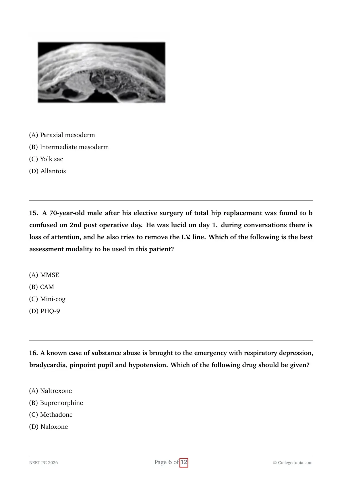
Kidney develops from which of the following parts?

View Solution
Concept:
During the third week of embryonic development, intraembryonic mesoderm on either side of the neural tube and notochord differentiates into three distinct structural zones: paraxial mesoderm, intermediate mesoderm, and lateral plate mesoderm.
Each mesodermal subdivision gives rise to specific organ systems.
Explanation:
Step 1: Mesodermal Subdivisions and Lineages:
$\bullet$ Paraxial Mesoderm: Forms somites, which differentiate into sclerotome (vertebral column, ribs), myotome (skeletal musculature), and dermatome (dermis of trunk).
$\bullet$ Intermediate Mesoderm: Forms the longitudinal nephrogenic cord and urogenital ridge, giving rise to all renal and internal reproductive organs.
$\bullet$ Lateral Plate Mesoderm: Gives rise to the cardiovascular system, body wall connective tissues, and viscera.
Step 2: Dual Origin of the Permanent Kidney:
The definitive kidney (metanephros) develops from two distinct structures derived from intermediate mesoderm:
1. Ureteric Bud: Outgrowth of the mesonephric duct that gives rise to the collecting system (ureter, renal pelvis, major/minor calyces, and collecting tubules).
2. Metanephric Blastema: Condensation of intermediate mesoderm that differentiates into the excretory nephrons (Bowman's capsule, proximal tubule, loop of Henle, and distal convoluted tubule).
Step 3: Conclusion:
Reciprocal induction between the ureteric bud and metanephric blastema creates the functional permanent kidney, originating entirely from intermediate mesoderm.
Final Answer:
The definitive kidneys and urogenital organs develop from the intermediate mesoderm.
$\bullet$ Paraxial Mesoderm: Somites $\rightarrow$ Axial skeleton, striated skeletal muscle, dermis.
$\bullet$ Intermediate Mesoderm: Urogenital system $\rightarrow$ Kidneys, ureters, gonads, internal ducts.
$\bullet$ Lateral Plate Mesoderm: Spleen, cardiovascular system, serous linings, limb bones.
A 70-year-old male after his elective surgery of total hip replacement was found to b confused on 2nd post operative day. He was lucid on day 1. during conversations there is loss of attention, and he also tries to remove the I.V. line. Which of the following is the best assessment modality to be used in this patient?
View Solution
Concept:
Postoperative delirium is an acute, fluctuating organic neuropsychiatric syndrome characterized by impaired attention, altered consciousness, and disorganized thinking following surgery or medical illness.
Standardized, rapid, and validated bedside diagnostic algorithms are required to promptly distinguish acute delirium from baseline chronic dementia.
Explanation:
Step 1: Clinical Recognition of Delirium:
The acute onset on postoperative day 2 following a lucid interval, accompanied by fluctuating inattention, agitation, and disorientation, is characteristic of hyperactive postoperative delirium.
Step 2: The Confusion Assessment Method (CAM):
The CAM algorithm is the most sensitive ($>94\%$) and specific ($>90\%$) validated bedside tool for diagnosing delirium.
A positive CAM diagnosis requires:
1. Acute onset and fluctuating course (Feature 1), AND
2. Inattention (Feature 2), AND
3. EITHER Disorganized thinking (Feature 3) OR Altered level of consciousness (Feature 4).
Step 3: Evaluation of Alternative Scales:
$\bullet$ MMSE and Mini-Cog: Primarily evaluate static cognitive impairment and chronic progressive dementia; they lack sensitivity for acute fluctuating inattention.
$\bullet$ PHQ-9: A depression screening scale, not applicable for acute organic delirium.
Final Answer:
The Confusion Assessment Method (CAM) is the most appropriate and validated assessment tool for diagnosing postoperative delirium.
$\bullet$ Delirium $\rightarrow$ Confusion Assessment Method (CAM / CAM-ICU).
$\bullet$ Dementia $\rightarrow$ MMSE / Mini-Cog / MoCA.
$\bullet$ Depression $\rightarrow$ PHQ-9 / Geriatric Depression Scale.
$\bullet$ Alcohol Withdrawal $\rightarrow$ CIWA-Ar scale.
A known case of substance abuse is brought to the emergency with respiratory depression, bradycardia, pinpoint pupil and hypotension. Which of the following drug should be given?
View Solution
Concept:
Acute opioid poisoning produces life-threatening respiratory depression due to potent agonism at $\mu$-opioid receptors located within the medullary respiratory center.
Emergency management centers on rapid restoration of adequate ventilation and oxygenation via administration of a rapid-acting competitive opioid receptor antagonist.
Explanation:
Step 1: Recognition of Opioid Overdose Triad:
The clinical triad of central nervous system depression (coma), severe respiratory depression ($<8\text{ breaths/min}$), and symmetrical pinpoint pupils (miosis) establishes acute opioid overdose.
Step 2: Pharmacodynamics of Naloxone:
Naloxone is a pure, competitive antagonist at $\mu$, $\kappa$, and $\delta$ opioid receptors with no intrinsic agonist activity.
Administered intravenously, intramuscularly, or intranasally ($0.4\text{--}2\text{ mg}$), it rapidly displaces opioids from receptors, restoring spontaneous respiration and consciousness within $1\text{--}2\text{ minutes}$.
Due to its short half-life ($30\text{--}90\text{ minutes}$), close monitoring is necessary to prevent recurrent hypoventilation from long-acting opioids.
Step 3: Inappropriateness of Other Agents:
$\bullet$ Naltrexone: An orally active, long-acting opioid antagonist used for maintenance therapy in detoxified patients; onset is too slow for acute resuscitation.
$\bullet$ Buprenorphine and Methadone: Opioid receptor agonists used in maintenance replacement therapy, contraindicated in acute overdose.
Final Answer:
Naloxone is the immediate first-line competitive antidote for acute opioid toxicity and life-threatening respiratory depression.
$\bullet$ Acute Overdose / Respiratory arrest $\rightarrow$ IV/Intranasal Naloxone (Rapid acting, short duration).
$\bullet$ Maintenance / Relapse prevention in detoxified patients $\rightarrow$ Oral Naltrexone.
$\bullet$ Classic Triad: Coma + Respiratory depression + Pinpoint pupils.
A 24-year-old male patient with bipolar disorder was taking lithium 900 mg/ day and quetiapine 50mg/day. He started having tremors, dysarthria and ataxia. Which of the following is correct statement related to the patient?
View Solution
Concept:
Lithium carbonate is a narrow therapeutic index mood stabilizer widely used in the treatment of bipolar affective disorder.
Therapeutic plasma levels are narrow ($0.6\text{--}1.2\text{ mEq/L}$), and slight elevations above the therapeutic window result in life-threatening multisystem toxicity.
Neurological manifestations are the most sensitive clinical indicators of escalating lithium blood concentrations.
Explanation:
Step 1: Therapeutic Range vs. Toxic Levels:
Lithium therapeutic concentrations range between $0.6\text{ and }1.2\text{ mEq/L}$.
At therapeutic levels, a fine postural hand tremor is a benign, expected side effect.
When serum levels exceed $1.5\text{ mEq/L}$, neurotoxicity develops.
Step 2: Clinical Grading of Lithium Toxicity:
$\bullet$ Moderate Toxicity ($1.5\text{--}2.0\text{ mEq/L}$): Coarse hand tremors, dysarthria (slurring of speech), hyperreflexia, nausea, vomiting, and muscle weakness.
$\bullet$ Severe Toxicity ($>2.0\text{ mEq/L}$): Gross cerebellar ataxia, myoclonus, seizures, delirium, coma, and acute renal failure.
The triad of coarse tremors, dysarthria, and ataxia confirms moderate-to-severe lithium toxicity.
Step 3: Clinical Management:
Lithium must be stopped immediately, hydration restored with $0.9\%$ saline to promote renal lithium clearance, and hemodialysis considered for levels $>4.0\text{ mEq/L}$ (or $>2.5\text{ mEq/L}$ with severe symptoms).
Quetiapine at $50\text{ mg}$ has minimal $D_2$ occupancy and is not the cause of these symptoms.
Final Answer:
Coarse tremors with dysarthria and ataxia are characteristic manifestations of lithium-induced toxicity.
$\bullet$ Therapeutic range: $0.6\text{--}1.2\text{ mEq/L}$.
$\bullet$ Fine postural hand tremor $\rightarrow$ Benign therapeutic effect (treat with Propranolol).
$\bullet$ Coarse tremor + Dysarthria + Ataxia $\rightarrow$ Lithium Toxicity!
$\bullet$ Toxicity precipitated by: Dehydration, Thiazides, NSAIDs, ACE inhibitors.
A 25-year-old male is presents with long standing pattern of social withdrawal. He has odd and eccentric behavior and hold some peculiar beliefs. Which of the following personality traits are seen here?
View Solution
Concept:
Personality disorders are categorized into three distinct clusters: Cluster A ("mad" / odd, eccentric), Cluster B ("bad" / dramatic, emotional), and Cluster C ("sad" / anxious, fearful).
Cluster A includes Paranoid, Schizoid, and Schizotypal personality disorders, which are distinguished primarily by cognitive distortions and patterns of social engagement.
Explanation:
Step 1: Clinical Features of Schizotypal Personality Disorder:
Schizotypal personality disorder (Cluster A) is characterized by pervasive social and interpersonal deficits, reduced capacity for close relationships, combined with cognitive or perceptual distortions and eccentricities of behavior.
Patients display magical thinking (e.g., belief in telepathy, clairvoyance), ideas of reference, odd speech, paranoid ideation, and eccentric grooming.
Step 2: Comparison with Schizoid Personality Disorder:
Patients with Schizoid personality disorder prefer solitary activities, show emotional detachment and flattened affect, but completely lack magical thinking, eccentric behavior, or perceptual distortions.
Step 3: Comparison with Other Clusters:
$\bullet$ Avoidant (Cluster C): Desires social relationships but avoids them due to intense fear of rejection or inadequacy.
$\bullet$ Antisocial (Cluster B): Disregard for the law, violation of rights of others, deceitfulness, and lack of remorse.
Final Answer:
The combination of social withdrawal with magical thinking, eccentric behavior, and odd beliefs is diagnostic of Schizotypal Personality Disorder.
$\bullet$ Schizoid $\rightarrow$ Socially distant, prefers solitude, flat affect, NO magical thinking.
$\bullet$ Schizotypal $\rightarrow$ Magical thinking, odd beliefs, eccentric dress/habits ("Schizo-type").
$\bullet$ Paranoid $\rightarrow$ Pervasive mistrust, hypersensitivity, interprets actions as malicious.
An adult male patient suffered head injury and during recovery, the patient was drinking 8liters of water per day, with intense third and make large dilute urine. There is no diabetes mellitus. Which hypothalamic nucleus and descending fibers are damaged?
View Solution
Concept:
Central (neurogenic) diabetes insipidus (DI) is caused by a deficiency in the synthesis, axonal transport, or release of Antidiuretic Hormone (ADH / Arginine Vasopressin).
ADH is synthesized within specialized magnocellular neurosecretory cells of specific hypothalamic nuclei and transported down the hypothalamohypophysial tract to the neurohypophysis.
Explanation:
Step 1: Synthesis and Axonal Transport of ADH:
ADH (arginine vasopressin) is synthesized by the magnocellular neurons located primarily in the supraoptic nucleus and paraventricular nucleus of the hypothalamus.
The synthesized hormone is packaged with neurophysin II and transported axonally down the hypothalamohypophysial tract to the posterior pituitary (neurohypophysis) for vascular release.
Step 2: Pathophysiology of Central Diabetes Insipidus:
Head injury, basal skull fractures, or pituitary stalk transection damage the supraoptic/paraventricular nuclei or disrupt axonal transport.
Loss of circulating ADH prevents the insertion of Aquaporin-2 channels into the apical membrane of principal cells in renal medullary collecting ducts.
Consequently, free water cannot be reabsorbed, producing profound polyuria ($>3\text{ L/day}$ of dilute urine, urine osmolality $<200\text{ mOsm/kg}$) and intense compensatory polydipsia.
Step 3: Normal Glycemia:
Normal fasting glucose excludes osmotic diuresis due to uncontrolled diabetes mellitus.
Final Answer:
ADH is synthesized in the supraoptic and paraventricular nuclei of the hypothalamus; trauma to these structures produces central diabetes insipidus.
$\bullet$ Supraoptic + Paraventricular $\rightarrow$ ADH and Oxytocin synthesis.
$\bullet$ Suprachiasmatic $\rightarrow$ Circadian rhythm / Biological clock.
$\bullet$ Ventromedial $\rightarrow$ Satiety center (destruction $\rightarrow$ Hyperphagia / Obesity).
$\bullet$ Lateral $\rightarrow$ Hunger center (destruction $\rightarrow$ Anorexia / Wasting).
An adult women is investigated for mild weakness, fatigue and occasional carpopedal spasm. Laboratory studies reveals, hypokalemia, metabolic alkalosis and high calcium in the urine. In which part of the nephron the defective channel is present?
View Solution
Concept:
Hereditary hypokalemic metabolic alkaloses are inherited renal tubulopathies classified according to the affected nephron segment and urinary calcium excretion.
The clinical and biochemical findings in this patient are diagnostic of Bartter syndrome.
Explanation:
Step 1: Ion Transport in the Thick Ascending Limb (TAL):
In the TAL of Henle's loop, the apical $Na^+\text{-}K^+\text{-}2Cl^-$ cotransporter (NKCC2) reabsorbs sodium, potassium, and chloride.
Potassium recycling via apical ROMK channels creates a lumen-positive electrical potential that drives paracellular reabsorption of calcium and magnesium.
Step 2: Pathophysiology of Bartter Syndrome:
Bartter syndrome is caused by mutations in the NKCC2 cotransporter, ROMK, or basolateral chloride channels (ClC-Kb/Barttin) in the Thick Ascending Limb, mimicking chronic loop diuretic administration.
Loss of the lumen-positive potential impairs calcium reabsorption, resulting in massive hypercalciuria and nephrocalcinosis.
Excessive solute delivery to the distal nephron stimulates renin and aldosterone, driving potassium and hydrogen ion excretion (hypokalemic metabolic alkalosis).
Step 3: Comparison with Gitelman Syndrome:
Gitelman syndrome is caused by mutations in the $Na^+\text{-}Cl^-$ cotransporter (NCCT) in the Distal Convoluted Tubule (DCT), which characteristically produces hypocalciuria (low urinary calcium).
Final Answer:
The clinical triad of hypokalemia, metabolic alkalosis, and hypercalciuria indicates a defect in the Thick Ascending Limb (TAL) of the Loop of Henle (Bartter syndrome).
$\bullet$ Bartter Syndrome $\rightarrow$ Thick Ascending Limb (NKCC2 defect) $\rightarrow$ Like Loop Diuretics $\rightarrow$ Hypercalciuria.
$\bullet$ Gitelman Syndrome $\rightarrow$ Distal Convoluted Tubule (NCCT defect) $\rightarrow$ Like Thiazides $\rightarrow$ Hypocalciuria.
A 28-year-old man presents with a painless testicular mass. On gross examination, the tumor is well-circumscribed, homogeneous, and gray-white, with a soft, fleshy appearance and minimal hemorrhage or necrosis. Histological examination shows sheets and nests of uniform polygonal cells with abundant clear cytoplasm containing glycogen and prominent nucleoli, separated by delicate fibrous septa infiltrated by lymphocytes. Immunohistochemistry shows tumor cells that are positive for CD117 (c-KIT) and OCT3/4. What is the most likely diagnosis?
View Solution
Concept:
Testicular germ cell tumors are broadly classified into seminomatous and non-seminomatous germ cell tumors (NSGCT).
Classic seminoma is the most common single pure germ cell tumor of the testis, typically presenting in young males in the third and fourth decades of life.
The diagnosis is confirmed through characteristic gross pathology, histology, and immunohistochemical profile.
Explanation:
Step 1: Gross Pathological Features:
Seminomas characteristically present as well-circumscribed, uniform, lobulated, fleshy, gray-white to tan masses.
In sharp contrast to non-seminomatous tumors, seminomas typically lack gross hemorrhage, necrosis, or cystic spaces.
Step 2: Histopathological Hallmarks:
$\bullet$ Microscopy: Shows uniform, large, polygonal cells arranged in sheets and cords.
$\bullet$ Cells possess distinct borders, clear glycogen-rich cytoplasm (PAS-positive), centrally placed hyperchromatic nuclei, and prominent nucleoli.
$\bullet$ The tumor nests are separated by thin fibrovascular septa containing a dense host T-lymphocytic infiltrate.
Step 3: Immunohistochemistry (IHC) Profile:
Seminoma cells are positive for CD117 (c-KIT), OCT3/4, SALL4, and PLAP, while negative for CD30 (marker for embryonal carcinoma) and AFP (marker for yolk sac tumor).
Final Answer:
The gross appearance and microscopic features of clear cells with lymphocytic septa and CD117/OCT3/4 positivity are diagnostic of classic Seminoma.
$\bullet$ Gross: Homogeneous, tan-white fleshy mass WITHOUT hemorrhage/necrosis.
$\bullet$ Micro: Clear glycogenated cells + Fibrous septa with T-lymphocytes.
$\bullet$ IHC: CD117 (c-KIT) positive, OCT3/4 positive, SALL4 positive.
$\bullet$ Highly radiosensitive and chemosensitive!
A 65-year-old chronic smoker presents with cough, weight loss, and progressive dyspnea. Chest imaging reveals a centrally located hilar lung mass. Histological examination shows sheets of small round blue cells with scant cytoplasm, hyperchromatic nuclei, a high nuclear-to-cytoplasmic ratio, nuclear molding, frequent mitotic figures, and areas of necrosis. Immunohistochemistry shows tumor cells that are positive for synaptophysin. What is the most likely diagnosis?
View Solution
Concept:
Primary lung malignancies are classified into Small Cell Lung Carcinoma (SCLC) and Non-Small Cell Lung Carcinomas (NSCLC).
SCLC is a high-grade, aggressive neuroendocrine neoplasm strongly linked to heavy cigarette smoking that arises centrally from pulmonary neuroendocrine (Kulchitsky) cells.
Explanation:
Step 1: Clinical and Anatomical Presentation:
SCLC typically occurs in older smokers, presenting centrally as a perihilar or hilar mass with rapid bronchial obstruction and early mediastinal lymphadenopathy.
Step 2: Microscopic Features:
$\bullet$ Small round, oval, or spindle-shaped cells with scant cytoplasm (high N:C ratio).
$\bullet$ Finely dispersed, granular "salt-and-pepper" chromatin with inconspicuous or absent nucleoli.
$\bullet$ Nuclear molding: Adjacent nuclei indent one another due to cell crowding and lack of intervening cytoplasm.
$\bullet$ High mitotic rate ($>10$ per HPF), extensive tumor necrosis, and crush artifact with encrustation of DNA into blood vessel walls (Azzopardi effect).
Step 3: Neuroendocrine Differentiation:
Tumor cells show strong immunohistochemical reactivity for neuroendocrine markers: Synaptophysin, Chromogranin A, CD56 (NCAM), and TTF-1.
Final Answer:
The small round blue cell morphology, nuclear molding, and neuroendocrine marker positivity confirm Small Cell Lung Carcinoma.
$\bullet$ Central = Small cell carcinoma and Squamous cell carcinoma (Strongly linked to Smoking).
$\bullet$ SCLC Features: Nuclear molding + Azzopardi effect + Small blue cells.
$\bullet$ Neuroendocrine Markers: Synaptophysin, Chromogranin, CD56.
A 45-year-old woman presents with a rapidly enlarging breast mass that is approximately twice the size of the opposite breast and has an irregular, nodular surface on gross examination. Histology shows a leaf-like pattern with increased stromal cellularity and stromal atypia. However, the mitotic index is low, with no significant stromal overgrowth or tumor necrosis. Which of the following is the most likely diagnosis?
View Solution
Concept:
Phyllodes tumors (cystosarcoma phyllodes) are uncommon biphasic fibroepithelial neoplasms of the breast derived from the intralobular specialized stroma.
They typically occur in women in their fourth to fifth decades ($40\text{--}50$ years), presenting as large, rapidly expanding breast masses that can clinically mimic giant fibroadenomas.
Explanation:
Step 1: Gross and Microscopic Architecture:
Phyllodes tumors present as large, fleshy, multinodular, lobulated masses with cystic spaces and clefts.
Microscopically, they are characterized by stromal hypercellularity forming broad, leaf-like papillary projections that protrude into epithelial-lined clefts (the term phyllon means "leaf").
Step 2: Histological Grading of Phyllodes Tumors:
The WHO grades phyllodes tumors into benign, borderline, and malignant based on stromal cellularity, cellular atypia, mitotic activity, margin status, and stromal overgrowth:
$\bullet$ Benign: Pushing margins, mild stromal cellularity, minimal nuclear atypia, and $<5$ mitoses per 10 HPF.
$\bullet$ Borderline: Moderate cellularity/atypia, 5--9 mitoses per 10 HPF.
$\bullet$ Malignant: Marked atypia, infiltrative margins, stromal overgrowth, $\ge 10$ mitoses per 10 HPF.
Step 3: Clinical Management:
Because of a low mitotic count ($<5/10$ HPF) and pushing borders, this case represents a benign phyllodes tumor.
Standard surgical management is wide local excision with $\ge 1\text{ cm}$ clear margins.
Final Answer:
A rapidly growing breast tumor with characteristic leaf-like stromal architecture is diagnostic of a Phyllodes tumor.
$\bullet$ Fibroadenoma: Young women ($20\text{--}30\text{ yrs}$), uniform bland stroma.
$\bullet$ Phyllodes Tumor: Older women ($40\text{--}50\text{ yrs}$), rapidly growing + Leaflike stromal projections + Stromal hypercellularity.
$\bullet$ Treatment: Wide local excision with $\ge 1\text{ cm}$ margins.
Calculate the GCS score of the patient speaking inappropriate words, doesn't open eyes to painful stimulus and there is withdrawal to pain:
View Solution
Concept:
The Glasgow Coma Scale (GCS) is a standardized scoring system used to objectively assess the level of consciousness and neurological impairment in patients with acute brain injury or trauma.
The scale scores three separate neurological domains: Eye Opening (E), Verbal Response (V), and Motor Response (M).
Explanation:
Step 1: Formula for GCS Calculation:
The overall GCS score is obtained by summing the scores of the three individual components:
\[ \text{Total GCS} = \text{Eye Opening (E)} + \text{Verbal Response (V)} + \text{Motor Response (M)} \]
Step 2: Scoring Individual Components:
$\bullet$ Eye Opening (E) (Range 1--4): No eye opening to pain = 1.
$\bullet$ Verbal Response (V) (Range 1--5): Speaks inappropriate words = 3.
$\bullet$ Motor Response (M) (Range 1--6): Normal flexion / withdrawal to pain = 4.
Step 3: Total Score Computation and Clinical Significance:
\[ \text{Total GCS} = 1 (\text{E}) + 3 (\text{V}) + 4 (\text{M}) = 8 \] A GCS score $\le 8$ signifies severe traumatic brain injury and mandates immediate airway protection via endotracheal intubation.
Final Answer:
The patient's total Glasgow Coma Scale score is $1 + 3 + 4 = 8$.
$\bullet$ E (4): 4-Spontaneous, 3-Voice, 2-Pain, 1-None.
$\bullet$ V (5): 5-Oriented, 4-Confused, 3-Inappropriate, 2-Incomprehensible, 1-None.
$\bullet$ M (6): 6-Obeys, 5-Localizes, 4-Withdraws, 3-Decorticate, 2-Decerebrate, 1-None.
$\bullet$ Severity: Severe TBI $\le 8$; Moderate TBI $9\text{--}12$; Mild TBI $13\text{--}15$.
An 65 old age man + Chronic Heart failure (HFrEF) + symptoms despite taking medication + now added Vericiguat + Mechanism by which Vericiguat acts ?
View Solution
Concept:
In heart failure with reduced ejection fraction (HFrEF), endothelial dysfunction and elevated oxidative stress deplete endogenous bioavailable Nitric Oxide (NO).
This reduces soluble Guanylyl Cyclase (sGC) activity and depletes cyclic guanosine monophosphate (cGMP), leading to myocardial stiffness, ventricular hypertrophy, coronary microvascular dysfunction, and systemic vasoconstriction.
Vericiguat is a first-in-class oral agent designed to directly restore the NO-sGC-cGMP signaling pathway.
Explanation:
Step 1: Direct Stimulation of Soluble Guanylyl Cyclase:
Vericiguat directly stimulates soluble guanylyl cyclase (sGC) by binding to a distinct non-heme catalytic site, generating cGMP independently of endogenous nitric oxide (NO).
Simultaneously, it enhances sGC sensitivity to residual endogenous NO by stabilizing the active NO-sGC binding conformation.
Step 2: Downstream Cellular Effects:
Increased intracellular cGMP activates protein kinase G (PKG), which induces:
$\bullet$ Systemic and pulmonary arterial vasodilation, reducing left ventricular afterload.
$\bullet$ Improved myocardial relaxation and compliance, reducing ventricular stiffness.
$\bullet$ Attenuation of pathological myocardial remodeling and interstitial fibrosis.
Step 3: Mechanism of Other Heart Failure Agents:
$\bullet$ PDE-5 inhibitors (e.g., Sildenafil): Inhibit cGMP breakdown rather than stimulating its synthesis.
$\bullet$ Neprilysin inhibitors (e.g., Sacubitril): Inhibit degradation of natriuretic peptides (ANP, BNP).
Final Answer:
Vericiguat directly stimulates soluble guanylyl cyclase (sGC), thereby enhancing intracellular cGMP generation.
$\bullet$ Vericiguat: Direct Soluble Guanylyl Cyclase (sGC) stimulator $\rightarrow$ $\uparrow$ cGMP.
$\bullet$ Sacubitril: Neprilysin inhibitor $\rightarrow$ $\uparrow$ Natriuretic peptides (BNP, ANP).
$\bullet$ Ivabradine: SA node $I_f$ (funny channel) blocker $\rightarrow$ slows heart rate.
$\bullet$ Dapagliflozin / Empagliflozin: SGLT2 inhibitors $\rightarrow$ renal glycosuria + osmotic diuresis.
A tumor cell does not express MHC class I molecules. Despite the presence of tumor-associated antigens, cytotoxic T lymphocytes are unable to recognize the tumor cell. What is the most likely mechanism by which the tumor escapes immune surveillance?
View Solution
Concept:
Cytotoxic T lymphocytes ($\text{CD8}^+$ CTLs) play a pivotal role in antitumor immunity by recognizing endogenous intracellular tumor-associated peptide antigens presented on the surface of tumor cells.
This antigen recognition is strictly restricted by Major Histocompatibility Complex (MHC) class I molecules (HLA-A, HLA-B, and HLA-C in humans).
Downregulation or complete loss of MHC class I expression is a classic immune evasion mechanism employed by evolving malignant clones.
Explanation:
Step 1: Mechanism of Cytotoxic T-Cell Recognition:
$\text{CD8}^+$ cytotoxic T lymphocytes utilize their T-cell receptor (TCR) along with the CD8 coreceptor to bind antigen peptides only when displayed within the groove of MHC class I molecules.
Endogenous neoantigens produced by mutated tumor cells are processed by the proteasome, transported into the endoplasmic reticulum via TAP (Transporter associated with Antigen Processing), and loaded onto newly synthesized MHC class I heavy chains complexed with $\beta_2$-microglobulin.
Step 2: Immune Evasion via MHC Class I Downregulation:
Malignant tumors often acquire genetic mutations (such as loss-of-function mutations in $\beta_2$-microglobulin or epigenetic silencing of HLA genes) that result in a complete absence of surface MHC class I expression.
Without MHC class I molecules, intracellular tumor-associated antigens cannot be presented on the cell membrane.
Consequently, $\text{CD8}^+$ cytotoxic T cells fail to recognize the tumor cell, allowing the malignancy to evade adaptive T-cell-mediated cytotoxicity.
Step 3: Role of Natural Killer (NK) Cells:
Although lack of MHC class I prevents $\text{CD8}^+$ CTL killing ("missing self"), it normally triggers Natural Killer (NK) cell activation unless the tumor develops additional immunosuppressive defenses.
Tumors do not evade CTL surveillance through antibody production against lymphocytes or excessive complement synthesis.
Final Answer:
The inability of cytotoxic T lymphocytes to recognize the tumor cell is directly caused by the failure of antigen presentation through MHC class I molecules.
$\bullet$ $\text{CD8}^+$ Cytotoxic T cells recognize peptides on MHC Class I molecules.
$\bullet$ $\text{CD4}^+$ Helper T cells recognize peptides on MHC Class II molecules.
$\bullet$ Loss of MHC Class I $\rightarrow$ Tumor escapes $\text{CD8}^+$ T cell killing (recognized instead by NK cells via the ``missing-self'' hypothesis).
A 6-year-old male with history of anal sexual assault is brought to hospital. Child presents with pain on defecation. Anal area is found normal on examination. The most appropriate method of examining passive agent in anal intercourse case is
View Solution
Concept:
In forensic medical jurisprudence, the clinical examination of a victim (passive agent) of suspected anal sexual assault or sodomy requires gentle inspection for both physical injuries and functional changes in sphincter tone.
Reflex anal dilatation (RAD) and relaxation of the external and internal anal sphincters on buttock separation are cardinal diagnostic signs of anal penetration.
Explanation:
Step 1: Clinical Approach to Suspected Anal Penetration:
Acute anal assault often causes superficial mucosal tears, perianal contusions, or painful fissures.
However, when the perianal skin appears superficially normal on initial inspection, evaluating sphincter function and dynamic reflex responses is crucial for forensic documentation.
Step 2: Buttock Separation and Reflex Anal Dilatation (RAD):
The standard forensic examination involves placing the patient in the lateral prone or knee-chest position and gently spreading/separating the buttocks laterally and anteriorly.
In a normal child, buttock separation triggers a transient reflex contraction of the external anal sphincter.
In cases of habituated or recent anal penetration, lateral traction on the buttocks reveals abnormal patency, effacement of anal radiating rugae, and prolonged reflex relaxation or dilatation of the anal orifice ($>10\text{--}20\text{ mm}$ without stool present).
Step 3: Evaluation of Other Methods:
$\bullet$ Invasive proctoscopic examination without anesthesia can worsen acute physical trauma and psychological distress in a pediatric victim.
$\bullet$ Routine lithotomy or isolated positioning without gentle dynamic buttock separation fails to reveal characteristic sphincter relaxation and funnel-shaped anal deformity.
Final Answer:
The most appropriate method of examining a passive agent in suspected anal intercourse is gentle lateral separation of the buttocks to assess for abnormal relaxation and reflex dilatation of the anus.
$\bullet$ Reflex Anal Dilatation (RAD): Abnormal opening/relaxation of anus upon lateral buttock traction.
$\bullet$ Chronic signs: Funnel-shaped anus, loss of radiating perianal folds/rugae, patulous sphincter.
$\bullet$ Preferred position for pediatric forensic exam: Left lateral (Sims') or Knee-chest position.
In Cholera diarrhoea, there is increased activity of which channel is primarily responsible for the increased watery intestinal secretion?
View Solution
Concept:
Vibrio cholerae produces an enterotoxin (cholera toxin, CTX) that causes profound secretory diarrhea ("rice-water stools") by permanently upregulating cyclic AMP-dependent apical ion channels in intestinal crypt enterocytes.
The massive luminal efflux of electrolytes creates an osmotic gradient that drives huge volumes of water into the bowel lumen.
Explanation:
Step 1: Molecular Mechanism of Cholera Toxin:
Cholera toxin consists of an $A\text{--}B_5$ subunit complex.
The $B$ subunits bind to the $GM_1$ ganglioside receptor on the apical membrane of intestinal mucosal cells, facilitating endocytosis of the active $A_1$ subunit.
The $A_1$ subunit catalyzes the ADP-ribosylation of the $\alpha$ subunit of the stimulatory $G$-protein ($G\alpha_s$), locking it in an irreversibly active, GTP-bound state.
Step 2: CFTR Chloride Channel Activation:
Persistent $G\alpha_s$ activation stimulates adenylate cyclase, resulting in massive intracellular accumulation of cyclic AMP (cAMP).
Elevated cAMP activates Protein Kinase A (PKA), which phosphorylates and constitutively opens the Cystic Fibrosis Transmembrane Conductance Regulator (CFTR) Chloride ($\text{Cl}^-$) channel on the apical luminal surface of intestinal crypt cells.
Step 3: Secretory Diarrhea Cascade:
Massive secretion of chloride ions ($\text{Cl}^-$) into the intestinal lumen creates a negative electrical potential that draws sodium ($\text{Na}^+$) and water paracellularly into the lumen, resulting in severe, dehydrating, isotonic secretory diarrhea.
Final Answer:
Increased activity of apical Chloride ($\text{Cl}^-$) channels (CFTR) is primarily responsible for the voluminous watery intestinal secretion in cholera.
Cholera Toxin $\rightarrow$ ADP-ribosylation of $G_s\alpha$ $\rightarrow$ $\uparrow$ Adenylate Cyclase $\rightarrow$ $\uparrow$ cAMP $\rightarrow$ Opens CFTR $\text{Cl}^-$ Channel $\rightarrow$ Massive luminal $\text{Cl}^-$, $\text{Na}^+$, and $\text{H}_2\text{O}$ loss.
$\bullet$ Treatment: Oral Rehydration Salts (ORS) utilizes intact $\text{Na}^+$-Glucose cotransporters (SGLT1).
A patient with overactive bladder is prescribed Merabagron suspended release tablet 25 mg OD. What is the mechanism of action of this drug?
View Solution
Concept:
Overactive Bladder (OAB) syndrome is characterized by urinary urgency, with or without urgency urinary incontinence, usually accompanied by urinary frequency and nocturia.
Pharmacotherapy aims to suppress involuntary detrusor contractions during the bladder filling (storage) phase.
Mirabegron represents a non-anticholinergic drug class designed to promote detrusor relaxation.
Explanation:
Step 1: Adrenergic Control of Micturition:
During the urine storage phase, sympathetic nerve fibers release norepinephrine, which acts on $\beta_3$-adrenergic receptors located throughout the human detrusor muscle.
Activation of $\beta_3$ receptors stimulates adenylate cyclase, increasing intracellular cyclic AMP (cAMP) and causing detrusor smooth muscle relaxation, which expands bladder storage capacity without increasing post-void residual volume.
Step 2: Mechanism of Action of Mirabegron:
Mirabegron (Merabagron) is a potent and selective $\beta_3$-adrenergic receptor agonist.
By stimulating $\beta_3$ receptors in the bladder wall, it relaxes the detrusor muscle during the storage phase, reducing the frequency of unstable detrusor contractions and increasing bladder storage capacity.
Step 3: Clinical Advantage Over Antimuscarinics:
Unlike traditional $M_3$ receptor antagonists (e.g., Oxybutynin, Tolterodine, Solifenacin), Mirabegron does not block parasympathetic cholinergic receptors.
Consequently, it avoids common antimuscarinic adverse effects such as dry mouth, constipation, blurred vision, and cognitive impairment in elderly individuals.
Its primary adverse effect is a mild increase in blood pressure and heart rate.
Final Answer:
Mirabegron functions as a selective Beta 3 receptor agonist to treat overactive bladder syndrome.
$\bullet$ Mirabegron / Vibegron: $\beta_3$-Adrenergic Agonist $\rightarrow$ Relaxes detrusor (No dry mouth).
$\bullet$ Solifenacin / Darifenacin: Selective $M_3$ Anticholinergic $\rightarrow$ Inhibits detrusor contraction.
$\bullet$ Botulinum Toxin A: Inhibits acetylcholine release at the neuromuscular junction.
A patient suffering from Giardia is resistant to Metronidazole therapy. What's the drug which can be used?
View Solution
Concept:
Giardiasis is a flagellated protozoan infection caused by Giardia lamblia (Giardia duodenalis), leading to malabsorptive watery/fatty diarrhea, abdominal bloating, and flatulence.
While Metronidazole and Tinidazole are standard first-line nitroimidazole therapies, treatment failures and clinical resistance occur, requiring alternative broad-spectrum antiprotozoal agents with different mechanisms of action.
Explanation:
Step 1: First-Line Agents and Resistance:
Metronidazole is a 5-nitroimidazole that requires reductive activation by pyruvate:ferredoxin oxidoreductase (PFOR) to generate toxic nitro radicals.
Downregulation of PFOR or altered ferredoxin oxidoreductase activity in Giardia trophozoites produces clinical resistance to metronidazole.
Step 2: Role and Mechanism of Nitazoxanide:
Nitazoxanide is a synthetic nitrothiazolyl-salicylamide derivative approved as a highly effective alternative for metronidazole-refractory giardiasis.
It acts by non-competitively inhibiting the pyruvate:ferredoxin oxidoreductase (PFOR) enzyme-dependent electron transfer reaction essential for anaerobic energy metabolism in the parasite.
Because its binding profile differs from nitroimidazoles, it retains high cure rates ($>85\text{--}90\%$) against metronidazole-resistant strains of Giardia lamblia and is also active against Cryptosporidium parvum.
Step 3: Evaluation of Other Options:
$\bullet$ Nifurtimox: Used for American trypanosomiasis (Chagas disease) and African trypanosomiasis.
$\bullet$ Miltefosine: Oral agent used for visceral and cutaneous leishmaniasis.
$\bullet$ Paromomycin: An aminoglycoside luminal amoebicide, less effective for refractory giardiasis.
Final Answer:
Nitazoxanide is the drug of choice for the treatment of Metronidazole-resistant Giardiasis.
$\bullet$ Drug of Choice: Tinidazole (single dose) or Metronidazole ($5\text{--}7\text{ days}$).
$\bullet$ Metronidazole-Resistant Giardia: Nitazoxanide, Albendazole, Quinacrine, or Combination therapy.
$\bullet$ Giardiasis in Pregnancy (1st trimester): Paromomycin (non-absorbable luminal agent).
Patient got 95% vitiligo he wants to have same colour through-out the skin. What is best management:
View Solution
Concept:
Vitiligo is an acquired autoimmune depigmenting disorder of the skin characterized by the progressive loss of functional melanocytes.
The therapeutic strategy is dictated by the total body surface area (BSA) involved: repigmentation therapy for localized/moderate disease vs. permanent depigmentation therapy for universal/extensive vitiligo.
Explanation:
Step 1: Clinical Rationale for Depigmentation Therapy:
When vitiligo involves $>80\text{--}90\%$ of total body surface area (Vitiligo Universalis) or when extensive patches fail repigmentation therapies, attempting to repigment the depigmented areas becomes impractical and cosmetically unsatisfactory.
In such patients seeking uniform skin coloring, depigmentation therapy is the treatment of choice to permanently remove residual islands of normally pigmented skin.
Step 2: Agent of Choice -- MBEH:
Monobenzyl ether of hydroquinone (MBEH / Monobenzone, 20\% cream) is the definitive medical agent approved for permanent depigmentation in extensive vitiligo.
MBEH induces selective, irreversible, cytotoxic destruction of remaining melanocytes through free-radical generation and immune-mediated clearance.
Topical application twice daily for $6\text{--}12\text{ months}$ produces permanent, uniform depigmentation matching the surrounding vitiliginous skin.
Step 3: Limitations of Other Modalities:
$\bullet$ Narrowband UVB (NBUVB) therapy and oral corticosteroids are intended for repigmentation in limited disease ($<20\text{--}50\%$ BSA) and have negligible efficacy in $95\%$ universal vitiligo.
$\bullet$ Standard Hydroquinone ($2\text{--}4\%$) produces only temporary, reversible inhibition of tyrosinase (used in melasma) and does not cause permanent melanocyte destruction.
Final Answer:
In a patient with 95\% extensive vitiligo desiring uniform skin color, the best management is permanent depigmentation using Monobenzyl ether of hydroquinone (MBEH).
$\bullet$ Localized ($<10\text{--}20\%$ BSA) $\rightarrow$ Topical Corticosteroids / Tacrolimus.
$\bullet$ Generalized ($20\text{--}80\%$ BSA) $\rightarrow$ Narrowband UVB (NB-UVB) phototherapy.
$\bullet$ Universal / Extensive ($>80\text{--}90\%$ BSA) $\rightarrow$ Permanent Depigmentation with Monobenzyl ether of hydroquinone (MBEH / Monobenzone).
A patient with sepsis develops fever, a rapidly spreading purpuric rash, profound hypotension, hyponatremia, hyperkalemia and shock. Imaging demonstrates bilateral adrenal hemorrhage. What is the most likely diagnosis?
View Solution
Concept:
Waterhouse–Friderichsen syndrome (fulminant meningococcemia / hemorrhagic adrenalitis) is a catastrophic complication of severe bacterial septicemia characterized by disseminated purpura, cardiovascular collapse, and acute primary adrenal insufficiency secondary to bilateral adrenal cortical hemorrhage.
Explanation:
Step 1: Clinical Presentation and Pathogenesis:
The classic triad comprises:
1. Severe sepsis (most commonly caused by Neisseria meningitidis, followed by Pseudomonas aeruginosa, Streptococcus pneumoniae, or Haemophilus influenzae).
2. Rapidly spreading petechial and coalescent purpuric rash (purpura fulminans) due to disseminated intravascular coagulation (DIC).
3. Refractory vasodilatory and cardiogenic septic shock.
Step 2: Acute Adrenal Crisis Mechanics:
Endotoxin release and severe microvascular thrombosis trigger widespread cortical necrosis and bilateral massive hemorrhage into both adrenal glands.
Destruction of the adrenal cortex leads to acute loss of cortisol and aldosterone.
Loss of aldosterone causes renal sodium wasting, potassium retention, and profound vascular collapse, manifesting as severe hyponatremia, hyperkalemia, and refractory hypotension.
Step 3: Comparison with Other Conditions:
$\bullet$ Addison's disease: Chronic autoimmune primary adrenal insufficiency that develops indolently over months without acute purpura fulminans or septic bilateral adrenal hemorrhage.
$\bullet$ Cushing syndrome: Characterized by chronic cortisol excess (hypertension, hypernatremia, hypokalemia).
$\bullet$ Primary hyperaldosteronism: Features hypertension, hypernatremia, and hypokalemia without shock.
Final Answer:
The presentation of severe sepsis, purpuric rash, shock, and imaging-confirmed bilateral adrenal hemorrhage is classic for Waterhouse–Friderichsen syndrome.
$\bullet$ Etiology: Fulminant Neisseria meningitidis septicemia.
$\bullet$ Pathology: Severe DIC + Bilateral Adrenal Hemorrhage.
$\bullet$ Clinical: Purpura fulminans + Refractory Shock + Hyponatremia + Hyperkalemia.
$\bullet$ Urgent Treatment: IV Ceftriaxone + Stress-dose IV Hydrocortisone + Aggressive fluid resuscitation.
Several students from the same school develop severe abdominal cramps and bloody diarrhea. Shiga toxin-producing Escherichia coli (STEC/EHEC) is suspected. Which reference method is considered the gold standard for diagnosis?
View Solution
Concept:
Enterohemorrhagic Escherichia coli (EHEC / STEC), notably serotype O157:H7, causes severe hemorrhagic colitis and hemolytic uremic syndrome (HUS) through the production of Shiga toxins (Stx1 and Stx2, also known as Verotoxins).
Accurate confirmation requires sensitive detection of functional toxin production.
Explanation:
Step 1: Pathophysiology of Shiga Toxins (Verotoxins):
Shiga toxins produced by STEC inhibit eukaryotic protein synthesis by cleaving adenine from 28S rRNA within the 60S ribosomal subunit, causing apoptotic cell death of microvascular endothelial cells.
Because these toxins produce profound, lethal cytotoxic effects on African green monkey kidney cells (Vero cells), they are also called Verotoxins.
Step 2: Gold Standard Reference Method:
The Vero-cell cytotoxicity neutralization assay (Vero-cell culture assay) is the definitive gold standard reference method for diagnosing STEC/EHEC.
In this assay, stool filtrates or bacterial culture supernatants are inoculated onto a Vero-cell monolayer.
The presence of functional toxin induces characteristic irreversible cytotoxic cytopathic changes (cell rounding, detaching, and death), which are specifically neutralized and confirmed using standard specific anti-Stx1 and anti-Stx2 neutralizing antibodies.
Step 3: Role of Other Diagnostic Modalities:
$\bullet$ Sorbitol MacConkey (SMAC) agar: A convenient phenotypic screening agar (EHEC O157:H7 is sorbitol non-fermenting, appearing colorless), but it misses non-O157 STEC strains.
$\bullet$ Enzyme-Linked Immunosorbent Assay (ELISA): A rapid immunoassay widely used in routine laboratories, but Vero-cell cytotoxicity remains the reference gold standard.
$\bullet$ Voges-Proskauer test: A biochemical test for acetoin production, unrelated to toxin detection.
Final Answer:
The Vero-cell cytotoxicity culture assay is the established gold standard reference method for detecting Shiga toxin-producing E. coli.
$\bullet$ Gold Standard Reference Test: Vero-cell cytotoxicity neutralization assay.
$\bullet$ Routine Clinical Screening: Sorbitol MacConkey (SMAC) agar (O157:H7 forms colorless/white colonies).
$\bullet$ Major Complication: Hemolytic Uremic Syndrome (HUS: Microangiopathic hemolytic anemia + Thrombocytopenia + Acute Renal Failure).
$\bullet$ Contraindicated: Antibiotics (may increase Shiga toxin release and worsen HUS risk).
A patient presented with muscle cramps. ECG showed ST depression and QT prolongation. Serum Potassium 2.8 meq/L. IV Potassium chloride was given. But there was no improvement in serum potassium over time. What can explain this?
View Solution
Concept:
Refractory hypokalemia is a frequent clinical challenge in electrolyte management.
Potassium homeostasis is biochemically coupled to magnesium levels because intracellular magnesium serves as a critical allosteric regulator of renal potassium transport channels.
Explanation:
Step 1: Physiological Role of Magnesium in Renal Potassium Handling:
Under normal physiological conditions, intracellular magnesium ($\text{Mg}^{2+}$) exerts an inhibitory brake on the Renal Outer Medullary Potassium (ROMK) channels located in the apical membrane of principal cells in the cortical collecting duct.
Intracellular magnesium blocks the pore of the ROMK channel from the cytoplasmic side, limiting excessive potassium excretion into the tubular lumen.
Step 2: Pathophysiology of Refractory Hypokalemia in Hypomagnesemia:
When a patient develops hypomagnesemia, intracellular magnesium concentrations drop, releasing the physiological inhibition on ROMK channels.
Uninhibited, open ROMK channels permit rapid, unregulated secretional leakage of potassium into the urine.
Additionally, magnesium is an obligatory cofactor for the $\text{Na}^+/\text{K}^+\text{-ATPase}$ pump, and its deficiency impairs cellular potassium uptake.
Consequently, any intravenously infused potassium is immediately excreted into the urine, resulting in hypokalemia that is completely refractory to potassium repletion until serum magnesium is corrected.
Step 3: ECG Correlation:
Hypomagnesemia itself causes QT interval prolongation and potentiates cardiac arrhythmias, compounding the ECG manifestations of hypokalemia (ST depression, T wave flattening, prominent U waves).
Final Answer:
Coexisting Hypomagnesemia explains the refractory hypokalemia that fails to improve with potassium chloride supplementation alone.
``You cannot correct Hypokalemia without correcting Hypomagnesemia first!''
$\bullet$ Mechanism: Low $\text{Mg}^{2+}$ removes inhibition on apical ROMK channels $\rightarrow$ massive urinary $\text{K}^+$ wasting.
$\bullet$ Action: Check and co-infuse IV Magnesium Sulfate ($\text{MgSO}_4$).
A 40 yr old hypertensive was found to have hypokalemia and met alkalosis. He was evaluated for primary hyperaldosteronism and was found to have high aldosterone and low Renin. The aldosterone renin ratio is >210. Saline loading test positive. Which of the following in further management of this patient
View Solution
Concept:
Primary Hyperaldosteronism (Conn's syndrome) is characterized by autonomous, renin-independent aldosterone overproduction leading to hypertension, hypokalemia, and metabolic alkalosis.
Diagnostic workup follows a strict sequential pathway: Screening $\rightarrow$ Confirmatory Testing $\rightarrow$ Subtype Differentiation (imaging and functional lateralization) to distinguish surgically curable unilateral aldosterone-producing adenoma (APA) from medically managed bilateral idiopathic adrenal hyperplasia (IHA).
Explanation:
Step 1: Confirmation of Primary Aldosteronism:
The patient has:
$\bullet$ Elevated screening Aldosterone-to-Renin Ratio ($\text{ARR} > 210$).
$\bullet$ Positive confirmatory test (failure of aldosterone suppression following intravenous saline loading).
These findings definitively establish autonomous primary hyperaldosteronism.
Step 2: Subtype Differentiation and Need for Lateralization:
The two primary etiologies are:
1. Unilateral Aldosterone-Producing Adenoma (APA, $\sim 35\text{--}40\%$) $\rightarrow$ Cured by unilateral laparoscopic adrenalectomy.
2. Bilateral Idiopathic Adrenal Hyperplasia (IHA, $\sim 60\%$) $\rightarrow$ Treated medically with mineralocorticoid receptor antagonists (Spironolactone / Eplerenone).
While cross-sectional imaging (CT abdomen) is initially performed, CT frequently misidentifies non-functioning adrenal incidentalomas in patients with bilateral hyperplasia or misses microadenomas ($<1\text{ cm}$).
Step 3: Gold Standard Lateralization Modality:
Adrenal Vein Sampling (AVS) is the established gold standard investigation to determine lateralization of aldosterone hypersecretion before offering curative adrenalectomy.
Catheters are positioned in both adrenal veins and the inferior vena cava (IVC) to measure aldosterone and cortisol concentrations. A lateralization gradient confirms unilateral disease amenable to surgery.
Adrenal biopsy is contraindicated due to risk of bleeding and hypertensive crisis (and cannot differentiate benign adenoma from hyperplasia).
Final Answer:
Following biochemical confirmation of primary aldosteronism, Adrenal Vein Sampling (AVS) is the definitive investigation for subtype classification and surgical lateralization.
1. Screening: Aldosterone-to-Renin Ratio (ARR $>20\text{--}30$).
2. Confirmatory Test: Saline Infusion Test or Oral Sodium Loading (fails to suppress aldosterone).
3. Anatomic Imaging: High-resolution CT of adrenal glands.
4. Gold Standard Lateralization: Adrenal Venous Sampling (AVS) prior to surgical adrenalectomy!
NEET PG 2026 Exam Pattern
| Particulars | Details |
|---|---|
| Conducting Body | National Board of Examinations in Medical Sciences (NBEMS) |
| Exam Mode | Computer-Based Test (CBT) |
| Exam Shift | Single Shift |
| Language / Medium | English only |
| Total Questions | 180 Multiple Choice Questions (MCQs) |
| Total Marks | 720 |
| Exam Duration | 3 hours 30 minutes (210 minutes) |
| Number of Sections | 5 time-bound sections (Group A, B, C, D & E) |
| Questions per Section | 36 |
| Time per Section | 42 minutes (strict; no going back to previous section) |
| Type of Questions | Single best response MCQs (4 options each) |
| Marking Scheme | +4 for correct answer −1 for incorrect answer 0 for unattempted |



Comments