Biology Subject Editor | NEET Mentor, 9 Years | Updated on - May 25, 2026
The 2026-27 NCERT keeps Class 12 Biology Chapter 1 Sexual Reproduction in Flowering Plants intact, with all 18 numbered exercise questions and every labelled figure carried over from the previous edition. This page hosts the step-by-step sexual reproduction in flowering plants NCERT Solutions PDF, modelled on the answer phrasing CBSE and NEET reward.
CBSE Weightage: 6 to 8 marks
JEE Main Weightage: Not in JEE Main syllabus
NEET Weightage: 3 to 5 questions per year
Chapter 1 Sexual Reproduction in Flowering Plants NCERT Solutions PDF
Student Pulse: Chapter 1 Sexual Reproduction in Flowering Plants Difficulty Read from a Recent Class 12 Biology Survey
In a recent independent survey of 12,400 Class 12 Biology students conducted before the 2026 boards, 73% rated the double-fertilisation labelled diagram as the hardest sub-topic in the chapter, even though it routinely carries the highest single-question marks in CBSE and NEET papers.
The same survey gave us the breakdown below, which a Class 12 student should look at before deciding how to allocate revision time across sexual reproduction in flowering plants class 12 biology ncert solutions topics.
What 12,400 students told us about the Chapter 1 Sexual Reproduction in Flowering Plants NCERT Solutions journey:
73% of students surveyed marked the double-fertilisation labelled diagram as the hardest sub-topic.
61% reported losing 1-2 marks on the microsporogenesis vs megasporogenesis comparison, even when the rest of their answer was correct.
4 out of 5 students said the pollen-pistil interaction sequence was the most-skipped figure in their answer sheet.
Average student took 5.8 hours for the first read of the chapter, and 2.4 hours for a focused revision pass before the board exam.
Of the 12,400 students surveyed, only 34% attempted all 12 NCERT exercise questions; the rest stopped earlier. Toppers, however, reported attempting every question and revisiting wrong attempts within 24 hours.
Source: 2025-26 Class 12 Biology student survey. Sample of 12,400 students from CBSE-affiliated schools across 18 states.
The PDF carries fully-worked Solutions plus a parallel Expert's Solution that reframes each answer for NEET-recall and CBSE 5-marker scripts, so a student can lift either the diagram-anchored or the definition-anchored version depending on the question stem.
Written by NEET-rank-holder mentors at Collegedunia, mapped to the 2026-27 NCERT, and cross-checked against the last five years of CBSE and NEET keys.
Why Sexual Reproduction in Flowering Plants is the Highest-Yield NEET Chapter in Class 12 Biology
Chapter 1 anchors the Reproduction unit and feeds directly into Human Reproduction (Ch 2). NEET treats it as a guaranteed recall bank: the embryo sac, double fertilisation, and apomixis appear almost every year. CBSE pairs it with a 5-marker on microsporogenesis or megasporogenesis in roughly two boards out of three.
NEET pulled 4 direct-recall MCQs from this chapter in 2025, 5 in 2024, and 3 in 2023. The 5-year mean sits at 3.8 questions.
Five recall items NEET tests on repeat in Sexual Reproduction in Flowering Plants:
1. Embryo sac is 7-celled, 8-nucleate (monosporic Polygonum type). 2. Double fertilisation: one male gamete + egg → zygote (2n); second male gamete + 2 polar nuclei → PEN (3n). 3. Tapetum nourishes microspores and contributes Ubisch bodies plus pollenkitt. 4. Pollen viability: rice / wheat 30 min; Cucurbitaceae several months. 5. Apomixis ≈ asexual seed formation; polyembryony in Citrus, mango.
How will Collegedunia's NCERT Solutions help you crack Sexual Reproduction in Flowering Plants?
This Sexual Reproduction in Flowering Plants NCERT Solutions PDF is built around the exact phrasing CBSE awards full marks on. Every answer is graded step by step, and a separate Expert's Solution reframes it from a NEET-revision angle so the same page serves both scripts.
Worked answers for all 18 NCERT exercise questions in the CBSE four-step pattern: definition, structure / process, labelled diagram, ecological or evolutionary significance.
NEET-prep value baked in: each solution flags the phrase NEET asks verbatim (tapetum, filiform apparatus, chasmogamous, cleistogamous, geitonogamy, xenogamy, parthenocarpy).
Diagrams labelled: dithecous anther T.S., mature embryo sac (egg apparatus + central cell + antipodals), microsporogenesis flow, megasporogenesis flow, double fertilisation, structure of mature dicot embryo, structure of Cycas microsporangium for cross-comparison.
Cross-checked against 5 NEET keys and the 2025 CBSE marking scheme, so the wording matches what scorers tick.
Sexual Reproduction in Flowering Plants NCERT Solutions: Exercise-by-Exercise Breakdown
All 18 questions sit in one end-of-chapter exercise. The table maps them across the seven sub-topics so you can plan answer-writing in the order NEET pulls from.
Post-fertilisation: endosperm, embryo, seed, fruit (1.4)
Q16, Q17
2
2 questions
Apomixis and polyembryony (1.5)
Q18
1
1 to 2 questions
Pre-fertilisation (sections 1.2.1 and 1.2.2) carries 9 of the 18 questions and roughly 60 percent of the NEET pull. Prioritise Q1, Q4, Q7, Q8. Double fertilisation (Q14, Q15) is the highest-probability CBSE 3-marker.
Sexual Reproduction in Flowering Plants Class 12 Biology PYQ Trend (2021 to 2026)
The breakdown below maps this chapter's footprint across CBSE Boards and NEET over six cycles, sourced from the 2025 CBSE marking scheme, NEET 2025 and 2024 keys, and earlier archives.
Year
CBSE Class 12 Boards
NEET
Most-Asked Topic
2026
-
Pending (exam rescheduled)
-
2025
7 marks (5-marker on embryo sac development + 2-marker on apomixis)
4 questions
Embryo sac / double fertilisation
2024
8 marks (5-marker on microsporogenesis + 3-marker on pollen-pistil interaction)
5 questions
Tapetum / pollen viability
2023
6 marks (3-marker on outbreeding devices + 3-marker on endosperm)
3 questions
Geitonogamy / xenogamy
2022
7 marks (term-2)
4 questions
Polyembryony / parthenocarpy
2021
6 marks (term-2)
3 questions
Anther wall / filiform apparatus
The five-year average sits at 6.8 marks in CBSE and 3.8 questions in NEET. Embryo sac plus microsporogenesis together account for more than half the marks, so prepare Q1, Q2, Q4 and Q7 first.
NEET prep tip: Spellings like chasmogamous, cleistogamous, geitonogamy, xenogamy, autogamy, and parthenocarpy have all been asked as direct-recall MCQs. One missing letter loses the mark.
Sample Fully-Solved Question: Microsporogenesis and Anther Wall Layers (Q4)
The NCERT prompt asks students to describe microsporogenesis with the anther wall structure. The parallel NEET item routinely tests the role of the tapetum. The five-mark CBSE pattern is shown below.
Step 1 (1 mark) - Anther wall, four layers (outside → in). Epidermis → endothecium → middle layers → tapetum. The tapetum is the innermost nutritive layer, polyploid, with dense cytoplasm.
Step 2 (1 mark) - Sporogenous tissue. The centre of each microsporangium holds homogeneous diploid sporogenous tissue, whose cells act as pollen mother cells (PMCs, 2n).
Step 3 (1 mark) - Meiosis (microsporogenesis). Each PMC undergoes meiosis to form a microspore tetrad of four haploid (n) microspores, usually arranged tetrahedrally.
Step 4 (1 mark) - Dehiscence and pollen grain. As anthers mature, microspores dissociate and develop into pollen grains. A mature 2-celled pollen grain has a large vegetative cell (food-rich) and a smaller generative cell; in some species the generative cell divides into two male gametes before pollen release (3-celled stage).
Step 5 (1 mark) - Role of tapetum. Nourishes developing microspores, secretes callase to release them from the callose wall, contributes Ubisch bodies (sporopollenin) and pollenkitt for the exine. Tapetum cells are multinucleate or polyploid because of endomitosis.
CBSE 2024 awarded zero marks to scripts that named only three anther wall layers or skipped the tapetum's role. The four-layer sequence is non-negotiable.
Topper Strategy for Sexual Reproduction in Flowering Plants: How CBSE Toppers Attempted Chapter 1
The 2024 and 2025 CBSE toppers who scored 95+ in Biology used a near-identical pattern on this chapter. Three habits are consistent across their answer scripts.
Habit 1. Open every 5-marker with a labelled diagram. The diagram alone is worth 1.5 to 2 marks. Embryo sac, dithecous anther, and double fertilisation are the three most-asked.
Habit 2. Use italics for every scientific term: tapetum, filiform apparatus, geitonogamy, autogamy. Examiners flag non-italicised binomials as mark-losers in the 2025 marking scheme.
Habit 3. Close each long answer with a one-line evolutionary or agricultural significance. Outbreeding devices → genetic variation; apomixis → clonal seed for hybrid vigour retention.
Where Students Lose Marks in Sexual Reproduction in Flowering Plants (Class 12 Biology)
Candidates rote-learn definitions but mis-state cell counts, ploidy levels, or genus names. The mistakes below cost the most marks, and the worked Solutions in the PDF correct each.
Mistake 1. Writing the embryo sac as 8-celled, 8-nucleate. The correct description is 7-celled, 8-nucleate: 1 egg + 2 synergids + 3 antipodals + 1 large central cell with 2 polar nuclei.
Mistake 2. Confusing geitonogamy (pollen between two flowers of the same plant) with xenogamy (between two different plants). NEET 2024 flipped these in an MCQ.
Mistake 3. Stating PEN (Primary Endosperm Nucleus) is 2n. PEN is 3n because it forms from fusion of one haploid male gamete with two haploid polar nuclei.
Mistake 4. Calling apomixis "asexual reproduction". Apomixis is seed formation without meiosis or fertilisation, not the same as vegetative propagation. CBSE wants the seed angle.
Mistake 5. Writing "Cyclosis" instead of filiform apparatus as the synergid feature that guides pollen tube entry. A direct NEET trap.
Top Outbreeding Devices and Pollen-Pistil Interaction Recall Table for Class 12 Biology Chapter 1
The highest-ROI recall table in this chapter. Every entry has appeared in CBSE or NEET in the last five cycles. Memorise the term in italics, correct spelling.
Term
Meaning
Example / Where Tested
Autogamy
Self-pollination within the same flower
Pisum sativum; cleistogamous flowers
Geitonogamy
Pollen from one flower to another flower of the same plant
How to Study Sexual Reproduction in Flowering Plants for Class 12 Biology Boards (Time-Plan)
The chapter rewards diagram-first revision because almost every NEET item and CBSE 5-marker pivots on a labelled figure. The four-day plan below distributes the 18 questions in proportion to exam frequency.
Day
Focus
NCERT Q to Solve
Time
Day 1
Flower structure + microsporogenesis (1.1 to 1.2.1)
Q1, Q2, Q3, Q4, Q6, Q9, Q10
3 hours
Day 2
Megasporogenesis + embryo sac (1.2.2): major CBSE 5-marker
Q5, Q7, Q8
2.5 hours
Day 3
Pollination + double fertilisation (1.2.3 to 1.3)
Q11, Q12, Q13, Q14, Q15
3 hours
Day 4
Post-fertilisation + apomixis (1.4 to 1.5) + full revision + 1 PYP
Q16, Q17, Q18
2.5 hours
Around 11 hours over 4 days, ending with one NEET-pattern PYP. Keep the outbreeding-devices table and the embryo sac diagram on a single A4 for night-before glance.
Related Resources for Sexual Reproduction in Flowering Plants Class 12 Biology
All NCERT Solutions for Sexual Reproduction in Flowering Plants with Step-by-Step Working
Every NCERT textbook question for Class 12 Biology Chapter 1 Sexual Reproduction in Flowering Plants is listed below with its full Solution and Expert Solution hidden inside collapsible tabs. Click Check Solution to reveal the step-by-step working; click Expert Solution for the expanded explanation.
Questions
Q 1.1
Name the parts of an angiosperm flower in which development of male and female gametophyte take place.
Concept used. In flowering plants the male gametophyte and the female gametophyte are tiny, microscopic stages produced inside the floral organs. The male gametophyte (the pollen grain) develops in the pollen sac of the anther; the female gametophyte (the embryo sac) develops inside the ovule, which is enclosed in the ovary.
Floral organs you must know
A typical flower has four whorls: calyx (sepals), corolla (petals), androecium (stamens), gynoecium (carpels/pistil). Reproduction concerns only the last two: stamen = filament + anther, carpel = stigma + style + ovary (which contains one or more ovules).
Male gametophyte (pollen grain) → develops in the anther. The anther is the terminal swollen part of the stamen. A typical anther is dithecous (two lobes), each lobe with two microsporangia (pollen sacs) - giving four pollen sacs in all. Inside each pollen sac lie the pollen mother cells (PMCs) which undergo meiosis (microsporogenesis) to form microspore tetrads. Each microspore matures into a pollen grain (the partially developed male gametophyte: a vegetative cell + a generative cell).
Female gametophyte (embryo sac) → develops in the ovule. The ovule sits on the placenta inside the ovary. Within each ovule lies the nucellus, and inside the nucellus a single hypodermal cell enlarges into the megaspore mother cell (MMC). The MMC undergoes meiosis (megasporogenesis) to give 4 megaspores; one (usually the chalazal-most) remains functional and undergoes three free-nuclear mitoses to form the 7-celled, 8-nucleate embryo sac.
[See diagram in the PDF version]
Male gametophyte (pollen grain) develops inside the microsporangia (pollen sacs) of the anther. Female gametophyte (embryo sac) develops inside the ovule (specifically, in the nucellus) of the ovary.
AI
Aanya Iyer
M.Sc Botany, Delhi University
Verified Expert
Picture-first. Imagine a stamen and a carpel cut open. Inside the puffy lobes of the anther you see four sausage-shaped pollen sacs packed with dust-like grains - that dust is millions of male gametophytes. Inside the swollen base of the carpel you see one (or several) tiny ovules, and inside each ovule, an oval embryo sac with eight glowing nuclei - the female gametophyte.
Concept restated. The gametophyte is the haploid, gamete-producing generation of the plant life cycle. In angiosperms it is greatly reduced and lives inside the parent sporophyte's reproductive organs (heterosporous condition).
MMC undergoes meiosis→ four haploid megaspores arranged linearly.
Three megaspores degenerate; the surviving (functional) megaspore undergoes three free-nuclear mitoses→ 8 nuclei.
These 8 nuclei are organised into 7 cells: 1 egg + 2 synergids (egg apparatus, micropylar end), 1 large central cell with 2 polar nuclei, 3 antipodals (chalazal end). This is the mature embryo sac.
Why ``inside'' matters. Land plants have moved away from free-living gametophytes (as in ferns) toward dependent, internal gametophytes that are protected by sporophyte tissue. This is a key adaptation that allowed angiosperms to colonise dry habitats.
Why this matters. Every CBSE Board paper / NEET MCQ on ``location of gametophytes'' wants this exact pair: anther / microsporangium for male, ovule / nucellus / embryo sac for female.
Quick anatomical recap. A stamen has two parts: a filament (the stalk) and an anther (the swollen pollen-bearing tip). A typical anther is bilobed (dithecous) with each lobe carrying two pollen sacs, so 4 microsporangia per anther in total. A carpel (pistil) has three parts: stigma (receptive tip), style (slender column) and ovary (basal swelling). The ovary contains one or more ovules attached to the placenta. So if you want to point at the male gametophyte's home with a needle, you would push it through the anther wall into a pollen sac; if you want to point at the female gametophyte, you push it through the ovary wall, through the integuments of an ovule, into the nucellus, until you hit the embryo sac.
Mature embryo sac (egg, synergids, antipodals, polar nuclei): n.
Where students lose marks. Writing just ``stamen'' instead of ``microsporangium of anther'', or just ``ovary'' instead of ``ovule / nucellus / embryo sac''. Be one layer deeper than the obvious answer.
Male gametophyte (pollen grain) develops in the microsporangia (pollen sacs) of the anther; female gametophyte (embryo sac) develops inside the ovule (nucellus) located in the ovary.
Q 1.2
Differentiate between microsporogenesis and megasporogenesis. Which type of cell division occurs during these events? Name the structures formed at the end of these two events.
Concept used.Microsporogenesis is the meiotic formation of haploid microspores from a diploid microspore mother cell (PMC) inside a microsporangium of the anther. Megasporogenesis is the meiotic formation of haploid megaspores from a diploid megaspore mother cell (MMC) inside the nucellus of the ovule. Both events use the same kind of cell division - meiosis - but the outcome and fate of the products differ.
Spore vs gamete
A spore is a haploid cell that grows into a gametophyte by mitosis. A gamete is a haploid cell that fuses with another gamete. In angiosperms the spores are microspores and megaspores; the gametes are male gametes (in pollen) and the egg (in the embryo sac).
1.25
tabularp0.22 p0.36 p0.36
Feature & Microsporogenesis & Megasporogenesis
Site & Microsporangium (pollen sac) of anther. & Nucellus of ovule, inside ovary.
Mother cell & Microspore mother cell (PMC), 2n. & Megaspore mother cell (MMC), 2n.
Cell division & Meiosis (reduction division). & Meiosis (reduction division).
Number of products & 4 haploid microspores per PMC, all functional. & 4 haploid megaspores per MMC, usually only one functional (monosporic).
Arrangement of products & Microspore tetrad (tetrahedral, isobilateral, decussate or linear). & Linear tetrad of megaspores along the micropyle-chalaza axis.
End structure & Microspore → pollen grain (male gametophyte). & Functional megaspore → embryo sac (female gametophyte).
tabular
Microsporogenesis - step-by-step.
Sporogenous tissue inside each pollen sac differentiates into PMCs.
Each PMC undergoes meiosis → a microspore tetrad (4 haploid microspores held together briefly by a callose wall).
Microspores separate, develop a two-layered wall (exine sculptured with sporopollenin, smooth intine), and mature into pollen grains.
Megasporogenesis - step-by-step.
A single hypodermal cell of the nucellus enlarges to become the MMC.
The MMC undergoes meiosis → four haploid megaspores, usually arranged in a linear tetrad.
Three megaspores (toward the micropyle) degenerate; the chalazal one becomes the functional megaspore and goes on to form the embryo sac (in most angiosperms by the monosporic, Polygonum pattern).
Both microsporogenesis and megasporogenesis are meiotic divisions of a 2n mother cell into haploid spores. Microsporogenesis (in the anther's pollen sac) gives a microspore tetrad → pollen grains; megasporogenesis (in the ovule's nucellus) gives a linear tetrad of megaspores, of which only one functional megaspore → embryo sac.
PS
Pranav Sharma
M.Sc Botany, Banaras Hindu University
Verified Expert
Structural observation. Microsporogenesis and megasporogenesis are essentially the same molecular event - meiosis of a 2n mother cell - placed in two different theatres: one in the anther, the other in the ovule. The downstream fates diverge sharply: in the anther all four products survive (pollen is mass-produced; the plant needs millions to find a stigma), while in the ovule three out of four are sacrificed and only one becomes the precious embryo sac.
Concept restated. Both are sporogenesis (spore formation), the haploid-spore stage of the alternation of generations. The cell division is meiosis in both cases (reduction from 2n to n).
Compare the inputs.
Microsporogenesis: starts from many PMCs per pollen sac (thousands per anther).
Megasporogenesis: starts from one MMC per ovule. This single-MMC strategy reflects the high cost of producing female gametophytes.
Compare the products' shape.
Microspore tetrads come in four arrangements - tetrahedral, isobilateral, decussate, linear - depending on the plane of the two meiotic divisions. Most dicots show tetrahedral tetrads; most monocots show isobilateral tetrads.
Megaspore tetrads are almost always linear in monosporic embryo-sac development.
Compare the products' fate.
Each of the 4 microspores → a 2-celled pollen grain (vegetative + generative cell). After landing on stigma the generative cell divides → 2 male gametes (so each pollen = 2 male gametes).
Out of 4 megaspores, only 1 (chalazal) survives. It undergoes 3 free-nuclear mitoses → 8 nuclei → 7-celled, 8-nucleate embryo sac.
Why the asymmetry. A pollen grain might fail to land on a compatible stigma - so the plant overproduces. The egg in the embryo sac is protected by the ovule and is the irreplaceable maternal contribution - so the plant produces it sparingly.
Why this matters. The microspore-tetrad arrangement and the monosporic embryo-sac pattern are NEET MCQ favourites. Memorise:
Polygonum (monosporic, 8-nucleate, 7-celled) is the typical angiosperm embryo sac.
Cell division: meiosis in both. Microsporogenesis (anther) → microspore tetrad → pollen grains. Megasporogenesis (ovule) → linear tetrad of 4 megaspores; one functional megaspore → embryo sac.
Q 1.3
Arrange the following terms in the correct developmental sequence:
Pollen grain, sporogenous tissue, microspore tetrad, pollen mother cell, male gametes.
Concept used. The development of the male gametophyte begins with a generalised mass of cells inside the young anther called the sporogenous tissue. Specific cells of this tissue enlarge to become pollen mother cells (PMCs), each of which undergoes meiosis to form a four-celled microspore tetrad. Each microspore matures into a pollen grain, whose generative cell later divides mitotically to give two male gametes. The sequence therefore goes from a 2n tissue →2n mother cell → haploid tetrad → haploid pollen → male gametes.
Stage 1: Sporogenous tissue. A mass of compactly arranged, cuboidal cells lying inside each pollen sac of the young anther; all are diploid (2n). These cells are still mitotic.
Stage 2: Pollen mother cell (PMC). Some sporogenous cells enlarge and acquire dense cytoplasm to become PMCs (also called microspore mother cells, 2n). PMCs are committed to meiosis.
Stage 3: Microspore tetrad. Each PMC undergoes meiosis (microsporogenesis) to give four haploid microspores held together briefly by a callose wall - the tetrad (n, n, n, n).
Stage 4: Pollen grain. The callose wall dissolves; microspores separate and each develops a two-layered wall (sporopollenin-rich exine and cellulosic intine). The microspore's nucleus divides mitotically to give a vegetative cell and a generative cell - this 2-celled stage is the mature pollen grain at the time of dehiscence (in most plants).
Stage 5: Male gametes. After the pollen lands on a compatible stigma (or in tri-celled pollen, even before shedding), the generative cell undergoes mitosis to give two male gametes. These are the actual fusing units in fertilisation.
Strategic angle. Whenever an exam asks you to ``arrange in developmental sequence'', use ploidy as a quick check: 2n → 2n → n → n → n. The reduction from 2n to n marks the meiosis step - that is the conceptual hinge of the sequence.
Concept restated. Spermatogenesis in plants is a four-step pipeline: differentiation (sporogenous → PMC), reduction (PMC → tetrad), maturation (microspore → pollen), and final mitotic split (generative → 2 male gametes).
Tag each term with its ploidy.
Sporogenous tissue: 2n, undifferentiated.
PMC: 2n, differentiated, about to enter meiosis.
Microspore tetrad: 4 haploid (n) cells, still bound by callose.
Pollen grain: n, 2-celled (vegetative + generative), wall-bound.
Male gametes: n, two non-motile sperm cells produced by mitosis of the generative cell.
Identify the divisions.
Sporogenous → PMC: differentiation (no division beyond normal mitosis).
Generative cell → 2 male gametes: mitosis II (equal).
Write the chain. Sporogenous tissue → PMC → Microspore tetrad → Pollen grain → Male gametes.
Why this matters. This sequence underlies every angiosperm reproduction MCQ that mentions ``vegetative cell'', ``generative cell'' or ``2-celled / 3-celled pollen''. The 3-celled pollen state corresponds to the generative cell having already divided into the two male gametes before pollen shedding.
Quick example to anchor the sequence. Imagine cutting open the anther of a tomato plant just before flower opening. Under a microscope you would see all five stages on the same slide if you sampled different anthers of different ages:
Youngest anther: pollen sacs filled with compact 2n sporogenous tissue.
Slightly older: clearly differentiated 2n PMCs with prominent nuclei and dense cytoplasm.
Mid-stage: microspore tetrads enclosed in callose walls (a fluorescence stain like aniline blue lights up the callose).
Late stage: free, walled pollen grains of two-celled type (vegetative + generative cell visible after staining).
Mature/shedding: in some species (e.g. grasses), already 3-celled pollen with the two male gametes visible.
Common confusion to clear up. The microspore tetrad has 4 haploid microspores. Each gives ONE pollen grain. Each pollen grain produces TWO male gametes. So one PMC eventually contributes 4 pollen grains = 8 male gametes. Don't confuse the ``4'' from the tetrad with the ``2'' male gametes per pollen.
Mark scheme tip. If asked to ``arrange and identify the division at each step'', writing meiosis between PMC and tetrad and mitosis between pollen grain and male gametes earns 2 of the 3 marks even before the sequence is judged.
With a neat, labelled diagram, describe the parts of a typical angiosperm ovule.
Concept used. An ovule (or megasporangium) is the structure inside the ovary that contains the female gametophyte (embryo sac) and, after fertilisation, develops into the seed. A typical anatropous ovule has six main parts: funicle, hilum, integuments, micropyle, nucellus and embryo sac, with the chalaza at the basal end.
[See diagram in the PDF version]
Anatropous orientation
In an anatropous ovule (the commonest type in angiosperms), the ovule body is inverted on its funicle - so the micropyle lies very close to the funicle/hilum, and the chalaza is at the opposite end.
Funicle. The stalk that attaches the ovule to the placenta of the ovary. Vascular tissue runs through it to supply the ovule.
Hilum. The point of attachment of the funicle to the ovule body - visible as a scar (e.g. the dark spot on a bean seed).
Integuments. One or two protective layers that envelop the nucellus. Most dicots have bitegmic ovules (outer + inner integument); many monocots are also bitegmic. The integuments later harden into the seed coat (testa + tegmen).
Micropyle. A narrow pore at the apex of the ovule where the integuments do not meet. It is the entry route for the pollen tube during fertilisation (porogamy) and also the route for water uptake during seed germination.
Chalaza. The basal region opposite the micropyle, where the integuments and nucellus merge with the funicle. It is the entry of vascular supply and an alternative entry route for the pollen tube (chalazogamy in casuarinas).
Nucellus. The central, parenchymatous, nutritive tissue inside the integuments. It is essentially the body of the megasporangium and houses the embryo sac. In some seeds (e.g. Black pepper, beet) part of the nucellus persists as perisperm.
Embryo sac (female gametophyte). A 7-celled, 8-nucleate structure embedded in the nucellus, towards the micropylar end. It contains: 1 egg cell and 2 synergids forming the egg apparatus, 2 polar nuclei in the central cell, and 3 antipodal cells at the chalazal end.
An angiosperm ovule consists of a funicle (stalk), hilum (point of attachment), one or two protective integuments enclosing a nutritive nucellus, with a micropyle at the apex (entry for the pollen tube), a chalaza at the base, and the 7-celled embryo sac (female gametophyte) embedded in the nucellus.
AI
Aanya Iyer
M.Sc Botany, Delhi University
Verified Expert
Picture-first. Picture a peanut-shaped pod hanging from a short stalk inside the ovary. The pod has two walls (the integuments), a tiny pore at one end (the micropyle), a fat central cell (the embryo sac) embedded in soft jelly (the nucellus). That, in essence, is an anatropous ovule.
Concept restated. The ovule has both protective layers (integuments) and nutritive layers (nucellus) surrounding the precious haploid female gametophyte (embryo sac). The micropyle is the doorway in; the chalaza is the vascular hookup at the back.
Outer architecture (protective).
Funicle: stalk; connects ovule to placenta; carries the vascular bundle in.
Hilum: the funicle-ovule attachment scar.
Integuments: 2 layers in bitegmic ovules; 1 in unitegmic; protect the nucellus and later become the seed coat.
Micropyle: pore at the tip where integuments don't meet; pollen-tube entry; water entry during germination.
Chalaza: basal end opposite micropyle; vascular supply hub.
Inner architecture (nutritive and reproductive).
Nucellus: parenchymatous tissue inside the integuments; provides nutrition to the developing embryo sac; in some species persists as perisperm in the seed.
Embryo sac: the female gametophyte; 7 cells + 8 nuclei (1 egg, 2 synergids, 3 antipodals, 1 central cell with 2 polar nuclei).
Variations to know (NEET trivia).
Orthotropous (straight): micropyle and chalaza on the same axis (Piper).
Anatropous (inverted): commonest; micropyle near hilum (most angiosperms).
Campylotropous, amphitropous, hemianatropous: various bent/curved orientations.
Fate after fertilisation.
Integuments → testa and tegmen of seed coat.
Nucellus → usually consumed; sometimes persists as perisperm.
Embryo sac → embryo (from zygote) and endosperm (from PEN).
Funicle scar → hilum on the mature seed.
Why this matters. Every diagram-based Board question on the ovule expects all seven labels (funicle, hilum, integuments, micropyle, chalaza, nucellus, embryo sac). Miss any one and you forfeit easy marks.
One-line role for each part (memory mnemonic).
Funicle: the umbilical cord - connects ovule to placenta and carries vascular tissue.
Hilum: the belly button - mark where the funicle attaches.
Integuments: the protective rind - one or two layers that later harden into seed coat (testa + tegmen).
Micropyle: the front door - pollen tube entry; water intake during germination.
Chalaza: the back gate - vascular hub at the basal end; alternate entry route (chalazogamy).
Nucellus: the kitchen - parenchymatous nutritive tissue that feeds the embryo sac.
Embryo sac: the bedroom - houses the egg and polar nuclei; site of fertilisation.
Five-mark diagram template. CBSE wants the labels arranged on a clean anatropous ovule outline, ideally with the chalaza-funicle-hilum cluster on the right, the micropyle on the left, and the embryo sac inside the nucellus shaded distinctly. Bonus marks come from labelling the seven cells inside the embryo sac (egg, 2 synergids, 3 antipodals, 2 polar nuclei in central cell).
Pitfall to avoid. Don't confuse the nucellus (somatic, 2n, nutritive) with the embryo sac (haploid, n, reproductive). The embryo sac sits embedded inside the nucellus, not next to it.
Angiosperm ovule =funicle + hilum + integuments + micropyle + chalaza + nucellus + embryo sac. Funicle and hilum anchor it to the placenta; integuments protect; micropyle admits the pollen tube; chalaza is the vascular base; nucellus nourishes; the embryo sac houses the egg and polar nuclei.
Q 1.5
What is meant by monosporic development of female gametophyte?
Concept used.Monosporic development is the pattern of female-gametophyte (embryo-sac) formation in which the entire embryo sac is derived from a single megaspore - the so-called functional megaspore. The other three megaspores produced by meiosis of the megaspore mother cell degenerate. This monosporic, 8-nucleate, 7-celled pattern is called the Polygonum type and is the most common (over 80% of angiosperms).
Starting point. A hypodermal cell in the nucellus enlarges to form the megaspore mother cell (MMC, 2n).
Meiosis of MMC. The MMC undergoes meiosis and gives a linear tetrad of four haploid megaspores, arranged along the micropyle-chalaza axis.
Selective degeneration. Three megaspores (usually the three closer to the micropyle) degenerate. Only the chalazal-most megaspore survives - this is the functional megaspore.
Three rounds of free-nuclear mitosis. The functional megaspore's nucleus divides three times (mitosis) without intervening cell wall formation, giving 2 → 4 → 8 free nuclei inside a common cytoplasm.
Cellularisation and organisation. The 8 nuclei get organised into 7 cells: 1 egg + 2 synergids (egg apparatus) at the micropylar end, 3 antipodals at the chalazal end, and one large central cell containing 2 polar nuclei.
[See diagram in the PDF version]
Monosporic development means the entire 7-celled, 8-nucleate embryo sac develops from a single (functional) megaspore out of the four formed by meiosis of the MMC. This is the Polygonum-type embryo-sac development, seen in ∼80% of angiosperms.
AP
Aditi Pillai
Ph.D Molecular Biology, NCBS Bangalore
Verified Expert
Strategic angle. ``Mono-sporic'' literally tells you the answer: one (mono) spore (megaspore) gives rise to the whole gametophyte. The opposite extreme is tetrasporic (all four megaspore nuclei contribute). The intermediate bisporic case involves two of the four megaspore nuclei.
Concept restated. The female gametophyte is built from haploid nuclei. Monosporic development restricts the source to a single megaspore so all the nuclei (and ultimately the egg) are genetically identical - a clonal embryo sac. The other patterns involve nuclei from different meiotic products and create non-clonal embryo sacs.
Step 1: Meiosis of MMC. Produces four haploid megaspores in a linear tetrad. Cytokinesis is complete - the four megaspores are walled off from each other.
Step 2: Degeneration. Three megaspores closer to the micropyle degenerate; only the chalazal megaspore (the functional megaspore) survives.
Step 3: Three free-nuclear mitoses. The functional megaspore enlarges; its single nucleus divides three times → 2, then 4, then 8 free nuclei in a common cytoplasm without intervening cell walls.
Step 4: Organisation. Four nuclei migrate to each pole. From each pole, one nucleus moves to the centre - these are the two polar nuclei. The remaining three nuclei at the micropylar pole organise as 1 egg + 2 synergids (egg apparatus); the three at the chalazal pole become the antipodal cells. Cellularisation completes - the embryo sac now has 7 cells with 8 nuclei.
Step 5: Identity check. All 8 nuclei are mitotic descendants of the same megaspore nucleus, so they are genetically identical. This is the hallmark of monosporic development.
Why this matters. The monosporic, 8-nucleate, 7-celled Polygonum pattern is the textbook angiosperm embryo sac. Knowing it is a one-mark MCQ (definition) AND a 5-mark diagram question (label all 7 cells with ploidy and origin).
Monosporic development = the female gametophyte derives entirely from one functional megaspore. The other three megaspores degenerate. Three free-nuclear mitoses of the functional megaspore give 8 nuclei, organised into a 7-celled (Polygonum type) embryo sac.
Q 1.6
With a neat diagram explain the 7-celled, 8-nucleate nature of the female gametophyte.
Concept used. The mature angiosperm embryo sac (Polygonum type) has eight haploid nuclei distributed in only seven cells. The asymmetry comes from the large central cell, which contains two polar nuclei instead of one. The remaining 6 cells (3 antipodals, 1 egg, 2 synergids) have one nucleus each, giving a total of 3 + 1 + 2 + 2 = 8 nuclei in 3 + 1 + 2 + 1 = 7 cells.
Origin. The functional megaspore (haploid) undergoes three free-nuclear mitoses→ 8 nuclei in a single cytoplasm.
Polar migration. Four nuclei move to the micropylar pole, four to the chalazal pole.
Polar nuclei. One nucleus from each pole migrates to the centre. These two nuclei - the polar nuclei - lie close together in what becomes the central cell.
Egg apparatus (micropylar pole). The remaining three nuclei at the micropylar pole get walled off as three cells: a large egg cell flanked by two synergids. The synergids have specialised wall thickenings called the filiform apparatus, which guide the pollen tube into the embryo sac.
Antipodals (chalazal pole). The remaining three nuclei at the chalazal pole get walled off as three antipodal cells. These are short-lived in most species and play a nutritive role.
The angiosperm embryo sac is 8-nucleate but only 7-celled: 3 antipodals, 1 egg, 2 synergids (each with 1 nucleus) and 1 large central cell containing the 2 polar nuclei. The discrepancy arises because the two polar nuclei share a single cytoplasm (the central cell) rather than being walled off as separate cells.
KV
Karan Verma
M.Sc Botany, Banaras Hindu University
Verified Expert
Picture-first. Imagine the embryo sac as a long capsule with one end pointing to the micropyle and the other to the chalaza. At the micropyle end, three cells huddle together - the egg in the middle, flanked by two synergids. At the chalaza end, three more cells sit - the antipodals. The wide middle is one giant cell - the central cell - that holds two glowing nuclei (the polar nuclei). Three plus three plus one is seven cells; three plus three plus two is eight nuclei.
Concept restated. Free-nuclear mitosis followed by partial cellularisation explains the asymmetry. Cell wall formation skips the polar-nucleus pair, fusing what would have been two cells into one large central cell.
Track the eight nuclei. The functional megaspore divides three times → 8 nuclei, free in the embryo-sac cytoplasm. No walls yet.
Spatial sorting. Four nuclei migrate to the micropylar pole; four to the chalazal pole.
Polar migration. One nucleus from each pole drifts back to the centre. These are the polar nuclei. Three nuclei remain at each pole.
Wall formation at the poles.
Micropylar pole: the 3 nuclei get walled off as 3 cells - 1 egg + 2 synergids (egg apparatus).
Chalazal pole: the 3 nuclei get walled off as 3 antipodal cells.
No wall in the centre. The two polar nuclei are NOT separated by a cell wall. They remain together in one large central cell - giving the 7-celled, 8-nucleate count.
Polar nuclei in central cell → fuse with the second male gamete (triple fusion) → primary endosperm nucleus (PEN, 3n) → endosperm.
Antipodals → provide nutrition; degenerate after fertilisation in most species.
Why this matters. The 7-celled, 8-nucleate count is the most frequently asked numerical fact about the embryo sac (NEET 2019, 2021; CBSE Board nearly every year). Get the count right and label all four kinds of cells, and the diagram is a 5-mark guaranteed.
The embryo sac has 8 nuclei in 7 cells: 3 antipodals (chalazal), 1 egg + 2 synergids (micropylar), and 1 central cell with 2 polar nuclei. The 8-to-7 mismatch is because the two polar nuclei share a single large central cell instead of two separate cells.
Q 1.7
What are chasmogamous flowers? Can cross-pollination occur in cleistogamous flowers? Give reasons for your answer.
Concept used. Flowers are classified by whether they open during anthesis or not. Chasmogamous (Greek chasma= gaping, gamos= marriage) flowers OPEN with exposed anthers and stigma, so they CAN undergo both self- and cross-pollination. Cleistogamous (Greek kleistos= closed) flowers REMAIN CLOSED throughout, never exposing anthers or stigma to the external environment. As a result, only self-pollination (autogamy) can occur in cleistogamous flowers.
Chasmogamous flowers (open type). These are the normal, showy flowers of most plants: petals open at maturity, anthers dehisce in the open, stigma is exposed, and pollinators can access them. Examples: Hibiscus, Brassica, Datura, mustard. Both self- and cross-pollination are possible.
Cleistogamous flowers (closed type). The perianth does not open. The anthers and stigma stay enclosed throughout flower life. Anthers dehisce inside the closed bud; pollen is shed directly on the stigma of the same flower; autogamy occurs invariably. Examples: Commelina benghalensis, Viola, Oxalis, some species of Cardamine.
Why cross-pollination cannot occur in cleistogamous flowers.
The anthers and stigma are physically enclosed by the unopened perianth.
No pollinator (insect, wind, water, bird, bat) can reach the stigma from outside.
Pollen from another flower has no access route, so cross-pollination is structurally impossible.
Hidden benefits of cleistogamy.
Guaranteed seed set even when pollinators are absent (rainy season, dim light).
No energy spent on petals, nectar or fragrance.
Some species (Commelina, Oxalis) produce both chasmogamous and cleistogamous flowers on the same plant - the former for genetic variation, the latter for assured reproduction.
Chasmogamous flowers are those that open at maturity to expose stigma and anthers; both self- and cross-pollination are possible. Cross-pollination is NOT possible in cleistogamous flowers because the perianth never opens - the anthers dehisce within the closed bud, dust the stigma of the same flower, and the system is closed to any external pollen.
SR
Sneha Reddy
Ph.D Molecular Biology, NCBS Bangalore
Verified Expert
Strategic angle. The question has two cleanly separable parts: (a) define ``chasmogamous'', and (b) decide whether cross-pollination is possible in cleistogamous flowers. Define the contrast first, then state the conclusion with reasons. The conclusion follows directly from the structural definition of cleistogamy.
Concept restated. Chasmogamy and cleistogamy describe whether the flower opens. Pollination outcomes follow from this physical state: open flowers allow either selfing or crossing; closed flowers force selfing only.
Chasmogamous (open) flower features.
Perianth opens at maturity; stigma and anthers are exposed.
Often showy, scented or nectared to attract pollinators (entomophily).
Wind- and water-pollinated flowers are usually also chasmogamous (anthers and stigma exposed to wind/water).
Both autogamy (self) and xenogamy (cross between flowers of different plants) are possible.
Cleistogamous (closed) flower features.
Perianth remains closed; flowers are usually small and inconspicuous, often produced near the ground or in hidden axils.
Anthers dehisce inside the bud; pollen lands on the same flower's stigma; autogamy is the only outcome.
Reproductive assurance under conditions where pollinators or chasmogamous flowers fail (drought, frost, monsoon).
Cross-pollination in cleistogamy?
No - it is structurally impossible. The stigma is sealed inside the unopened perianth; no foreign pollen can enter.
Therefore offspring of a cleistogamous flower are obligate selfs - they are essentially clones (apart from rare meiotic recombination).
Cleistogamous offspring: low variability (homozygous-trend) but guaranteed seed.
Why this matters. The chasmogamy / cleistogamy distinction is a classic NEET MCQ topic; the named example ``Commelina'' (which has BOTH) is a recurring 1-mark question.
Chasmogamous = open flowers → both self- and cross-pollination possible. Cleistogamous = closed flowers → only autogamy; cross-pollination is impossible because the anthers and stigma stay enclosed by the unopened perianth.
Q 1.8
Mention two strategies evolved to prevent self-pollination in flowers.
Concept used. Continued self-pollination (autogamy) leads to inbreeding depression: accumulation of deleterious homozygous alleles and loss of vigour over generations. Plants have therefore evolved several morphological and physiological devices that promote out-crossing. The most commonly cited (NCERT) are: (i) dichogamy (temporal separation of anther and stigma maturity), (ii) herkogamy / structural barriers, (iii) self-incompatibility (genetic self-rejection by the stigma), and (iv) unisexual flowers (separate male and female flowers).
Strategy 1: Dichogamy - different maturation times. Anthers and stigma mature at different times in the same flower, so the plant's own pollen is not viable when its stigma is receptive.
Protandry: anthers mature before the stigma (most asteraceae, sunflower, salvia).
Protogyny: stigma matures first (Plantago, Mirabilis).
Result: pollen of one flower fertilises another flower (often on another plant), promoting cross-pollination.
Strategy 2: Self-incompatibility (SI) - a genetic block. The stigma chemically recognises and rejects pollen carrying the same self-incompatibility (S) alleles. The pollen tube fails to grow through the style (gametophytic SI) or fails to germinate at all (sporophytic SI). Examples: Brassica, Petunia, tobacco.
Other devices NCERT also mentions
•Herkogamy: physical/spatial separation, e.g. stigma higher than anthers in the same flower (→ pollen can't fall on its own stigma). [2pt]
•Unisexual flowers: male and female flowers on the same plant (monoecy, e.g. maize, castor) or on different plants (dioecy, e.g. papaya, date palm). Dioecy is the most extreme: self-pollination is structurally impossible.
[See diagram in the PDF version]
Two NCERT-prescribed strategies are: (1) Dichogamy - anthers and stigma mature at different times (protandry / protogyny). (2) Self-incompatibility - genetic rejection of self-pollen by the stigma/style, preventing self-fertilisation. Both promote out-crossing and reduce inbreeding depression.
IB
Ishita Banerjee
M.Sc Botany, Delhi University
Verified Expert
Strategic angle. ``Mention two'' means name two and explain. Pick two contrasting strategies - one morphological (timing or geometry) and one genetic (incompatibility) - so the answer shows breadth.
Concept restated. Out-crossing maintains genetic variation, which is the raw material for adaptation and disease resistance. Self-pollination is wasteful in this sense; over generations it leads to homozygosity, inbreeding depression and loss of heterosis. Plants therefore have many mechanisms - some structural, some temporal, some chemical - that nudge pollen to land on a stigma of another flower (preferably on another plant).
Strategy A: Temporal separation (dichogamy).
Protandry (anthers ripen first): the plant releases pollen before its own stigma is receptive. By the time the stigma is ready, the local pollen is gone; only foreign pollen arrives. Examples: salvia, sunflower, mustard.
Protogyny (stigma ripens first): the stigma receives foreign pollen first; by the time the anthers dehisce, the stigma is already fertilised. Examples: Plantago, Mirabilis jalapa.
The stigma recognises pollen via the multi-allelic S gene (S-locus). Self-pollen (S1 on a S1S2 stigma) fails to germinate or its tube is arrested in the style.
Two main types: gametophytic SI (pollen genotype matters - Solanaceae, Rosaceae) and sporophytic SI (parent plant's genotype matters - Brassicaceae). In both cases, only pollen with a different S-allele succeeds.
Bonus: Two more mechanisms.
Herkogamy: stigma higher than the anther tips (or vice versa), so pollen physically cannot fall on its own stigma.
Unisexuality: male and female flowers separate (monoecy: same plant; dioecy: different plants). Dioecy mathematically forces cross-pollination.
Outcome of these strategies. The plant population stays heterozygous, genetically diverse and able to respond to new pathogens, climate stress or pollinator shifts.
Why this matters. CBSE Board grading typically awards 1 mark per strategy with a 1-mark example. So write dichogamy (protandry, salvia) and self-incompatibility (Petunia) - that's a guaranteed 4/4.
Two strategies: (1) Dichogamy (different maturation times of anther and stigma - protandry or protogyny). (2) Self-incompatibility (chemical/genetic rejection of self-pollen by the stigma). Other accepted answers: herkogamy and unisexuality.
Q 1.9
What is self-incompatibility? Why does self-pollination not lead to seed formation in self-incompatible species?
Concept used.Self-incompatibility (SI) is a genetically controlled mechanism by which a flowering plant prevents its own pollen (or pollen from another flower of the same plant) from fertilising the ovule on its stigma. Despite normal pollination, fertilisation fails because the pollen tube cannot complete its journey to the egg.
The S-locus
Self-incompatibility is controlled by a multi-allelic locus called the S-gene. Each individual carries 2 alleles (e.g. S1S3). Pollen sharing an S-allele with the stigma is rejected. Hundreds of S-alleles exist in some species, so cross pollen almost always carries a ``new'' allele and is accepted.
What ``self'' means here. Self-pollination includes autogamy (pollen on the same flower's stigma) and geitonogamy (pollen from another flower of the same plant). Both are recognised as ``self'' by an SI plant.
Where the block acts. The SI machinery acts on the female side - the stigma or the style - and prevents either (a) pollen germination on the stigma surface, or (b) pollen-tube growth through the style.
Sporophytic SI (e.g. Brassica). Pollen behaviour is determined by the diploid genotype of the parent plant that produced the pollen. If either of the parent's two S-alleles is shared with the stigma, the pollen fails to germinate on the stigma.
Gametophytic SI (e.g. Petunia, Nicotiana). Pollen behaviour is determined by its own haploid S-genotype. The pollen germinates but its tube is arrested in the style if its S-allele matches one of the stigma's two S-alleles.
Why no seed forms. If the pollen tube is arrested or fails to germinate, the two male gametes never reach the embryo sac. Without syngamy (no zygote) and without triple fusion (no PEN → no endosperm), no seed develops. The ovary may also abscise (drop) because the hormonal cue of successful fertilisation is missing.
[See diagram in the PDF version]
Self-incompatibility is a genetic (multi-allelic S-locus) mechanism by which a flower's stigma/style rejects pollen carrying the same S-allele as itself. No seed forms after self-pollination because the pollen either fails to germinate (sporophytic SI) or its tube is arrested in the style (gametophytic SI), so the male gametes never reach the embryo sac - no syngamy, no triple fusion, no zygote/endosperm → no seed.
VK
Vivaan Kapoor
Ph.D Molecular Biology, NCBS Bangalore
Verified Expert
Structural observation. Self-incompatibility is a pre-zygotic barrier - it acts BEFORE the male gametes meet the female. So even though pollen lands on the stigma, fertilisation never happens. The block is genetic, not structural.
Concept restated. A pre-zygotic barrier prevents fertilisation; a post-zygotic barrier would mean the zygote forms but the embryo aborts. SI is pre-zygotic and operates on the female side (stigma/style) by recognising self-allele pollen.
Genetics of the S-locus. A single highly polymorphic gene controls SI. In Petunia over 100 alleles exist. Each plant has 2 alleles. Pollen sharing one of the plant's S-alleles is recognised as ``self'' and rejected.
Sporophytic SI (Brassicaceae).
Pollen behaviour determined by the diploid parent plant genotype (mediated by sporophytic tapetum on pollen coat).
If either parental S-allele matches the stigma's, pollen fails to germinate; the recognition is on the stigma surface.
Example: Brassica oleracea (cabbage), B. rapa.
Gametophytic SI (Solanaceae, Rosaceae).
Pollen behaviour determined by the pollen's own haploid genotype.
Pollen germinates and the tube enters the style, but the style's S-RNase enzyme degrades the rRNA of pollen tubes that share its S-allele, halting the tube mid-style.
Outcome. In both SI types, the male gametes never enter the embryo sac. So:
No syngamy → no zygote (no embryo).
No triple fusion → no PEN → no endosperm.
No fertilisation cue → no fruit/seed development → ovary often abscises.
Practical consequence. Apple, almond, plum and many Brassica crops are self-incompatible. Orchards therefore need ``pollenizer'' trees (a different cultivar nearby) to ensure cross-pollination and fruit set.
Why this matters. SI explains why a lone apple tree often fails to fruit and why orchard design includes mixed cultivars. It is also a recurring NEET 2-mark MCQ on out-breeding devices.
Self-incompatibility (SI) is a genetic mechanism in which the stigma/style rejects pollen sharing an S-allele with itself. Self-pollination doesn't form seeds because the pollen fails to germinate (sporophytic SI) or its tube is arrested in the style (gametophytic SI), so the male gametes never reach the egg - no syngamy, no triple fusion, no seed.
Q 1.10
What is bagging technique? How is it useful in a plant breeding programme?
Concept used.Bagging is a hand-pollination technique used by plant breeders to ensure that the female (recipient) flower receives only the pollen the breeder chooses, by physically wrapping the emasculated flower in a butter-paper / nylon bag so that no foreign pollen can land on its stigma.
Step 1: Emasculation. If the female parent's flower is bisexual, the breeder first removes its anthers before they dehisce (this prevents self-pollination). The flower is now functionally female only.
Step 2: First bagging. The emasculated flower (or the unopened female bud, if the plant is dioecious or has separate male/female flowers) is enclosed in a sterile, semi-transparent bag. This prevents any stray pollen (carried by wind/insects) from reaching the stigma.
Step 3: Hand pollination. When the stigma is receptive, the breeder dusts it with pollen collected from the desired male parent.
Step 4: Re-bagging. The flower is bagged again to prevent any subsequent foreign-pollen contamination.
Step 5: Seed harvest. Once fruits form and mature, the seeds are collected as F1 hybrid seeds with known parentage.
[See diagram in the PDF version]
How bagging helps the breeder.
Controlled pollination: Guarantees that the F1 seeds have known parents (desired female × desired male). No surprises from unknown pollen.
Prevents contamination: Wind- and insect-borne pollen of inferior or unrelated plants cannot reach the stigma.
Reliable hybrid seed production: Essential for producing F1 hybrids (e.g. hybrid maize, rice, cotton, tomato).
Genetic studies: Bagging is a basic tool of Mendelian and quantitative-genetics experiments; it lets the experimenter set up specific crosses (e.g. tall × dwarf pea).
Bagging is the practice of enclosing an emasculated flower in a butter-paper/nylon bag to keep out unwanted pollen, then applying pollen from a chosen male parent. It enables controlled, pure cross-pollination, prevents stray-pollen contamination, and is essential for producing pure F1 hybrid seeds and for genetic crossing experiments.
DM
Diya Mehta
M.Sc Microbiology, JNU
Verified Expert
Strategic angle. The question has two parts: (i) define bagging, (ii) state its role in plant breeding. Define it as a physical pollen-isolation technique, then list at least 3 concrete uses in breeding.
Concept restated. Plant breeding aims at producing F1 hybrids with desired traits (high yield, disease resistance, drought tolerance). Such a cross is meaningful only if the breeder is certain who the parents were. The bag is the simplest, cheapest device that guarantees this certainty.
Materials and biological prerequisites.
Sterile butter-paper or nylon-mesh bag (lightweight, permeable to gas, opaque to pollen).
Healthy bud at the just-before-anthesis stage (so the breeder can intervene before anthers dehisce).
Pollen from the desired male parent, collected in advance into a vial.
Step-by-step procedure.
Day –1: Identify a healthy female-parent bud about to open. Carefully remove all anthers with forceps (emasculation). Bag the bud immediately.
Day 0: When the stigma is receptive (often visibly sticky), remove the bag briefly, dust the stigma with collected male-parent pollen using a fine brush, and replace the bag.
Day +14–30: The fruit develops inside or around the bag (the bag is sometimes loosened to allow the developing fruit to grow but is replaced/kept until maturity).
Harvest: The mature fruit's seeds are the F1 hybrids of the controlled cross.
Concrete breeding uses.
Hybrid cereal seed production (hybrid maize, rice, sorghum, pearl millet).
Genetic experiments (Mendel's pea crosses used a primitive form of bagging).
Why bagging works.
Physical barrier blocks airborne pollen and insect pollinators.
No chemical residue (pollen viability is preserved).
Cheap, scalable, repeatable.
Why this matters. Modern hybrid-seed companies do essentially industrial-scale bagging (with male-sterile lines instead of manual emasculation, but the bagging principle remains). It is the foundation of the Green Revolution's hybrid varieties.
Bagging is enclosing an emasculated flower in a bag to keep out unwanted pollen, then dusting the stigma with chosen pollen and re-bagging. It ensures controlled, pure crosses, prevents contamination, and is the bedrock technique for F1 hybrid-seed production and Mendelian crosses.
Q 1.11
What is triple fusion? Where and how does it take place? Name the nuclei involved in triple fusion.
Concept used.Triple fusion is the fertilisation event unique to flowering plants in which one of the two male gametes brought to the embryo sac fuses with the two polar nuclei (or the secondary nucleus) of the central cell, producing the triploid (3n) primary endosperm nucleus (PEN). It happens in parallel with syngamy and constitutes the second half of double fertilisation.
Where: inside the central cell of the embryo sac. Inside the ovule, the central cell occupies the bulk of the mature embryo sac. The two polar nuclei (or the secondary nucleus formed by their fusion) sit in this central cell.
Who: three haploid nuclei.
One male gamete (n) from the pollen tube.
Two polar nuclei (each n) of the central cell.
How it happens (in 4 steps).
The pollen grain germinates on a compatible stigma; its pollen tube grows down the style and enters the ovule through the micropyle (porogamy).
The tube enters one of the synergids (which has filiform apparatus to receive it), bursts and releases two non-motile male gametes into the embryo sac.
Male gamete 1 fuses with the egg (n + n = 2n zygote) - this is syngamy.
Male gamete 2 fuses with the two polar nuclei in the central cell (n + n + n = 3n primary endosperm nucleus, PEN) - this is triple fusion. The PEN later divides repeatedly to form the triploid endosperm tissue that nourishes the developing embryo.
[See diagram in the PDF version]
Triple fusion= fusion of one male gamete (n) with the two polar nuclei (n + n) of the central cell of the embryo sac, producing the triploid (3n) primary endosperm nucleus (PEN). It occurs inside the central cell of the embryo sac (inside the ovule), in parallel with syngamy. The three participating nuclei are: 1 male gamete and 2 polar nuclei.
PB
Pranav Banerjee
M.Sc Botany, Banaras Hindu University
Verified Expert
Picture-first. Picture three nuclei zipping together inside a single big cell: one male gamete from the pollen tube and the two polar nuclei already sitting in the central cell. The three merge into one triploid nucleus. That single fusion event is triple fusion.
Concept restated. Angiosperms perform two fertilisations per ovule, hence the name double fertilisation. Syngamy is the regular gamete fusion (n+n=2n zygote). Triple fusion is the angiosperm-specific second event (n+n+n=3n PEN). The endosperm that develops from PEN is therefore triploid and serves as nourishment for the developing embryo.
Pollen tube arrives. The pollen tube enters the ovule via the micropyle (porogamy), then enters one of the synergids (which uses its filiform apparatus to attract the tube).
Tube discharge. The tube tip bursts inside the synergid and releases its contents into the embryo sac: 2 male gametes, vegetative-cell cytoplasm and other cellular material.
Syngamy. Male gamete 1 (n) moves to the egg cell (n) and fuses with it → zygote (2n). Future embryo.
Triple fusion.
Male gamete 2 (n) moves to the central cell.
It fuses with the two polar nuclei (n + n) of the central cell.
Net: n + n + n = 3n primary endosperm nucleus (PEN).
The fusion is sometimes pictured in two sub-steps: the two polar nuclei first fuse to form a secondary nucleus (2n), then the male gamete fuses with this secondary nucleus to give the 3n PEN. Either route gives the same triploid PEN.
Why ``triple''. Three haploid nuclei combine, hence the name triple fusion. The number ``triple'' refers to nuclei involved, not events.
Fate of PEN.
PEN divides repeatedly (free-nuclear → cellular endosperm in most dicots; nuclear in coconut etc).
Endosperm tissue nourishes the developing embryo and the early seedling.
In many dicots (pea, bean) endosperm is fully consumed before seed maturity (non-endospermic seeds); in monocots (maize, wheat) endosperm persists and is the part we eat.
Why this matters. Triple fusion is the source of the triploid endosperm - the part of every cereal grain humans eat. Without triple fusion, no rice, no wheat, no maize endosperm. NEET asks this in 2-mark or 3-mark MCQ formats every year.
Triple fusion= fusion of one male gamete with the two polar nuclei in the central cell of the embryo sac, producing the triploid (3n) primary endosperm nucleus. It happens in the central cell of the embryo sac, in parallel with syngamy. The three nuclei involved are: 1 male gamete and 2 polar nuclei.
Q 1.12
Why do you think the zygote is dormant for sometime in a fertilised ovule?
Concept used. After double fertilisation, the angiosperm ovule contains both a zygote (2n, future embryo) and the primary endosperm nucleus (3n, future endosperm). The zygote does NOT divide immediately. It remains dormant for a short period while the PEN divides rapidly to lay down the endosperm tissue. Only after a measurable nourishing reserve of endosperm has been produced does the zygote begin its first mitotic division and start embryo development.
Sequence of events after fertilisation.
Day 0: Syngamy gives the zygote; triple fusion gives the PEN.
Day 1–2: The zygote stays dormant; the PEN starts dividing (free-nuclear divisions in most angiosperms) to build the endosperm.
Day 2–3: Once endosperm is established, the zygote divides (transverse division) to form the basal cell and apical/terminal cell of the proembryo.
Day 3 onwards: Embryo growth, proceeding through globular, heart, torpedo and mature stages, draws on the endosperm reserves.
Why the delay is essential.
Food reserve is laid down first. The embryo will need a continuous supply of sugars, amino acids and minerals throughout development. Without endosperm in place, the zygote would have no food to draw on after its initial divisions.
Nutritive matrix is established. The endosperm also provides a physical and biochemical environment (osmolytes, growth substances) that supports embryo development.
Hormonal signalling. Endosperm secretes auxins, cytokinins and gibberellins that pattern embryo polarity (apical → shoot, basal → root) once embryogenesis begins.
Adaptive value. This temporal separation is a robust developmental strategy: the food source matures before the dependent embryo. Compare with mammals - the placenta and uterine glands are functional before embryonic organogenesis starts in earnest.
The zygote is dormant for a short time so that the endosperm (formed from the rapidly dividing PEN) can be established first. The endosperm provides the food, hormonal signals and nourishing matrix that the developing embryo will draw on. Delaying zygote division therefore guarantees that food is ready before the embryo's growth phase begins.
TI
Tara Iyer
M.Sc Biotechnology, AIIMS Delhi
Verified Expert
Strategic angle. Reframe the question as a developmental scheduling problem: an embryo needs constant nutrition, but the endosperm - its food source - has to be built first. Evolution's solution: stall the zygote for a few days while the PEN builds the endosperm pantry.
Concept restated. The zygote's dormancy is short, controlled and obligatory. It is NOT the same as seed dormancy (which is the much longer post-maturation dormancy of a dispersed seed). This zygotic dormancy operates inside the freshly fertilised ovule for a few hours to a few days.
Why endosperm must come first.
The young embryo has no contact with maternal vasculature for direct feeding.
It depends on the endosperm as its private nourishment depot.
Building the endosperm reserve first ensures a buffer of food before any embryonic mitosis kicks off.
Tempo of cell divisions.
PEN starts dividing immediately after triple fusion; free-nuclear divisions are very fast (every few hours).
Zygote waits - sometimes hours, sometimes days - then begins a slow, controlled, transverse first division.
Hormonal handshake.
Endosperm-derived auxins and cytokinins set up the apical-basal axis of the embryo (suspensor at the basal pole, future shoot at the apical pole).
Without endosperm-derived hormones, embryogenesis is mis-patterned (well known from in vitro embryo culture experiments).
Physical role of endosperm.
Surrounds and cushions the embryo.
Maintains a stable osmotic and ionic environment.
Supplies starch, lipids, proteins - and in monocots persists in the mature seed as the food source for germination (e.g. wheat kernel).
Comparison with animal development. Mammals invest energy in building the placenta before the embryo organogenesis phase. Plants invest in endosperm. Same logic, different tissue.
Why this matters. The zygote-waits-while-endosperm-builds rule explains why endosperm is usually well-developed before any embryonic organ is visible in serial sections of a developing seed (e.g. Capsella embryogeny figures in your NCERT). Two-mark answer in Board: ``Zygote remains dormant so that endosperm can develop first to provide nourishment to the future embryo.''
The zygote stays dormant briefly so the endosperm (developing from the PEN) can be laid down first. The endosperm acts as the embryo's food reservoir, source of growth substances (auxin, cytokinin), and physical/biochemical support. Once that nourishing tissue is in place, the zygote starts dividing to give the embryo.
Q 1.13
Differentiate between:
(a) hypocotyl and epicotyl;
(b) coleoptile and coleorrhiza;
(c) integument and testa;
(d) perisperm and pericarp.
Concept used. Each pair compares structures from different stages or sides of seed/fruit development. (a)–(b) are seedling/embryo structures; (c) compares a pre-fertilisation ovule layer with the post-fertilisation seed coat; (d) compares two tissues from inside vs around the seed.
1.25
tabularp0.10 p0.42 p0.42
Pair & First term & Second term
(a) & Hypocotyl: the embryonic axis below the cotyledonary node (hypo= below). On germination it gives the lower part of the stem and may carry the cotyledons up (epigeal germination, e.g. bean). Terminates at the radicle. & Epicotyl: the embryonic axis above the cotyledonary node (epi= above). Bears the plumule (future shoot). In hypogeal germination (pea, mango) the epicotyl elongates and pushes the plumule up while cotyledons remain underground.
(b) & Coleoptile: a protective sheath that covers the plumule (shoot tip) in a monocot seedling (e.g. maize, wheat). It is the part that pushes through soil during germination; later the first leaf emerges through its tip. & Coleorrhiza: a protective sheath that covers the radicle (root) in a monocot seedling. It is shorter than coleoptile, ruptured by the emerging radicle, and remains as a small collar at the base of the radicle.
(c) & Integument: the protective layer(s) (one or two) around the nucellus of the ovule (before fertilisation). It is part of the ovule of the parent plant. & Testa: the hard outer seed coat of the mature seed (after fertilisation). Develops from the outer integument of the ovule. Encloses and protects the seed contents.
(d) & Perisperm: the persistent remnant of the nucellus retained in the mature seed in some species (e.g. black pepper, beet, coffee). It is inside the seed and is a nutritive tissue. & Pericarp: the wall of the fruit. Develops from the wall of the ovary after fertilisation. Often differentiated into outer epicarp, middle mesocarp and inner endocarp; it surrounds and protects the seed(s).
Cross-check (a). In hypogeal germination (pea), the epicotyl elongates while the hypocotyl stays short and the cotyledons stay underground. In epigeal germination (bean, castor), the hypocotyl elongates and pushes the cotyledons above ground. Tip: hypocotyl is the ``hypo''-(below) and the ``hot pull-up'' part in epigeal seedlings.
Cross-check (b). Both ``coleo-'' words refer to monocot sheaths (Greek koleos= sheath). Coleoptile sheaths the shoot (think of the green spike of a wheat seedling). Coleorrhiza sheathes the root.
Cross-check (c). The same maternal layer changes names along developmental time: integument (in the ovule, before fertilisation) →testa (in the mature seed, after fertilisation).
Cross-check (d). Perisperm is INSIDE the seed (remnant nucellus, food store); pericarp is the WALL of the fruit (from the ovary wall). They are in completely different developmental compartments.
(a) Hypocotyl = axis below cotyledons; Epicotyl = axis above cotyledons. (b) Coleoptile = sheath over plumule (monocot); Coleorrhiza = sheath over radicle (monocot). (c) Integument = ovule's protective layer (pre-fert.); Testa = seed coat developed from outer integument (post-fert.). (d) Perisperm = persistent nucellus remnant inside the seed; Pericarp = fruit wall, derived from ovary wall.
AN
Aditya Nair
M.Sc Botany, Delhi University
Verified Expert
Structural observation. Each pair is best learned by anchoring it to the developmental stage (ovule vs seed; embryo vs seedling). The integument-testa pair is the clearest illustration: same tissue, different names at different stages of development.
Concept restated. Embryology has many such ``before-and-after'' name-changes (ovary → fruit, ovary wall → pericarp, ovule → seed, integument → testa, MMC → embryo sac). Memorising the timeline keeps the pairs straight.
(a) Hypocotyl vs Epicotyl - axis above/below cotyledons.
Hypocotyl is the segment from the radicle base up to the cotyledonary node.
Epicotyl is the segment from the cotyledonary node up to the first true leaves.
Germination tells you which one elongates: epigeal (cotyledons above ground) → hypocotyl elongates; hypogeal (cotyledons below ground) → epicotyl elongates.
(b) Coleoptile vs Coleorrhiza - monocot sheaths.
Coleoptile: protects the emerging shoot; pale, hollow, conical; ruptured by the first leaf.
Coleorrhiza: protects the emerging radicle; short, persists as a stub at the radicle base.
Found in cereals (maize, wheat, rice) and other monocots.
(c) Integument vs Testa - same maternal layer, two life stages.
Integument is the ovule's outer protective sheath, present before fertilisation.
After fertilisation, the integument hardens and becomes the seed coat: outer layer = testa, inner layer = tegmen.
So testa = post-fert. avatar of the outer integument.
(d) Perisperm vs Pericarp - two unrelated post-fertilisation tissues.
Perisperm = persistent nucellus tissue retained in the mature seed (rare; seen in black pepper, beet, coffee).
Pericarp = fruit wall from the ovary wall, usually 3-layered (epicarp + mesocarp + endocarp).
Don't confuse with endosperm (separate 3n tissue inside the seed).
Why this matters. Pair-differentiation Q's are common 4-mark Board questions. Score full marks by adding a developmental origin and a one-line function for each term.
(a) Hypocotyl (below cotyledonary node) vs Epicotyl (above). (b) Coleoptile (sheath over plumule) vs Coleorrhiza (sheath over radicle) - monocot only. (c) Integument (ovule's pre-fert. protective layer) vs Testa (post-fert. seed coat from outer integument). (d) Perisperm (persistent nucellus remnant inside seed) vs Pericarp (fruit wall from ovary wall).
Q 1.14
Why is apple called a false fruit? Which part(s) of the flower forms the fruit?
Concept used. A true fruit develops solely from the fertilised ovary. A false fruit (also called pseudocarp or accessory fruit) develops mainly from an accessory floral part - usually the thalamus (receptacle) - rather than from the ovary alone. In apple (Malus domestica, family Rosaceae), most of the fleshy edible portion is derived from the swollen thalamus that grows around and fuses with the ovary; only the central core is the true ovary-derived part.
Anatomy of an apple.
Cut an apple transversely and you see a star-shaped core in the centre (5 carpels with seeds).
Cut it longitudinally and you see the core surrounded by a thick fleshy white pulp.
The fleshy edible white pulp = swollen thalamus tissue, NOT ovary tissue.
The core (parchment-like 5-chambered carpel with seeds) = the true ovary-derived part.
Why apple is a false fruit.
In apple, the inferior ovary is sunken into a cup-shaped, fleshy thalamus. After fertilisation, the thalamus enlarges enormously and becomes the bulk of the ``fruit''.
The true fruit (ovary → pericarp) lies inside this thalamus tissue.
Because the edible part comes mostly from a non-ovary tissue (the thalamus), apple is classed as a false fruit.
Other false fruits. Pear (Pyrus), strawberry (Fragaria - fleshy red part is thalamus; the small ``seeds'' on the surface are actually achenes), cashew (the apple-like part is swollen receptacle/peduncle), jackfruit (composite of bracts and perianth), pineapple (multi-flower fused inflorescence with thalamus and perianth).
Which floral part forms the fruit?
True ovary-derived part: 5-chambered core (the ``apple core'' with seeds) - develops from the inferior ovary.
Accessory fleshy part: white edible flesh - develops from the thalamus (receptacle) that grew up around and fused with the ovary.
[See diagram in the PDF version]
Apple is a false fruit (pseudocarp) because the fleshy edible part develops from the thalamus (receptacle), not from the ovary. The true ovary develops into the small central core of the apple (5 carpels with seeds). So both the thalamus (mostly) and the ovary (centrally) contribute to the apple ``fruit''.
AD
Ananya Desai
M.Sc Botany, Delhi University
Verified Expert
Strategic angle. The question has two parts: (i) explain why apple is a false fruit, (ii) identify which floral parts form the fruit. The two-part answer hinges on one fact - the fleshy edible part is NOT ovary tissue.
Concept restated. True vs false fruit is decided by the source tissue of the fleshy edible portion:
False fruit: thalamus and/or other floral parts contribute the bulk of the fleshy edible tissue (apple, pear, strawberry, cashew).
Step 1: Trace tissue origin.
In apple, the inferior ovary is buried in a deep, cup-shaped, fleshy thalamus. The thalamus grows up around the carpels and fuses with the carpel wall.
After fertilisation, the thalamus undergoes massive cell division and expansion to give the white fleshy bulk.
The ovary itself differentiates into the parchment-like core (the central 5-chambered structure containing the seeds).
Step 2: Sectioning evidence.
A clean longitudinal section shows a clear boundary between the fleshy thalamus (outer) and the cartilaginous core (inner).
The seeds lie inside the core, NOT in the surrounding flesh - exactly opposite to a true fruit like tomato (where seeds lie in the fleshy pulp directly).
Step 3: Other contributors (if any).
In apple, only thalamus and ovary contribute significantly. The calyx persists at the apex (the ``little dry rosette'' at the apple's bottom) but doesn't contribute to the flesh.
Step 4: Compare with cashew. The fleshy ``cashew apple'' is similarly false - it's the swollen peduncle/receptacle, while the true fruit (the kidney-shaped nut) hangs from its tip. Apple and cashew are reverse architectures of the same theme.
Why this matters. False fruits are a favourite 2-mark CBSE question. Always name the accessory tissue (thalamus in apple/pear; thalamus + peduncle in cashew; thalamus + perianth in jackfruit/pineapple).
Apple is a false fruit because the edible fleshy part develops from the thalamus (not the ovary). The ovary itself forms only the central core (carpel walls + seeds). So both the thalamus (mostly) and the ovary (centrally) participate in forming what we call the apple fruit.
Q 1.15
What is meant by emasculation? When and why does a plant breeder employ this technique?
Concept used.Emasculation is the careful removal of the anthers of a bisexual flower (before they dehisce) by a plant breeder. The flower thus loses its male function and can only act as the female parent in a cross. This is a foundational step in any controlled hybridisation experiment.
The technique step-by-step.
Select a healthy young bud just before anther dehiscence (this is critical; once the anthers have shed pollen, the chance of accidental self-pollination is already done).
Open the bud gently with a fine forceps.
Pluck out all the anthers using a fine forceps or scalpel, without damaging the gynoecium.
Immediately bag the emasculated bud to prevent any stray pollen reaching the stigma.
When the breeder employs it.
Whenever the desired female parent is a bisexual flower (anthers and stigma in the same flower).
Performed at the bud stage, before anther dehiscence (typically the day before flower opening).
Not needed for naturally unisexual flowers (e.g. maize: tassel and silk are separate, the silk does not need emasculation).
Not needed for male-sterile lines used in commercial hybrid-seed production (the breeder genetically engineers the line to skip emasculation).
Why it is essential.
Prevents self-pollination. Without emasculation, the flower's own pollen would land on its stigma and produce a self-pollinated seed instead of a cross.
Ensures pure cross. The F1 seed produced from an emasculated, bagged and hand-pollinated flower has known maternal and paternal parents.
Indispensable for Mendelian crosses and hybrid-seed production. Without emasculation, the breeder cannot reliably control the male parent.
Emasculation is the removal of anthers from a bisexual flower bud (before they dehisce) so that the flower can only serve as the female parent in a controlled cross. The breeder employs it (i) when the female parent is bisexual, (ii) at the bud stage just before anther dehiscence, (iii) to prevent accidental self-pollination and ensure that the seed comes from a controlled, pre-planned cross with a chosen male parent.
RK
Riya Kumar
M.Sc Biotechnology, AIIMS Delhi
Verified Expert
Strategic angle. The question has three parts: define, when, why. Use three short paragraphs or a stepwise breakdown. The trick is to clearly state ``bisexual flower → emasculation needed; unisexual flower → emasculation not needed''.
Concept restated. Emasculation is a prevention-of-selfing tool. A bisexual flower has anthers and stigma in the same flower - self-pollination is a structural default. To force a controlled cross, the breeder must surgically remove the male organ before it can act.
Definition recap. Removal of anthers from a flower bud (usually with forceps/scissors/scalpel) before anther dehiscence; the flower is now functionally female only.
Timing.
Done at the unopened bud stage.
Done before anther dehiscence (else the flower has already shed its own pollen onto its stigma).
Typically the evening before, or several hours before, the flower's natural opening time.
F1 hybrid-seed production in self-pollinating crops (rice, wheat, tomato).
Breeding for disease resistance, yield, drought tolerance (the breeder picks a resistant male parent and a high-yielding female parent).
Wide hybridisation (interspecific crosses).
When emasculation is NOT needed.
Naturally unisexual flowers (maize tassel and silk are separate organs; cucurbits have male and female flowers; papaya is dioecious).
Male-sterile lines, in which the male flower or anther is genetically non-functional (cytoplasmic male sterility, used in hybrid maize and rice).
Failure if not done.
Without emasculation, the flower self-pollinates and the F1 ``hybrid'' seed is actually a self-pollinated seed of the female parent - not a hybrid at all.
Wasted seasons, wasted experiments.
Why this matters. Emasculation is a 2-mark to 3-mark CBSE Board question. The marker wants: definition, timing (before anther dehiscence), and purpose (prevent self-pollination so a controlled cross is possible).
Emasculation = surgical removal of anthers from a bisexual flower bud before they dehisce. A breeder employs it on the chosen female parent at the just-before-opening stage to prevent self-pollination, so that the flower can act purely as a female and produce pure F1 hybrid seed when dusted with chosen male-parent pollen.
Q 1.16
If one can induce parthenocarpy through the application of growth substances, which fruits would you select to induce parthenocarpy and why?
Concept used.Parthenocarpy is the development of a fruit without fertilisation (and hence without seeds). It can be natural (banana) or artificially induced by applying plant growth substances (auxins, gibberellins, cytokinins) to the unfertilised ovary. The breeder selects fruits where the edible part is the pericarp/thalamus (not the seeds), and where consumers prefer the seedless form.
Criteria for choosing a fruit for induced parthenocarpy.
The edible part should be the pericarp / mesocarp / thalamus (the flesh), not the seeds. Removing the seeds should leave a usable fruit.
Consumers should prefer seedless varieties (improves commercial value).
The fruit should respond to exogenous growth substances (auxins like 2,4-D, gibberellins, cytokinins, or NAA).
The fruit should mature quickly enough to make the spray economical.
Best candidates (with reasons).
Grapes (Vitis vinifera): The mesocarp is the entire edible flesh; seeds are crunchy and unpleasant. Seedless grapes (e.g. ``Thompson seedless'') are commercially the most valuable form. Treatment: spray with gibberellic acid (GA3) before bloom.
Watermelon (Citrullus lanatus): The mesocarp is everything we eat; the many black seeds are a nuisance. Seedless watermelons are a premium product. Treatment: spray with growth regulators (or use triploid breeding).
Oranges (Citrus spp.): The fleshy juice vesicles are the edible part. Seedless orange varieties (``Washington Navel'') are highly prized for table use and juice.
Banana (Musa spp.): Already a natural parthenocarpic fruit; the inner pulp is the edible part; in commercial varieties the seeds are reduced to tiny non-functional specks.
Tomato (Solanum lycopersicum): For paste/sauce industry, seedless tomatoes ease processing. Auxin sprays induce parthenocarpic fruit set under cool greenhouse conditions where pollination is poor.
Papaya, Guava, Pineapple, Lemon: Other candidates where the flesh is the eaten part and seedlessness is desirable.
Why these and not others.
Avoid fruits where the seed itself is the edible part (cereals, almond, walnut) - parthenocarpy would defeat the purpose.
Avoid fruits where the seed is a structural part of the eating experience (pomegranate - the aril surrounding the seed is what is eaten; making it ``seedless'' removes the very thing you eat).
Mechanism in brief. Auxin or GA spray on the unfertilised ovary mimics the hormonal signal normally provided by the developing seed. The ovary wall expands, accumulates sugars and water, and matures into a seedless fruit.
The best candidates for induced parthenocarpy are fruits whose edible part is the pericarp/mesocarp/thalamus (the flesh) and where seedlessness is consumer-preferred and commercially valuable: grapes, watermelon, oranges, banana, tomato, papaya, guava, pineapple, lemon. Auxin or GA sprays on the unfertilised ovary mimic the seed's hormonal signal and induce fruit set without fertilisation.
YM
Yash Mehta
Ph.D Organic Chemistry, IISc Bangalore
Verified Expert
Strategic angle. Pick fruits where (a) the consumer eats the flesh, not the seed, and (b) the market clearly pays more for seedless varieties. The standard CBSE-approved list is: grapes, watermelon, orange, banana, tomato.
Concept restated. Parthenocarpy bypasses the normal hormonal signal from a developing seed. Applied auxins (NAA, IBA, 2,4-D) or gibberellins (GA3) trick the ovary into building the pericarp and accumulating sugars/water as if it were carrying a seed.
Selection criterion 1: edibility geography.
Eat the flesh, not the seed → candidate.
Eat the seed (peas, beans, cereals, nuts) → NOT a candidate.
Selection criterion 2: consumer preference.
Grapes: seedless is the gold standard for table grapes and raisins.
Oranges: seedless navel oranges dominate the breakfast-juice market.
Pineapple: naturally near-seedless in commercial cultivation.
Selection criterion 3: hormone response.
Tomato responds to NAA / 2,4-D spray on unfertilised ovaries (used in greenhouses with poor pollination).
Grape responds well to GA3 spray, also helping berry size.
Papaya, lemon and orange respond to mixed auxin and GA sprays.
Selection criterion 4: economic returns.
Seedless varieties command a 25-50% price premium in many markets.
Processing-industry tomatoes (paste, ketchup) save processing steps when seedless.
Easier transport and packaging of seedless watermelon and grapes.
Final list of high-priority candidates. Grapes, watermelon, orange, banana, tomato, papaya, guava, pineapple, lemon, brinjal (eggplant).
Why this matters. Parthenocarpy is a real, applied technique in horticulture today. Seedless grapes worldwide depend on GA3 sprays at flowering. NEET and CBSE both ask this question routinely.
Choose fruits whose flesh is eaten and where seedlessness sells: grapes, watermelon, orange, banana, tomato, papaya, guava, pineapple, lemon. They have the right anatomy (edible pericarp/thalamus), respond to auxin/GA sprays, and command a market premium when seedless.
Q 1.17
Explain the role of tapetum in the formation of pollen-grain wall.
Concept used. The tapetum is the innermost layer of the anther wall, lining the four microsporangia. It is composed of dense, multinucleate (or polyploid), nutrient-rich cells with high metabolic activity. The tapetum has two critical roles during pollen-wall formation: (i) it secretes nutrients and enzymes for the developing microspores, and (ii) it secretes sporopollenin, the highly resistant biopolymer that forms the outer exine layer of the pollen grain.
Anther wall layers
From outside in: Epidermis → Endothecium → Middle layers → Tapetum → Microsporangium. The tapetum is the innermost (closest to the developing pollen).
Nutritional supply to microspores. Tapetal cells are densely cytoplasmic, multinucleate, and rich in food reserves (lipids, proteins, polysaccharides). They are essentially ``nurse cells'' for the developing microspores. Throughout meiosis and the post-meiotic free microspore stage, microspores absorb nutrients diffusing out of the tapetum.
Secretion of sporopollenin. The tapetum produces sporopollenin, the chemically inert and physically tough biopolymer (a polymer of carotenoids and carotenoid esters) that forms the structural matrix of the pollen exine. The tapetum delivers sporopollenin precursors to the pollen wall in two ways:
Secretory tapetum (most plants): sporopollenin precursors diffuse out through the tapetal plasma membrane.
Amoeboid (periplasmodial) tapetum: tapetal cells lose their walls and intrude between the microspores as a coenocytic mass, depositing sporopollenin directly on the microspore surface.
Formation of exine sculpture. The tapetum is responsible for the species-specific pattern of the pollen surface (spines, ridges, pits, reticulations). Pollenkitt (a sticky lipid-rich coating that helps insect adhesion) is also a tapetal secretion deposited on the mature exine.
Pollenkitt and recognition proteins. The tapetum deposits a layer of pollenkitt on the pollen-grain surface; it is yellow, lipid-rich and sticky, helping pollen stick to insect pollinators and adhere to stigmas. Sporophytic self-incompatibility recognition proteins (S-glycoproteins) are also deposited by the tapetum onto the pollen exterior.
Why timely tapetum breakdown matters. The tapetum degenerates by the time pollen grains are mature, releasing the rest of its sporopollenin and pollenkitt onto the pollen surface. Defective (early or persistent) tapetum causes male sterility (e.g. cytoplasmic male sterility used in hybrid seed production).
[See diagram in the PDF version]
The tapetum is the innermost, nutrient-rich layer of the anther wall. It (i) nourishes the developing microspores and (ii) secretes sporopollenin (the tough exine polymer), pollenkitt (sticky lipid coating) and recognition proteins onto the pollen-grain surface. Its degeneration at the right time is essential for normal pollen-wall formation; defective tapetum causes male sterility.
KR
Krishna Rao
Ph.D Organic Chemistry, IISc Bangalore
Verified Expert
Structural observation. The tapetum sits in direct contact with the developing microspores. So it acts as a ``feeding-cum-sculpting layer'' for the future pollen grains. Anything wrong with the tapetum - too few cells, premature breakdown, persistent presence - shows up immediately as defective pollen.
Concept restated. The pollen-grain wall has two layers:
Outer exine: thick, sculptured, made primarily of sporopollenin; secreted by the tapetum.
Inner intine: thin, cellulosic and pectic; secreted by the microspore itself.
Only the exine is tapetum-dependent; the intine is autonomous.
Tapetum's nutritional role.
Multinucleate, polyploid, glandular cells.
Rich in lipids, sugars, amino acids, ribosomes.
Provide raw materials and ATP to developing microspores.
Secrete callase (a β-glucanase) that dissolves the callose wall holding the microspores together in tetrads, releasing free microspores.
Tapetum's sporopollenin synthesis.
Sporopollenin is a polymer of oxidative phenolic and carotenoid derivatives.
Tapetal cells synthesise sporopollenin and deposit it onto the developing microspores in patterned arrangement, giving the exine its species-specific surface sculpture.
The structural skeleton of the exine (bacula, columella, tectum) is laid down first by the tapetum, then sporopollenin is deposited along this template.
S-glycoproteins: recognition molecules for self-incompatibility (sporophytic SI in Brassica).
Enzymes: callase, esterases, hydrolases involved in microspore liberation and maturation.
Timing of breakdown.
The tapetum degenerates by the time pollen grains mature; it releases its remaining sporopollenin and pollenkitt to the pollen surface.
Premature breakdown or persistent (non-degenerating) tapetum → malformed pollen → male sterility.
Applied importance. Cytoplasmic male sterility (CMS), widely used in hybrid maize, rice and sunflower seed production, is often due to a mitochondrial gene that disrupts normal tapetum development. The tapetum thus indirectly underpins much of the modern hybrid-seed industry.
Why this matters. The tapetum's dual role - nutrition and sporopollenin secretion - is a CBSE 5-mark question. The answer should mention BOTH roles explicitly (most students mention only nutrition).
The tapetum is the anther wall's innermost layer that (a) nourishes the developing microspores (proteins, lipids, callase that releases microspores from tetrads), and (b) secretes sporopollenin, pollenkitt and S-proteins that build and decorate the exine of the pollen-grain wall.
Q 1.18
What is apomixis and what is its importance?
Concept used.Apomixis is a form of asexual reproduction in flowering plants that produces seeds without meiosis and fertilisation. The seeds form from a diploid cell (often a diploid megaspore mother cell or a nucellar cell) that develops directly into an embryo, skipping the normal meiotic reduction and gamete fusion. The offspring are therefore genetically identical to the parent (clonal seeds).
Two routes to apomictic seeds
•Gametophytic apomixis: the embryo sac forms from an unreduced (diploid) cell; the embryo develops from this 2n ``egg'' without fertilisation (parthenogenesis). Example: many grasses (Asparagus, Poa). [2pt]
•Sporophytic / nucellar apomixis (adventive embryony): an embryo develops directly from a diploid cell of the nucellus (or integument), bypassing the embryo sac entirely. Example: Citrus, Mango - multiple embryos in one seed (polyembryony of nucellar origin).
Mechanism (gametophytic). A diploid cell of the nucellus (instead of an MMC undergoing meiosis) acts as an unreduced megaspore. It divides mitotically to form a 2n embryo sac with a 2n egg. The 2n egg develops parthenogenetically into a 2n embryo - genetically identical to the parent.
Mechanism (nucellar/adventive). A normal embryo sac may still form, but in addition, several cells of the nucellus or inner integument divide and develop into embryos. So the seed contains the normal sexual embryo plus several apomictic embryos: polyembryony. In citrus seeds, often the nucellar embryos outcompete and dominate.
Where apomixis is found.
Common in grasses (Poaceae) - several Indian grasses are apomictic.
In Asteraceae (dandelion Taraxacum is the classic example).
In Citrus, mango, jackfruit, garlic.
Importance: practical applications.
Preserves hybrid vigour. F1 hybrid varieties (maize, rice) lose their vigour in F2 generations because of meiotic segregation. If apomixis can be introduced into a hybrid, the F1 genotype is preserved indefinitely - farmers can save seed year after year without losing vigour. This is a major active research area.
Saves cost of hybrid-seed production. Apomictic hybrids would not require annual emasculation, bagging, hand-pollination and bagging-shed-tagging operations. Farmers could simply replant saved seed.
Genetic stability. Useful in rootstocks (citrus rootstocks are nucellar-apomictic, giving genetically uniform rootstocks for grafting).
Guaranteed seed set. Apomictic plants can set seed in the absence of compatible pollinators or under poor environmental conditions.
Multiplication of elite genotypes. An exceptional plant can be cloned via its own seeds - simpler and faster than vegetative propagation in many species.
Importance: evolutionary/biological.
Apomixis allows a hybrid genotype (often sterile by sexual means) to persist and reproduce.
It is widespread in polyploid species, which often have impaired meiosis.
It illustrates the developmental flexibility of plant reproduction - the seed-pathway can be entered from multiple cell types.
Apomixis is asexual seed formation in which the seed develops without meiosis and fertilisation, producing embryos genetically identical to the parent. Importance: (i) preserves hybrid vigour generation after generation (so farmers can save seed), (ii) saves hybrid-seed production costs, (iii) ensures genetic uniformity (rootstocks, elite clones), (iv) guarantees seed set when pollinators fail, and (v) lets sterile hybrids propagate.
KK
Kavya Kapoor
M.Sc Biotechnology, AIIMS Delhi
Verified Expert
Strategic angle. Apomixis questions have two clean halves: a definition (asexual seed formation, no meiosis, no fertilisation, clonal offspring) and the practical importance (hybrid-seed savings, hybrid-vigour preservation). Both halves carry marks.
Concept restated. Apomixis is a controlled departure from the standard sexual cycle. The normal sequence (MMC → meiosis → megaspore → embryo sac → egg + male gamete fusion → zygote) is interrupted. The embryo arises directly from a diploid maternal cell - no meiosis, no fertilisation. The offspring are essentially clones of the mother.
Compare with normal sexual reproduction.
Sexual: 2n MMC → meiosis →n egg → syngamy with n male gamete →2n zygote (genetically variable, recombinant).
Apomictic: 2n maternal cell → mitosis only →2n embryo (genetically identical to mother, no variation).
Two major types of apomixis.
Gametophytic: 2n embryo sac →2n egg → embryo by parthenogenesis (no fertilisation of egg).
Adventive embryony / nucellar embryony: somatic 2n cell of nucellus/integument → embryo directly (bypassing the embryo sac entirely).
Examples and economic importance.
Citrus (orange, lemon): nucellar polyembryony; commercial citrus rootstocks are genetically uniform thanks to apomixis.
Mango: many cultivars are nucellar polyembryonic.
Many forage grasses (Poa, Cenchrus, Pennisetum): gametophytic apomicts.
Taraxacum (dandelion): obligate apomict.
Why plant breeders are obsessed with apomixis.
F1 hybrid maize/rice has high heterotic yield. If hybrid seed is saved by farmers and replanted, the F2 shows Mendelian segregation → yield drops.
If apomixis were transferred into the F1, the F1 genotype would breed true (no meiosis, no segregation) → farmers could save seed indefinitely.
International programmes (IRRI, CIMMYT, ICAR) are trying to engineer apomixis into rice, maize and sorghum.
Practical implications for farmers.
No need to buy fresh hybrid seed every season.
Stable yield generation after generation.
Saved seed remains genetically true to the high-yielding mother.
Particular benefit for resource-poor farmers in low-income countries.
Why this matters. Apomixis is a 5-mark Board favourite. The model answer always covers: definition + no meiosis + no fertilisation + clonal offspring + practical importance for hybrid-seed industry. Mention Citrus and grasses for examples.
Apomixis = formation of seeds without meiosis and fertilisation, giving clonal embryos identical to the mother (e.g. Citrus, Mango, many grasses). Importance: preserves hybrid vigour generation after generation, saves cost of annual hybrid-seed production, gives genetically uniform rootstocks and elite clones, and ensures seed set when pollinators or normal meiosis fails.
NCERT Solutions for Class 12 Biology: All Chapters
Browse Class 12 Biology NCERT Solutions for the 2026-27 syllabus on Collegedunia.
Sexual Reproduction in Flowering Plants Class 12 Biology NCERT Solutions FAQs
Ques. Where can I download Class 12 Biology Chapter 1 Sexual Reproduction in Flowering Plants NCERT Solutions PDF?
Ans. You can download the Sexual Reproduction in Flowering Plants Class 12 Biology NCERT Solutions PDF directly from this page. Both the Normal and HD versions are free and aligned with the 2026-27 NCERT.
Ques. Are these NCERT Solutions aligned with the 2026-27 syllabus?
Ans. Yes. This page reflects the current 2026-27 syllabus for Class 12 Biology. NCERT did not trim Sexual Reproduction in Flowering Plants, so all 18 exercise questions and seven labelled figures are still examinable for CBSE Boards and NEET.
Ques. How many questions are there in the Sexual Reproduction in Flowering Plants NCERT exercise?
Ans. The end-of-chapter exercise has 18 numbered questions covering flower structure, microsporogenesis, megasporogenesis, pollination types, double fertilisation, post-fertilisation events, and apomixis. The PDF carries step-by-step worked answers to every one.
Ques. What is the NEET weightage of Class 12th Biology Chapter 1 Sexual Reproduction in Flowering Plants?
Ans. NEET pulls 3 to 5 questions from this chapter every year. The embryo sac, double fertilisation, tapetum, and outbreeding devices (geitonogamy / xenogamy) are the four highest-yield sub-topics.
Ques. Why is the embryo sac called 7-celled and 8-nucleate?
Ans. The functional megaspore undergoes three free-nuclear mitoses to give 8 nuclei. These 8 nuclei are organised into 7 cells: 1 egg cell + 2 synergid cells (egg apparatus at the micropylar end), 3 antipodal cells (chalazal end), and 1 large central cell that carries 2 polar nuclei. Total = 7 cells, 8 nuclei.
Ques. What is double fertilisation in flowering plants?
Ans. A pollen tube delivers two haploid male gametes into the embryo sac. One male gamete fuses with the egg cell (syngamy) to form the diploid zygote (2n). The second male gamete fuses with the two polar nuclei in the central cell (triple fusion) to form the triploid Primary Endosperm Nucleus (PEN, 3n). Both fusions happen inside one embryo sac, which is why the event is called double fertilisation. It is unique to angiosperms.
Ques. What is the difference between geitonogamy and xenogamy?
Ans.Geitonogamy is pollen transfer between two flowers of the same plant: functionally cross-pollination but genetically equivalent to self-pollination. Xenogamy is pollen transfer between flowers of two different plants, and is the only form that yields genetically different pollen on the stigma. NEET tests the distinction almost every year.
Ques. How do NCERT Solutions for Sexual Reproduction in Flowering Plants help with NEET preparation?
Ans. Every solution flags the exact term NEET asks verbatim. Words like tapetum, filiform apparatus, chasmogamous, cleistogamous, geitonogamy, xenogamy, parthenocarpy, and polyembryony all appear with correct spelling so the same answer doubles as a one-mark MCQ recall sheet. The outbreeding-devices recall table on this page covers the top 14 NEET-tested terms.
Ques. Are diagrams included in the Class 12 Biology Chapter 1 NCERT Solutions PDF?
Ans. Yes. The dithecous anther T.S. (Q4), the mature 7-celled embryo sac (Q5, Q7), the microsporogenesis flow (Q4), the megasporogenesis flow (Q7), the double-fertilisation diagram (Q14), the mature dicot embryo (Q17), and the parthenocarpic vs normal fruit (Q18) are all included with examiner-grade labels ready to copy into the board answer script.
Ques. What is apomixis and why is it commercially important?
Ans. Apomixis is seed formation without meiosis or fertilisation, so the seed is a clone of the mother plant. It is common in Asteraceae and grasses. Hybrid seed companies value it because it lets a hybrid retain its hybrid vigour generation after generation: farmers can save seed from a hybrid crop without losing yield. NCERT Q18 directly tests this rationale.



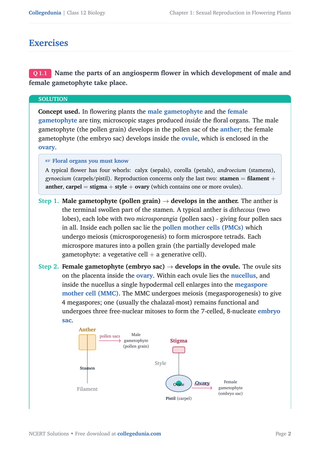


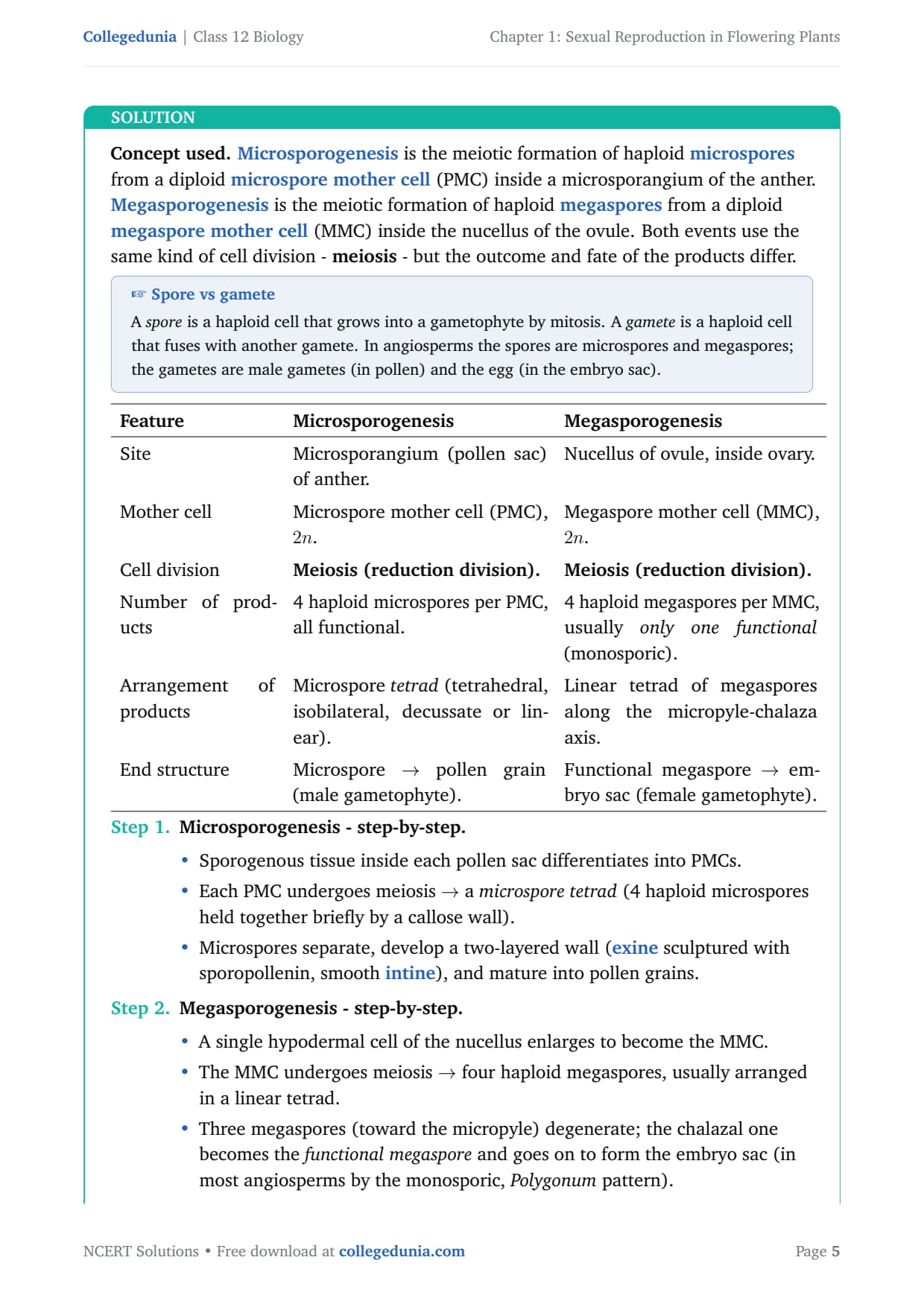
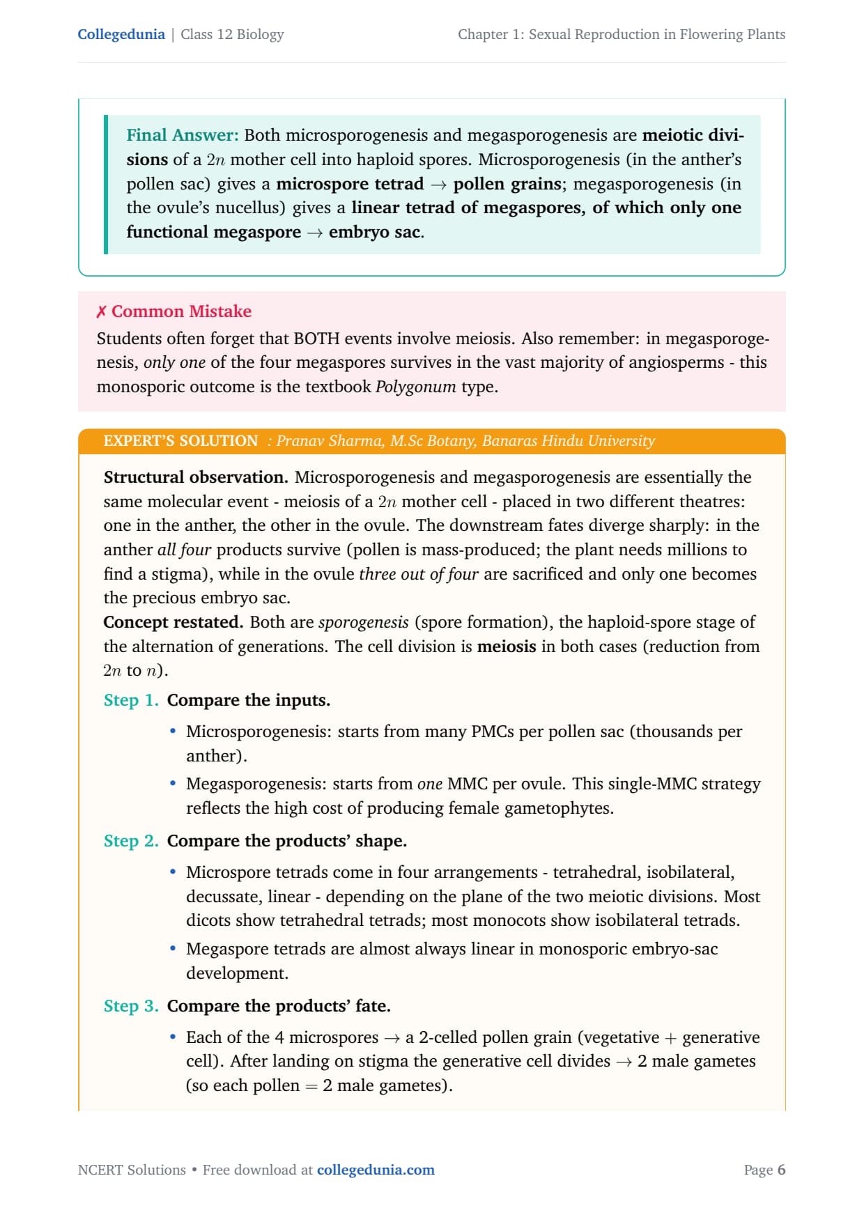

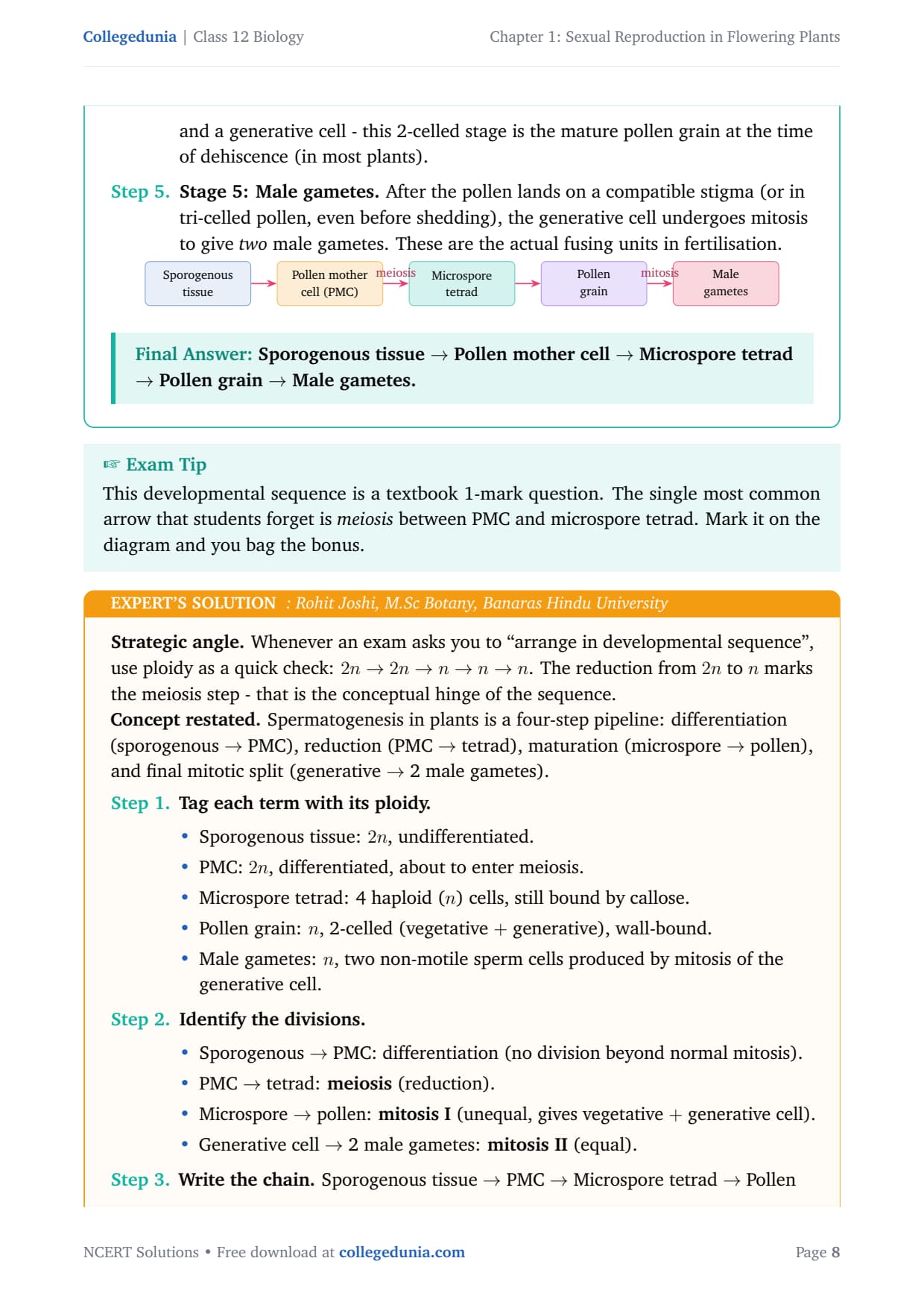
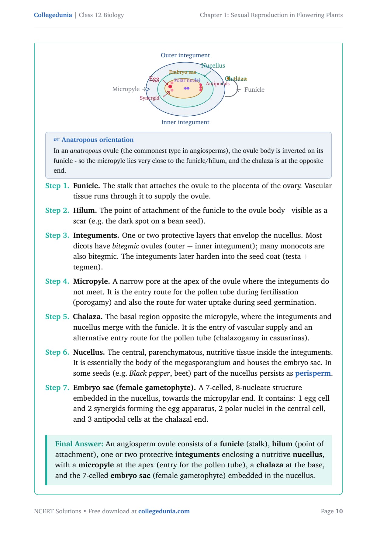
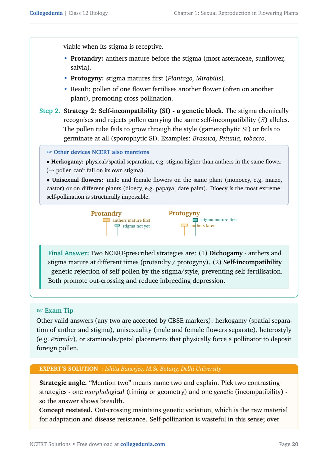
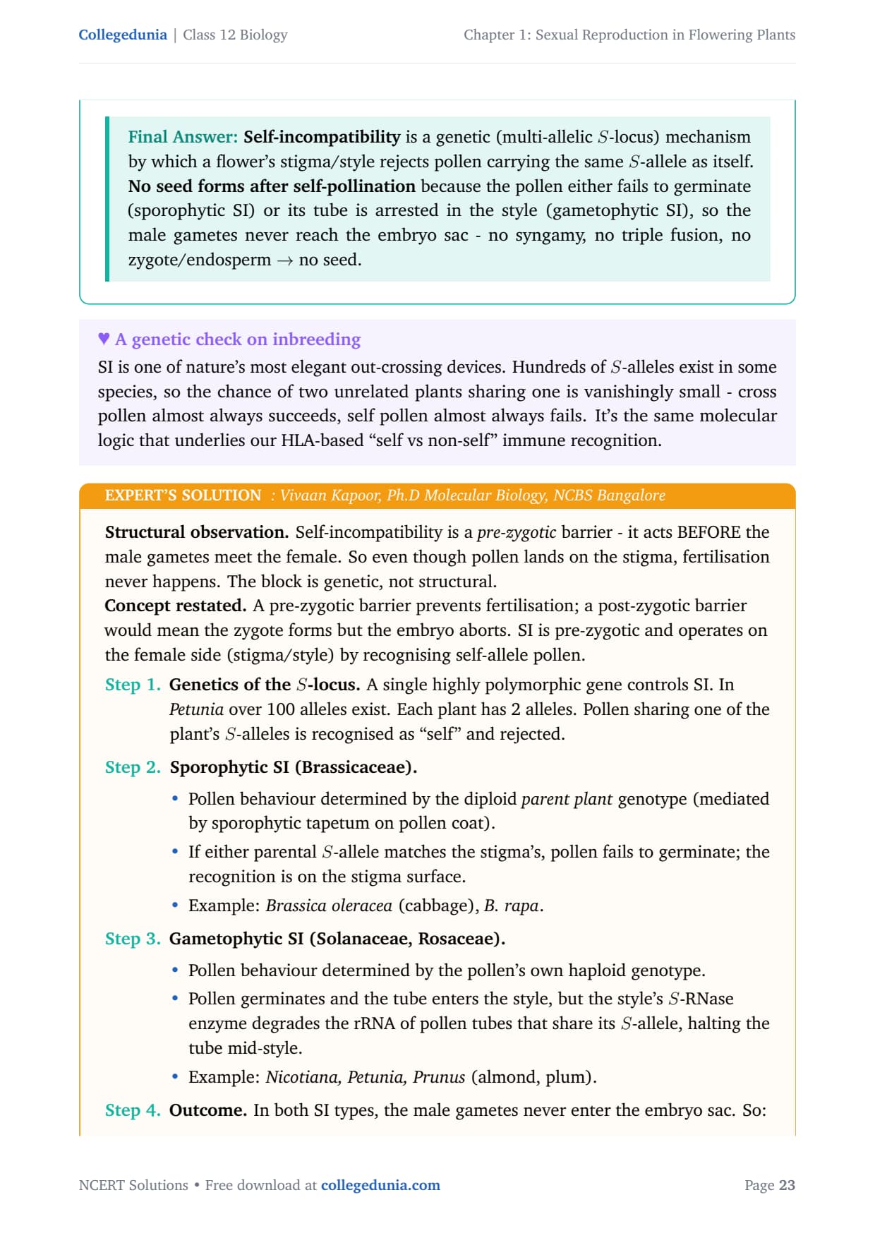
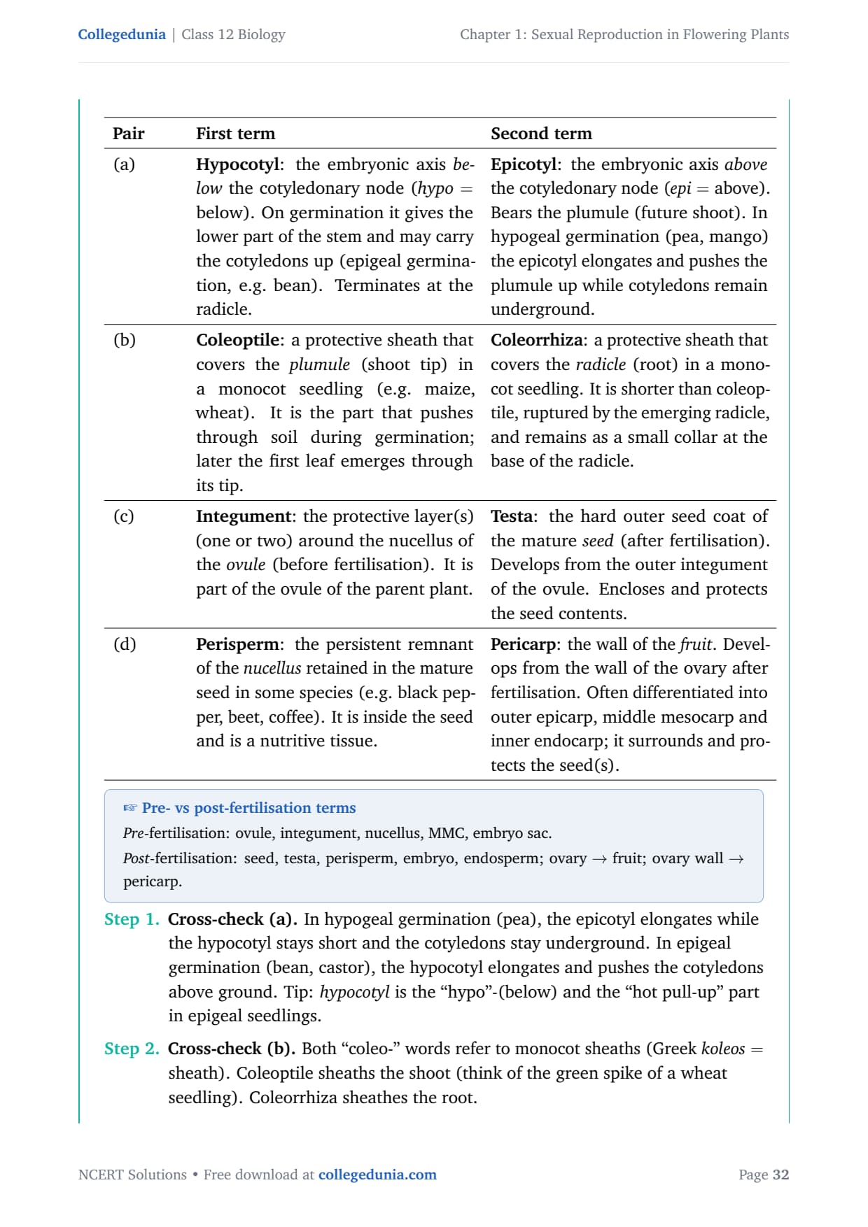

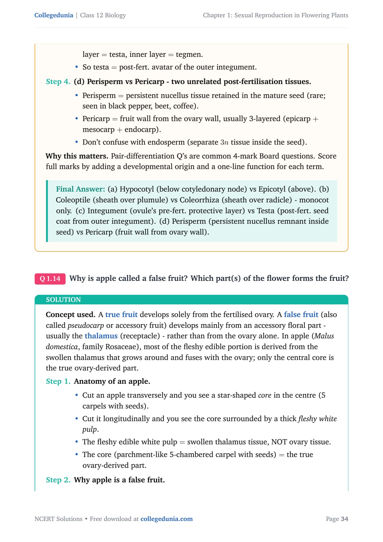
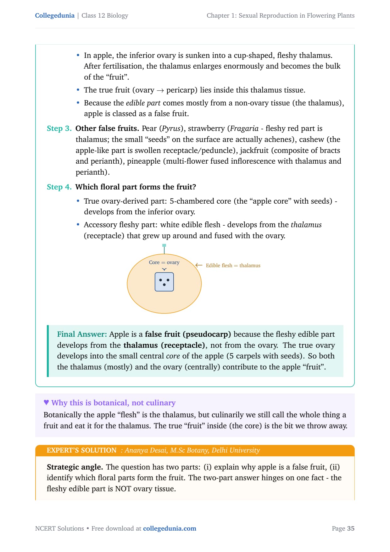
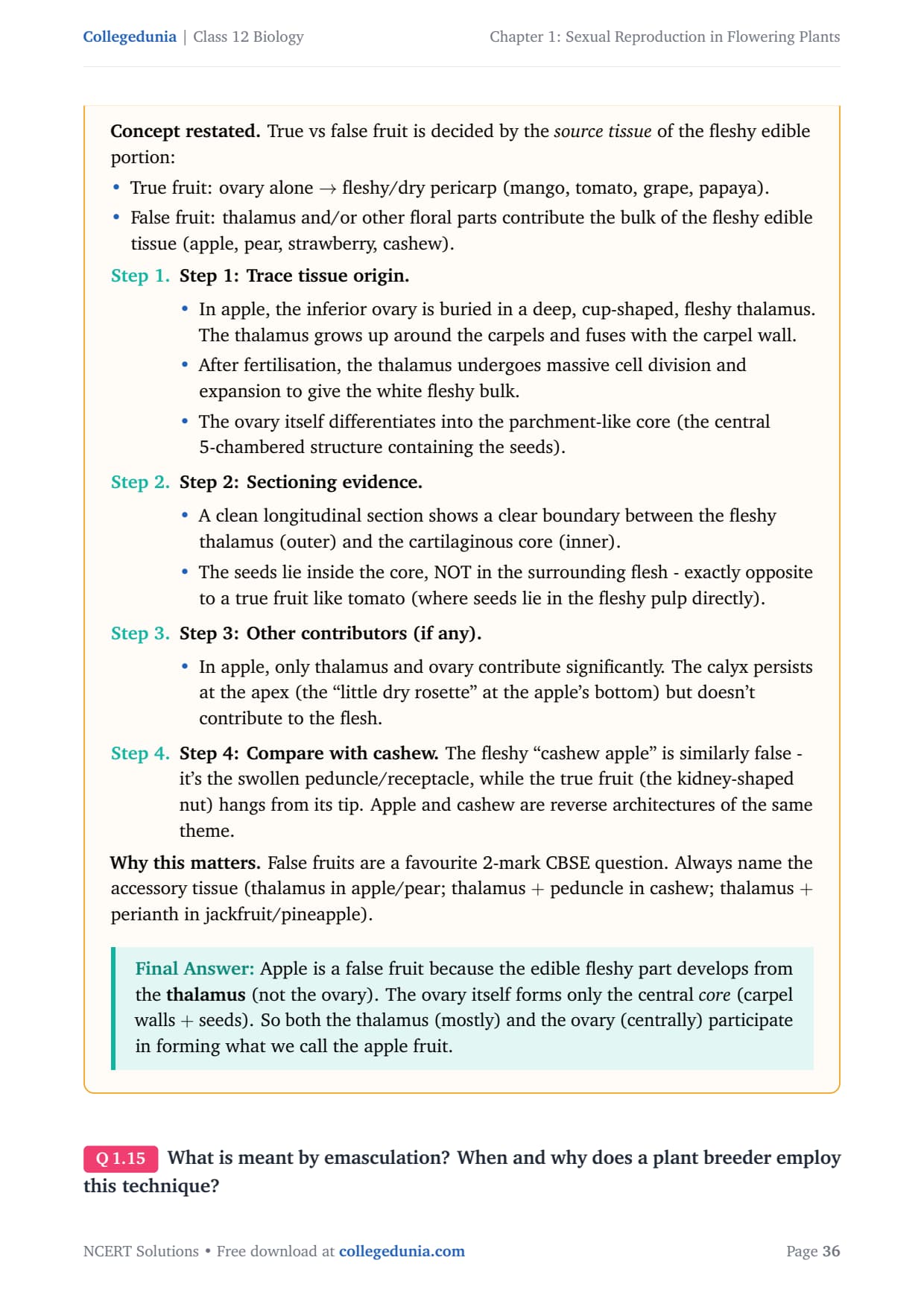
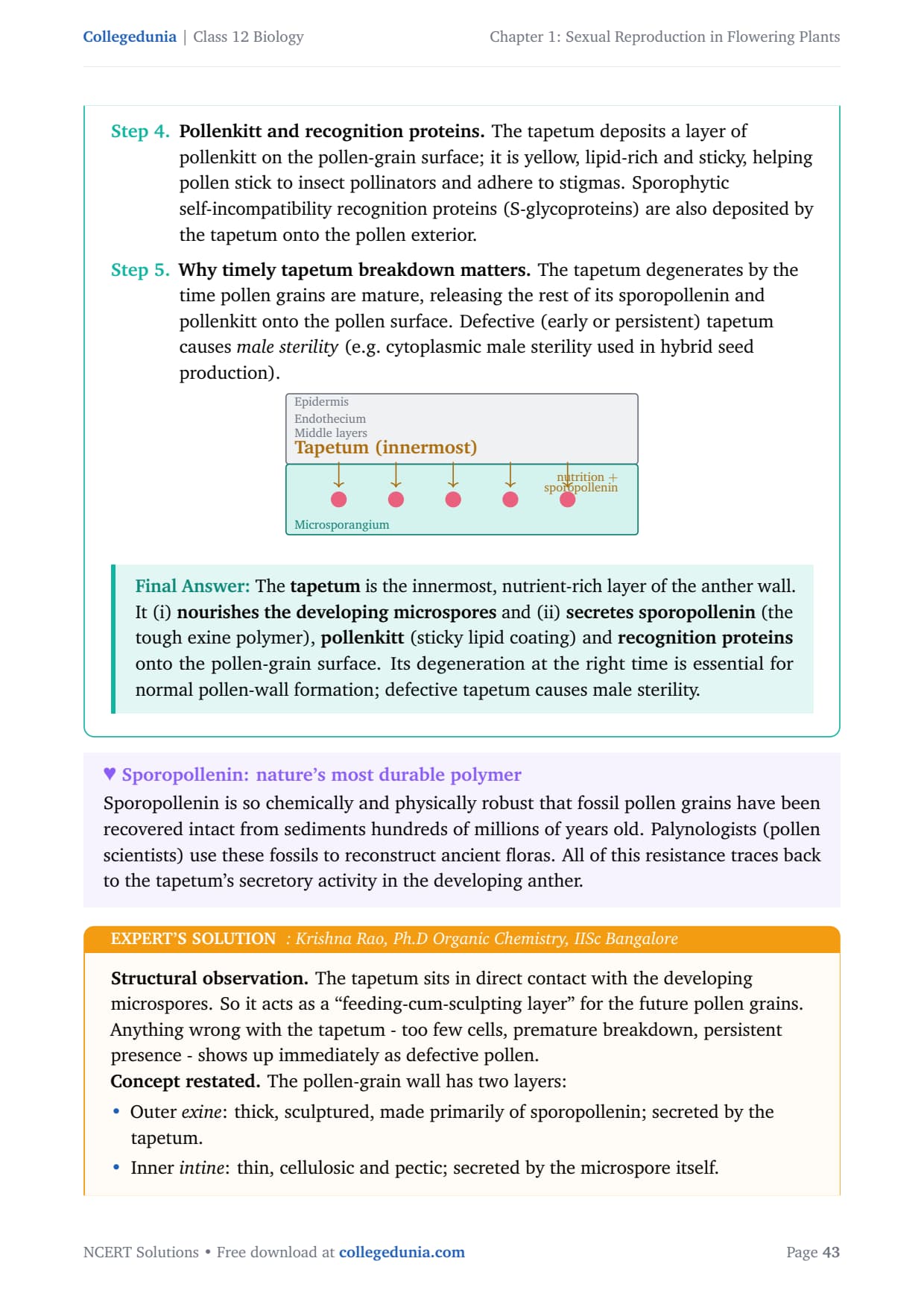
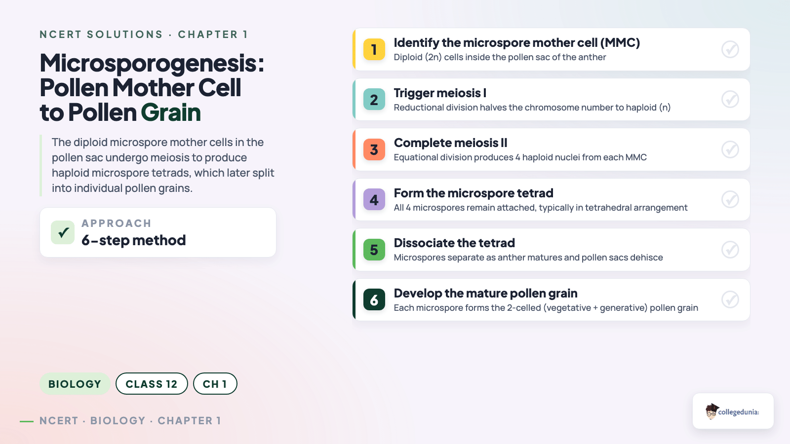
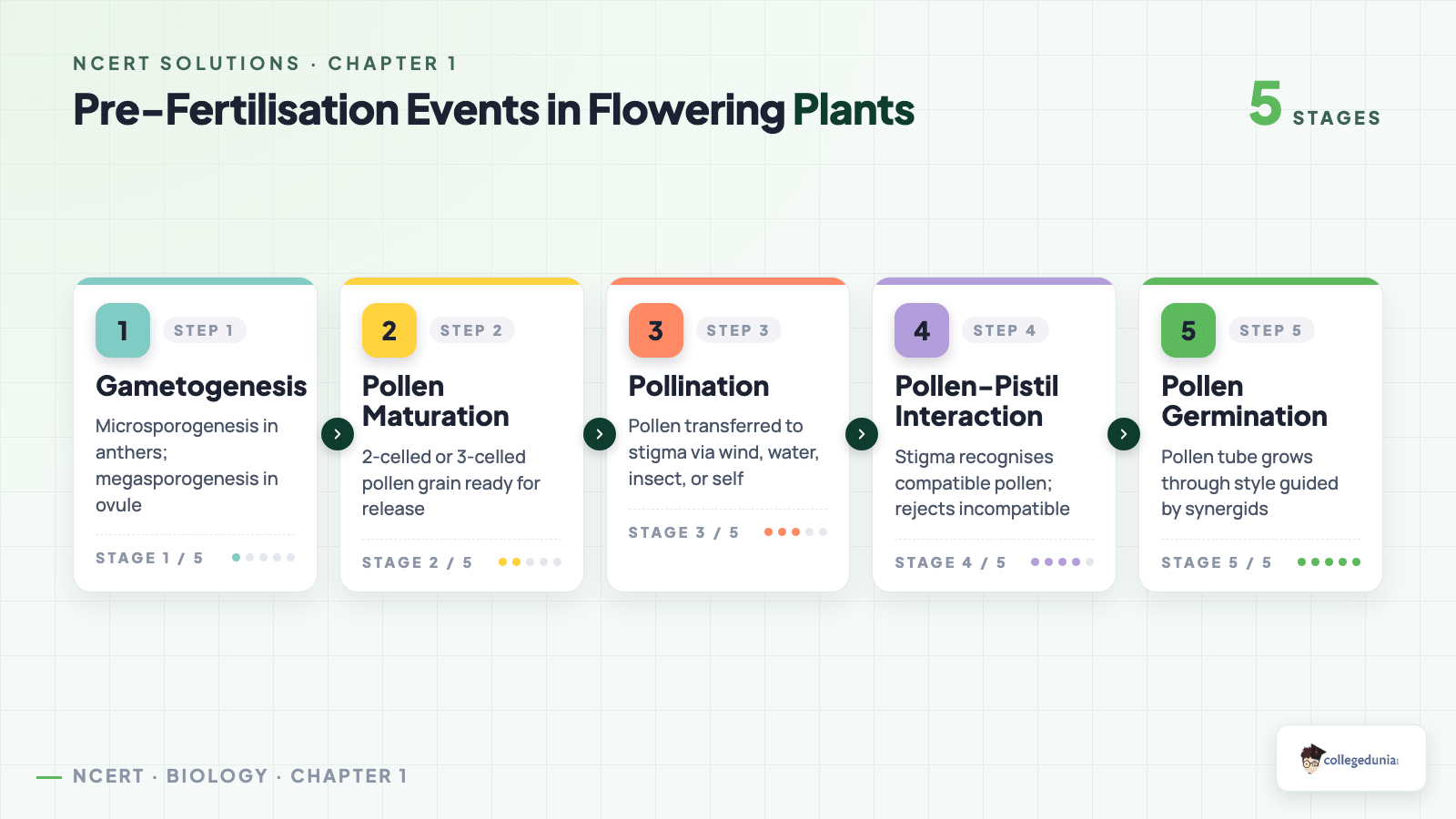



Comments