



The NCERT Class 11 Biotechnology Handwritten Notes Chapter 3 Biomolecules are scanned notebook pages written for one-sitting revision against the 2026-27 syllabus. They cover the four classes of biomolecules, carbohydrate classification with D and L sugars, anomers and epimers, fatty acids and lipids, the twenty standard amino acids, the peptide bond and all four levels of protein structure, and nucleic acids from base pairing to the Watson-Crick double helix.
- Length: 37 handwritten pages covering all five sections of the chapter, the textbook figures and every one of the 30 exercise questions.
- Must-learn pair: alpha helix against beta pleated sheet, and A pairing with T against G pairing with C, the two comparisons this chapter is built around.
- Also on this page: a classification tree of the four biomolecule classes, a peptide bond breakdown, a quick recall table and links to sibling chapters.
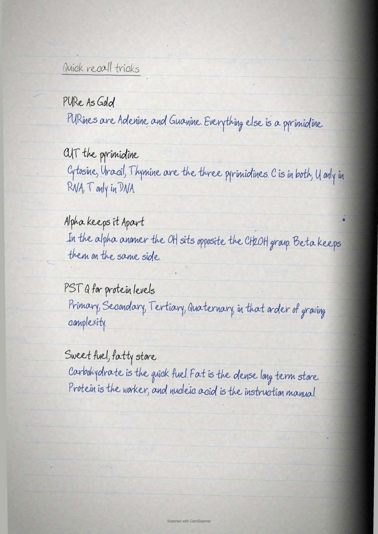
Every page in these Biomolecules notes is written by hand, checked against the 2026-27 NCERT Biotechnology print, and matched to the way Class 11 papers ask about sugar isomerism, amphipathic lipids, protein folding and DNA base pairing.
Student Feedback: In a Collegedunia survey of 9,460 Class 11 Biotechnology students, 71% of students said Chapter 3 feels harder than it is because four unrelated chemistries are packed into one chapter. Toppers reported the fix that works: revise one class of biomolecule per sitting, and always finish a sitting by drawing its structure from memory rather than rereading it.
Source: 2026-27 Class 11 Biotechnology student survey. Sample of 9,460 students from schools across 14 states.
What Biomolecules Are and Why This Chapter Follows Cellular Organelles
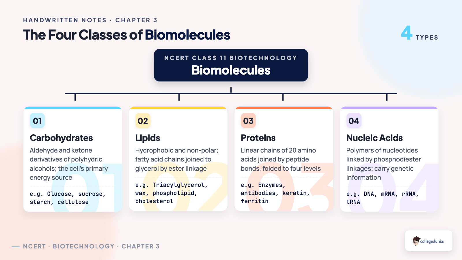
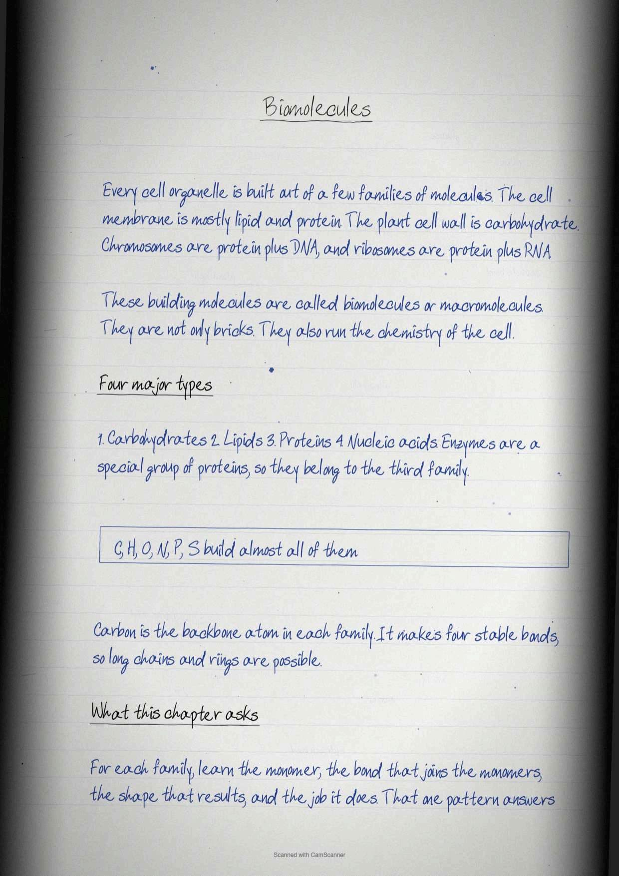
The chapter opens by looking back at the organelles. The cell membrane is built from lipids and proteins, the cell wall from carbohydrates, chromosomes largely from proteins and DNA, and ribosomes from proteins and RNA. Strip any organelle down and the same short list of ingredients appears.
Those ingredients are called macromolecules or biomolecules, and there are four major types: carbohydrates, proteins, lipids and nucleic acids. They are not only structural. Each one also runs a job inside the cell, which is why the chapter treats structure and function together rather than separately.
| Class of biomolecule | Building block | Where the chapter meets it |
|---|---|---|
| Carbohydrates | Monosaccharides | Cell wall of bacteria and plants, energy supply, part of DNA and RNA |
| Lipids | Fatty acids and glycerol | Cell membrane, energy store in adipose tissue, steroid hormones |
| Proteins | Amino acids | Catalysts, antibodies, transporters, hormones, muscle fibres |
| Nucleic acids | Nucleotides | Nucleus, mitochondria and chloroplast, inheritance of information |
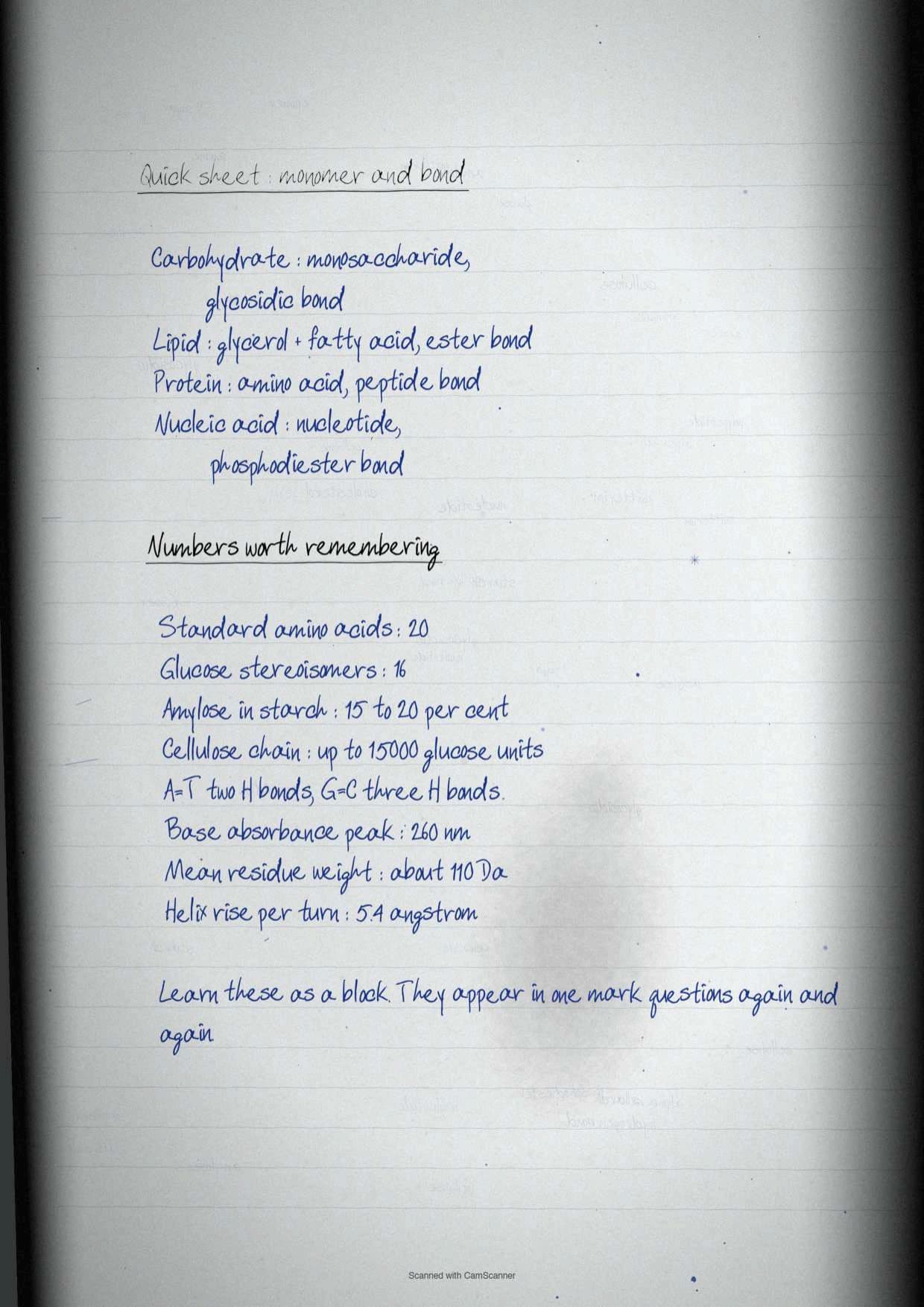
One line in this section is worth learning word for word because it explains the whole chapter. Carbohydrates are aldehyde and ketone derivatives of the polyhydric alcohols, lipids are hydrophobic and non-polar, proteins are linear chains of 20 amino acids, and nucleic acids are polymers of nucleotides linked through phosphodiester linkages. Four sentences, four classes, and the rest of the chapter is detail hanging off them.
Carbohydrates: Roles in the Cell and the Three-Way Classification
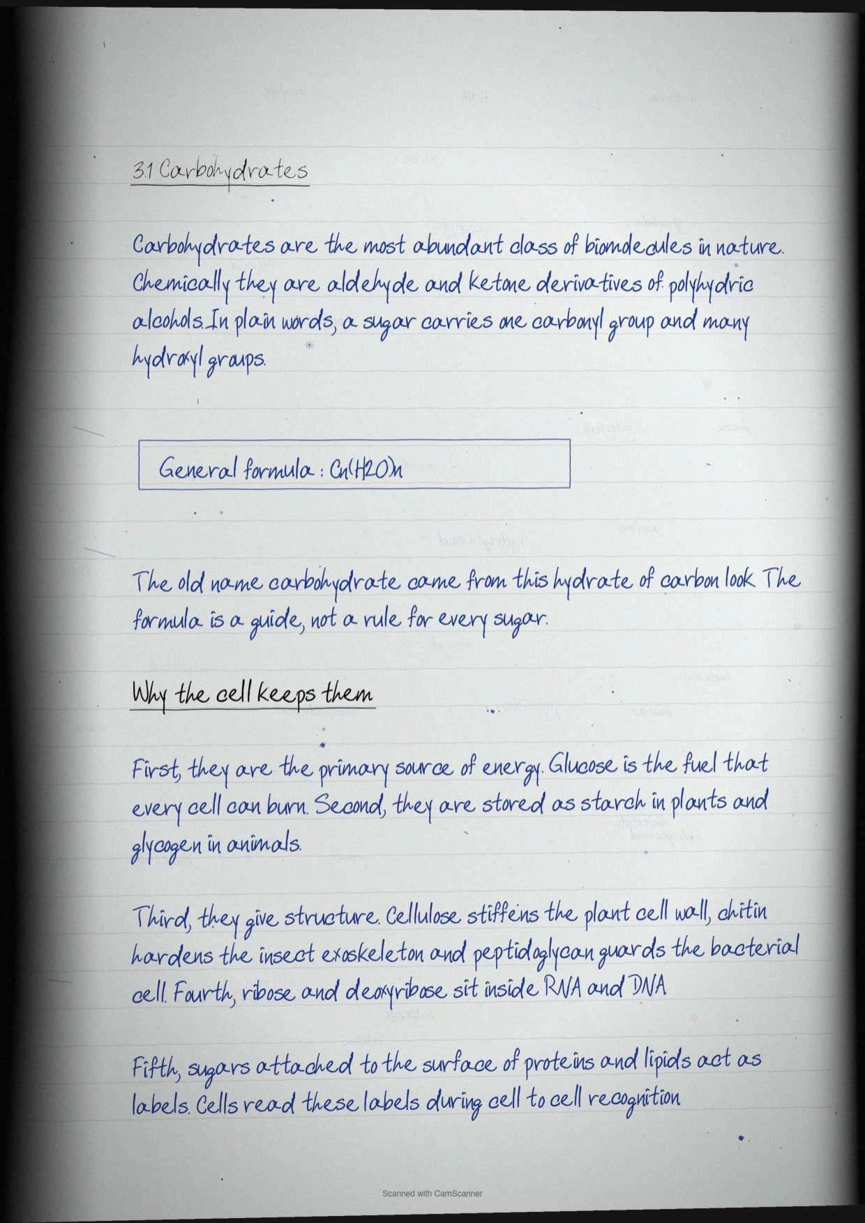
Carbohydrates are described as one of the most abundant classes of biomolecules in nature and are found in every life form. Their major role is to act as a primary source of energy, but the chapter lists four more jobs that a five-mark question expects to see.
- Energy stores and metabolic intermediates inside the cell.
- Structural components of the bacterial and plant cell wall, both of which are polymers of carbohydrates.
- Parts of DNA and RNA, since the pentose sugar of every nucleotide is a carbohydrate.
- Informational materials, linked to the surfaces of proteins and lipids so that cells can recognise one another and interact with their environment.
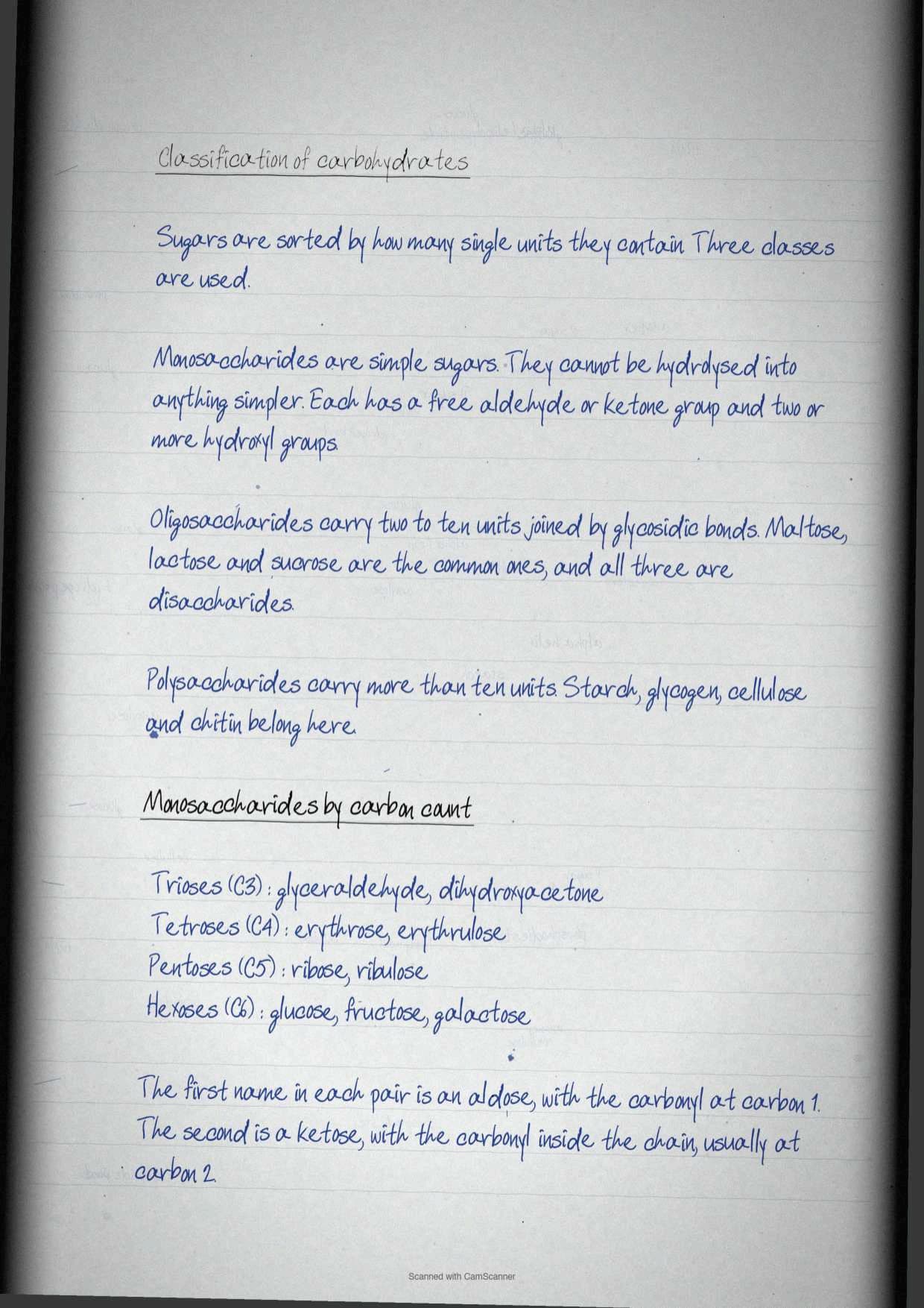
The classification itself has exactly three categories, and the chapter is strict about the boundaries between them.
| Category | Definition | Examples |
|---|---|---|
| Monosaccharides | Simple sugars that cannot be hydrolysed into simpler forms, with free aldehyde or ketone groups and two or more hydroxyl groups, general formula Cn(H2O)n | Glucose, fructose, ribose, glyceraldehyde |
| Oligosaccharides | Two to ten monosaccharide units joined by glycosidic bonds | Maltose, lactose, sucrose, raffinose |
| Polysaccharides | More than ten monosaccharide units joined by glycosidic linkages | Starch, glycogen, cellulose, chitin |
Carbohydrates also join proteins and lipids to form glycoconjugates, and the three names there are easy marks. If protein dominates the pairing it is a glycoprotein; if carbohydrate dominates it is a proteoglycan; and when carbohydrate joins a lipid it is a glycolipid.
Tip: the chapter summary at the end of the textbook says carbohydrates fall into four classes because it counts disaccharides separately from oligosaccharides. The main text says three. Write the three-way split, then add a line saying disaccharides are the commonest oligosaccharides, and both versions are covered.
Monosaccharides: Trioses to Hexoses, Aldoses and Ketoses
Monosaccharides are sorted twice over, once by how many carbon atoms they carry and once by which functional group sits on them. Table 3.1 of the chapter crosses the two, and reproducing that grid is usually the whole answer to a short question.
| Number of carbons | General formula | Aldose example | Ketose example |
|---|---|---|---|
| Trioses | C3H6O3 | Glyceraldehyde, an aldotriose | Dihydroxyacetone, a ketotriose |
| Tetroses | C4H8O4 | Erythrose | Erythrulose |
| Pentoses | C5H10O5 | Ribose | Ribulose |
| Hexoses | C6H12O6 | Glucose | Fructose |
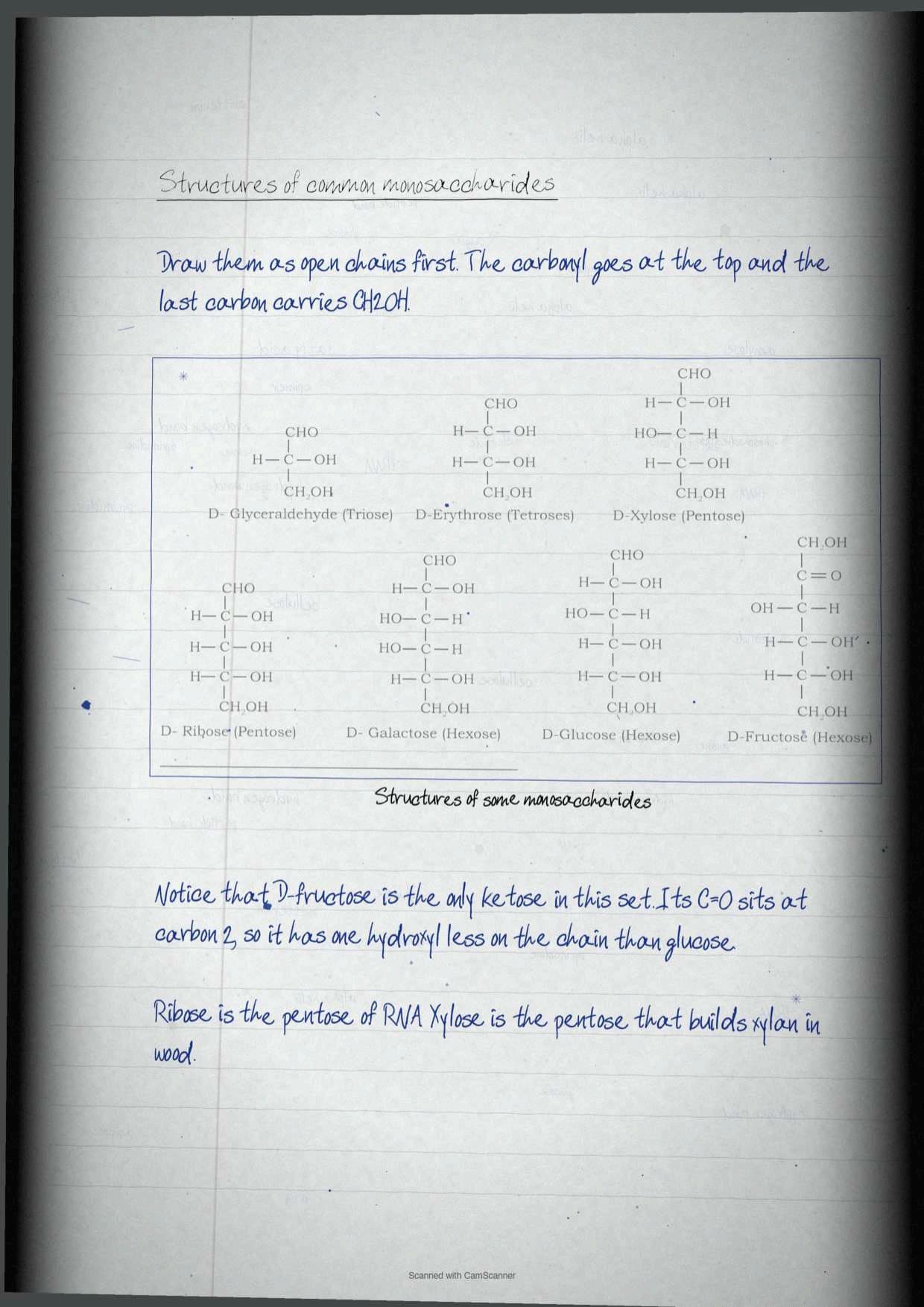
An aldose carries a free aldehyde group written as -CHO, and a ketose carries a ketone group written as >C=O. Glucose and fructose have the identical formula C6H12O6 and differ only in which of those two groups they carry, which is the cleanest one-line answer to a question on functional group isomerism.
Two of the pentoses matter far beyond this section. Ribose and its relative 2'-deoxy-D-ribose are the sugars of RNA and DNA, so the last section of the chapter depends on this table being learnt properly first.
D and L Sugars, Chirality and Why Most Biological Sugars Are D
This is the section that separates a confident answer from a vague one, because it rests on one rule that can be stated in a single sentence.
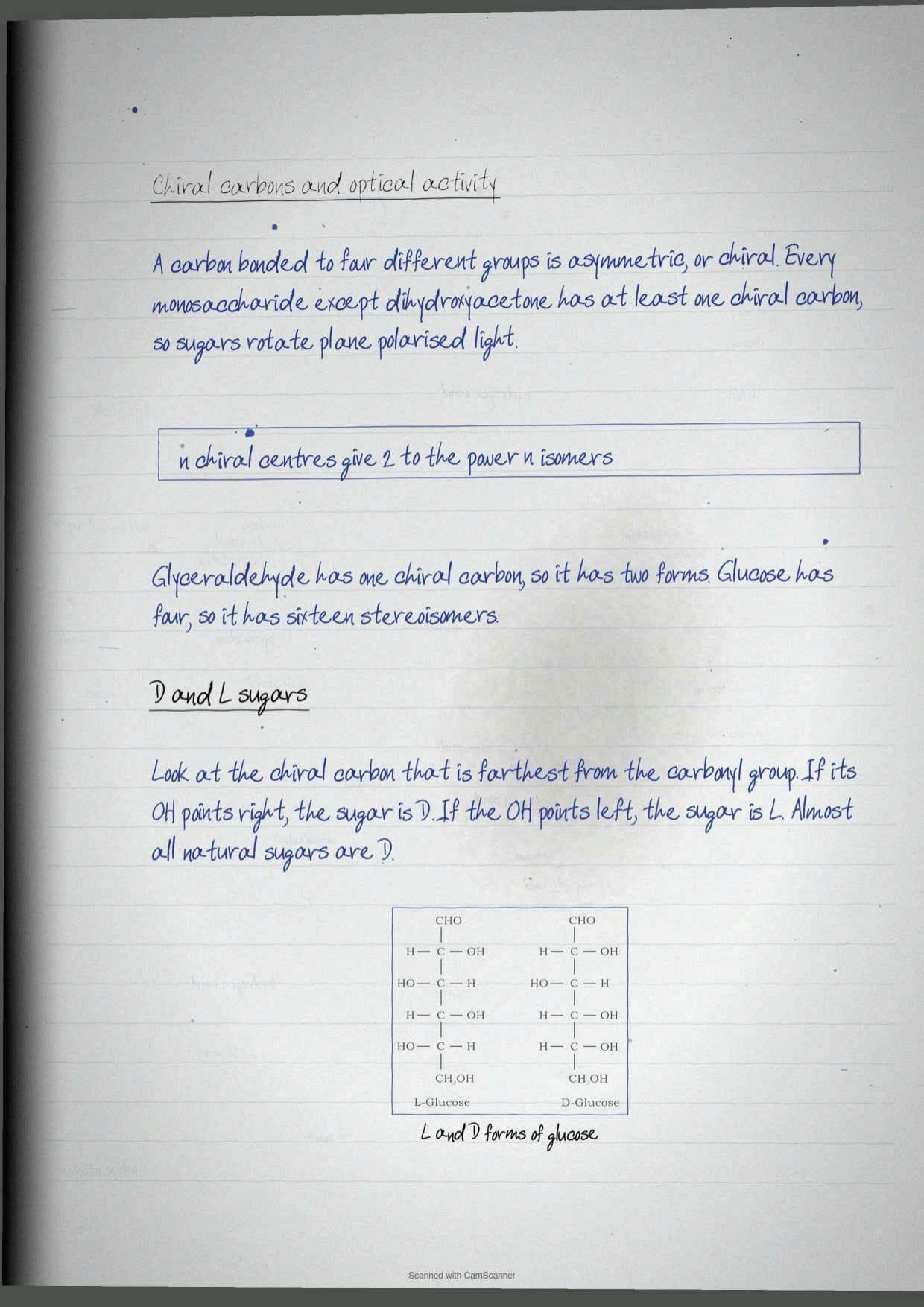
The orientation of the hydroxyl group that is most distant from the carbonyl carbon decides whether the sugar is D or L. Hydroxyl on the right gives the D isomer, hydroxyl on the left gives the L isomer. Most sugars in biological systems are D sugars.
Behind that rule sits chirality. Every monosaccharide except dihydroxyacetone contains one or more asymmetric or chiral carbon atoms, meaning a carbon bound to four different groups, so these sugars are optically active isomers called enantiomers.
- A molecule with n chiral centres can have 2n stereoisomers.
- Glyceraldehyde has one chiral centre, so 21 = 2 stereoisomers.
- Glucose has four chiral centres, so 24 = 16 stereoisomers.
Exercise question 2 of the chapter asks students to differentiate between the D and L forms of glucose, and Figure 3.3 is the model answer. Draw the two open chains side by side, mark carbon 5 in each, and put the hydroxyl on the right for D-glucose and on the left for L-glucose. Nothing else is needed.
Straight Chain Against Ring Form, Anomers, Epimers and Mutarotation
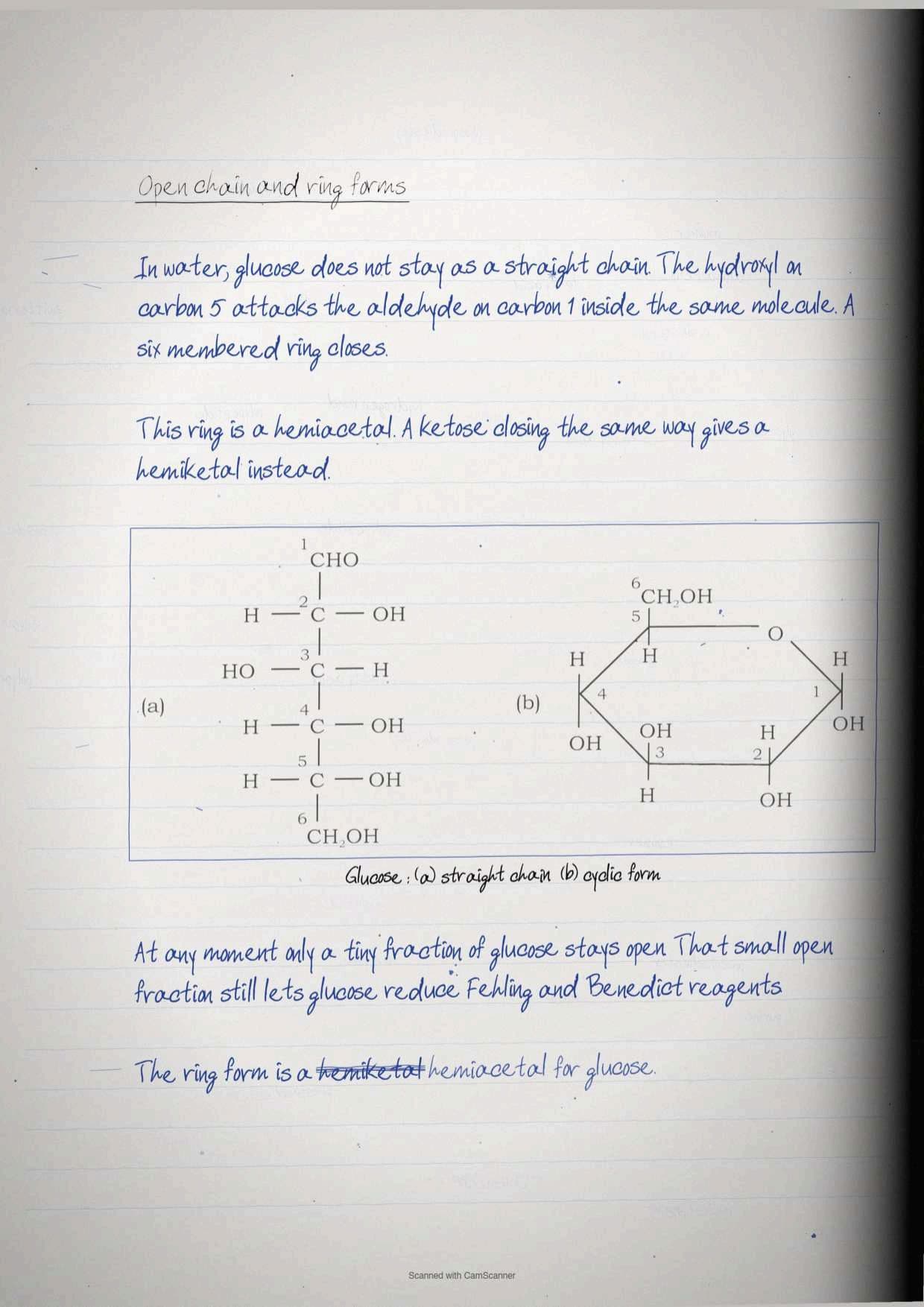
Glucose exists both as a straight chain and as a cyclic structure. The ring is not a different molecule, it is the same molecule folded onto itself. Cyclic structures are the result of hemiacetal formation by an intramolecular reaction between the carbonyl group and a hydroxyl group of the same sugar.
Closing the ring creates a brand new asymmetric carbon, and everything else in this section follows from that.
| Term | What it means | Worked example |
|---|---|---|
| Hemiacetal | Formed when the alcoholic group reacts with the aldehyde group of a monosaccharide | Ring closure in glucose, an aldose |
| Hemiketal | Formed when the alcoholic group reacts with the keto group of a monosaccharide | Ring closure in fructose, a ketose |
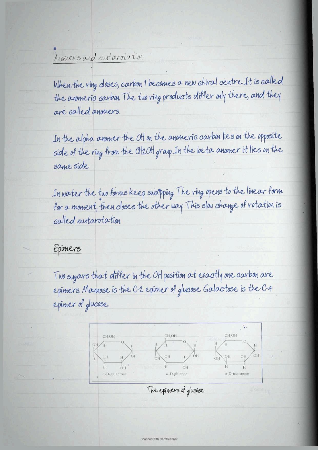
| Anomeric carbon | The carbonyl carbon that becomes chiral once the ring closes | C-1 of glucose |
| Anomers | Isomers differing only in configuration about the hemiacetal or hemiketal carbon | α-D-glucopyranose and β-D-glucopyranose |
| Mutarotation | Interconversion of α and β anomers in aqueous solution, the ring opening briefly to the linear form and closing again | A fresh glucose solution slowly changing its optical rotation |
| Epimers | Isomers with a different hydroxyl configuration at exactly one carbon atom | Mannose at C-2 and galactose at C-4, both epimers of glucose |
The alpha anomer is defined precisely in the chapter. In the alpha form, the hydroxyl group of the anomeric carbon sits on the opposite side of the sugar ring from the CH2OH group at the chiral centre that sets the D or L configuration, which is C-5 in glucose. The other form is the beta anomer.
Tip: exercise question 6 asks students to describe isomerisation in monosaccharides. A full answer needs four words in order: enantiomers, anomers, mutarotation, epimers. Define each one, give the glucose example for each, and the answer is complete.
Oligosaccharides and the Glycosidic Bond
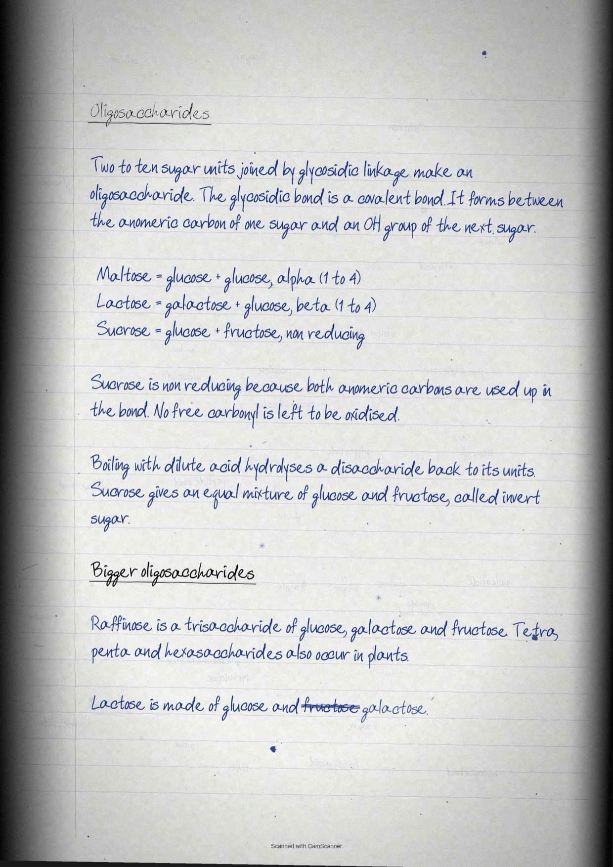
An oligosaccharide is two to ten monosaccharides joined by a glycosidic linkage, and the definition of that bond is examinable on its own. A glycosidic bond is a covalent bond formed by joining the hydroxyl group of one monosaccharide with the anomeric carbon of the other sugar unit.
| Oligosaccharide | Made from | Note from the chapter |
|---|---|---|
| Maltose | Two D-glucose residues | The textbook forms it in Figure 3.6 from two glucose molecules |
| Lactose | D-galactose and D-glucose | Written in full as beta-D-galactopyranosyl (1→4) D-glucopyranose |
| Sucrose | Glucose and fructose | Hydrolysis with dilute acid yields the mixture of both, and sucrose is the non-reducing disaccharide of question 24 |
| Raffinose | Glucose, galactose and fructose | A trisaccharide, the standard example beyond disaccharides |
Disaccharides can be hydrolysed back to their constituent monosaccharides simply by boiling with dilute acid. Beyond trisaccharides the chapter names tetra, penta and hexasaccharides without giving separate examples, so those are worth one line each and nothing more.
Exercise question 3 asks for the structure of a disaccharide made from glucose and fructose. That is sucrose, and the notebook draws the two rings joined through the anomeric carbons with the glycosidic oxygen bridging them.
Polysaccharides: Starch, Glycogen, Cellulose, Chitin and Peptidoglycan
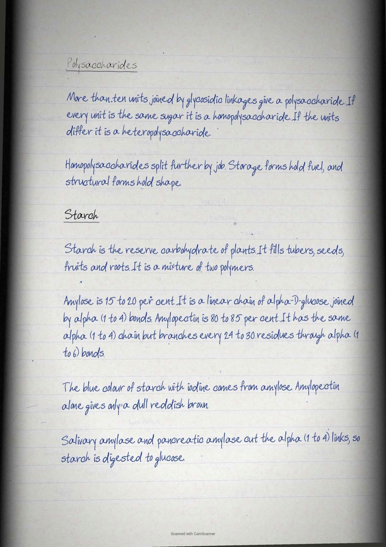
Polysaccharides carry more marks than any other part of the carbohydrate section, because they can be asked as definitions, as comparisons or as diagrams. They are classified in three ways: by the type of repeating unit into homopolysaccharides and heteropolysaccharides, by the degree of branching, and by the type of glycosidic linkage.
Homopolysaccharides split again by job. Storage polysaccharides hold monosaccharide for use as fuel, and structural polysaccharides build the plant cell wall and the animal exoskeleton. Heteropolysaccharides do something different again, providing extracellular support and forming the matrix that holds animal tissue cells together.
| Name | Constituent monosaccharide | Biological significance |
|---|---|---|
| Starch | α-D-glucose | Storage of energy in plants |
| Glycogen | α-D-glucose | Storage of energy in bacteria and animals |
| Cellulose | β-D-glucose | Structural role, gives rigidity and strength to the cell wall |
| Chitin | β-N-acetyl-D-glucosamine | Structural role, gives rigidity to the exoskeleton of insects |
| Inulin | β-D-fructose | Storage of energy in plants |
| Pectin | α-D-galacturonic acid | Structural, holds cellulose fibrils together in plant cell walls |
| Dextran | α-D-glucose | Structural, acts as an extracellular adhesive in bacteria |
| Xylan | β-D-xylose | Storage and supporting roles in plants |
Starch occurs in tubers, seeds, fruits and roots as reserve carbohydrate, and it is a mixture of two polymers. Amylose makes up 15 to 20 per cent and is a linear polymer of alpha-D-glucose linked by α(1→4) bonds. Amylopectin makes up 80 to 85 per cent, uses the same α(1→4) linkage along the chain but is highly branched, with branch points every 24 to 30 glucose residues joined by α(1→6) linkages. The characteristic blue colour of starch with iodine comes from amylose alone, since amylopectin gives only a dull reddish brown colour. Salivary amylase and pancreatic amylase hydrolyse the α(1→4) linkages and digest starch to glucose.
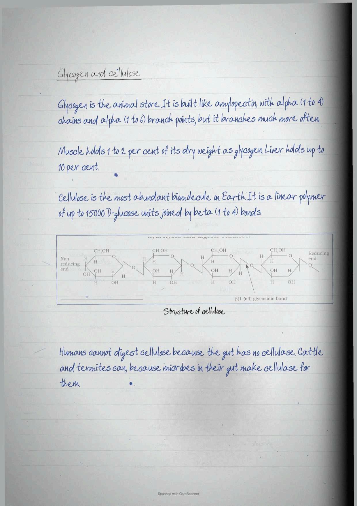
Glycogen is the animal equivalent and is even more extensively branched. It uses the same α(1→4) chain with α(1→6) branch points. Muscle cells hold glycogen at 1 to 2 per cent of their dry weight and liver cells up to 10 per cent.
Cellulose is the single most quotable molecule in the chapter. It is the most abundant extracellular structural polysaccharide in plants and the most abundant of all biomolecules in the biosphere. It is a linear polymer of up to 15,000 D-glucose units joined by β(1→4) glycosidic bonds. Humans cannot digest it because the human gut lacks cellulase, the enzyme that hydrolyses that bond. Cattle and termites manage it only because symbiotic microorganisms in their gut secrete cellulase for them.
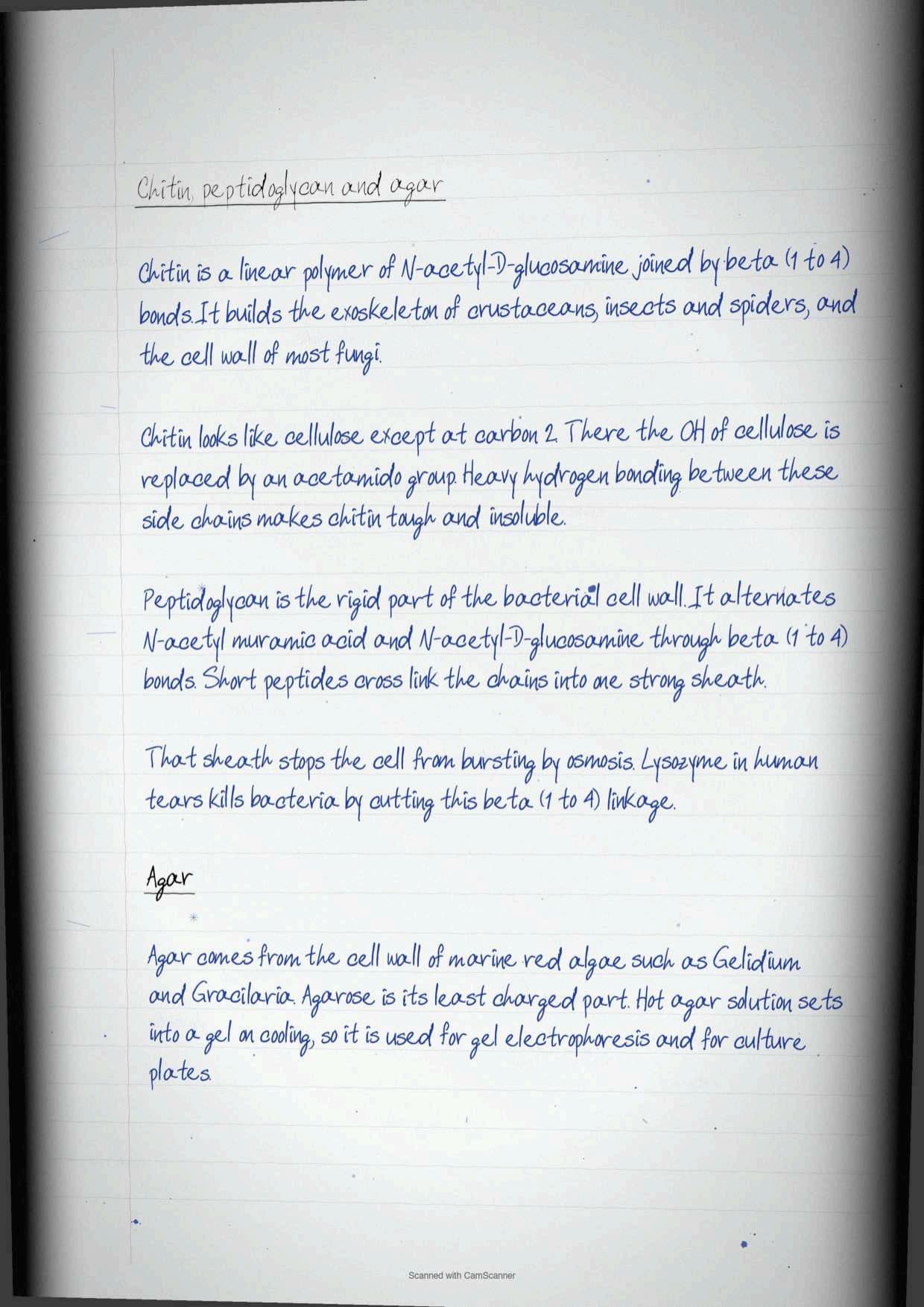
- Chitin is a linear polymer of β(1→4) linked N-acetyl-D-glucosamine. It builds the exoskeleton of crustaceans, insects and spiders and the cell wall of most fungi. It differs from cellulose only in that the hydroxyl at the second carbon is replaced by an acetamido group, and extensive hydrogen bonding of the N-acetyl side chains makes it tough and insoluble.
- Peptidoglycan is the rigid component of the bacterial cell wall, a heteropolysaccharide of alternating β(1→4) linked N-acetyl muramic acid and N-acetyl-D-glucosamine residues, cross linked by short peptides that weld the chains into a sheath and prevent osmotic rupture. Lysozyme in human tears kills bacteria by hydrolysing exactly this linkage.
- Agar, from Box 1, is a gelatinous heteropolysaccharide from the cell wall of marine red algae such as Gelidium, Gracilaria and Gigartina. Agarose is its lightly charged component, molecular weight 80,000 to 1,40,000, used as inert support for the electrophoretic separation of nucleic acids and as a surface for bacterial and plant tissue culture.
Biomolecules Explained in Simple Language
Source: Magnet Brains on YouTube
Fatty Acids: Saturated, Unsaturated and the Delta Notation
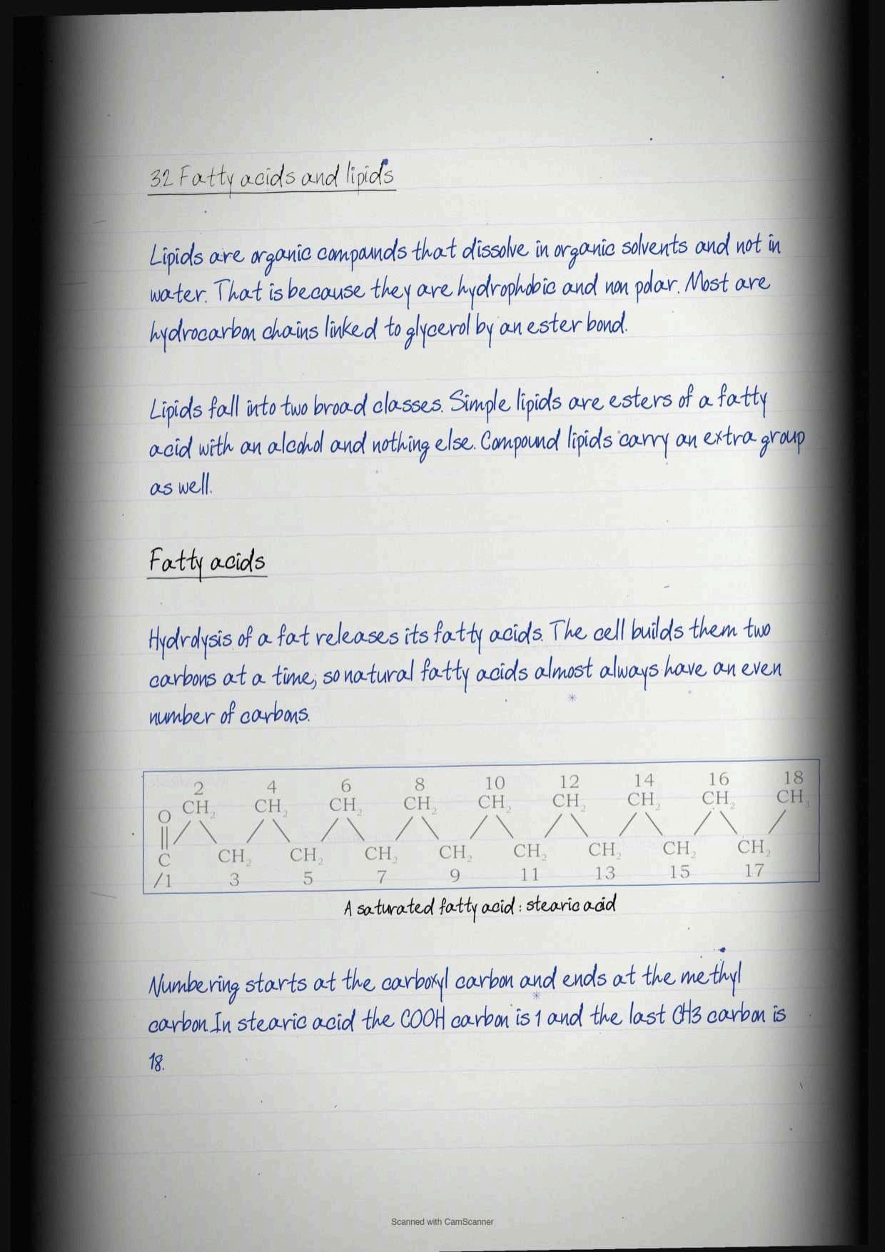
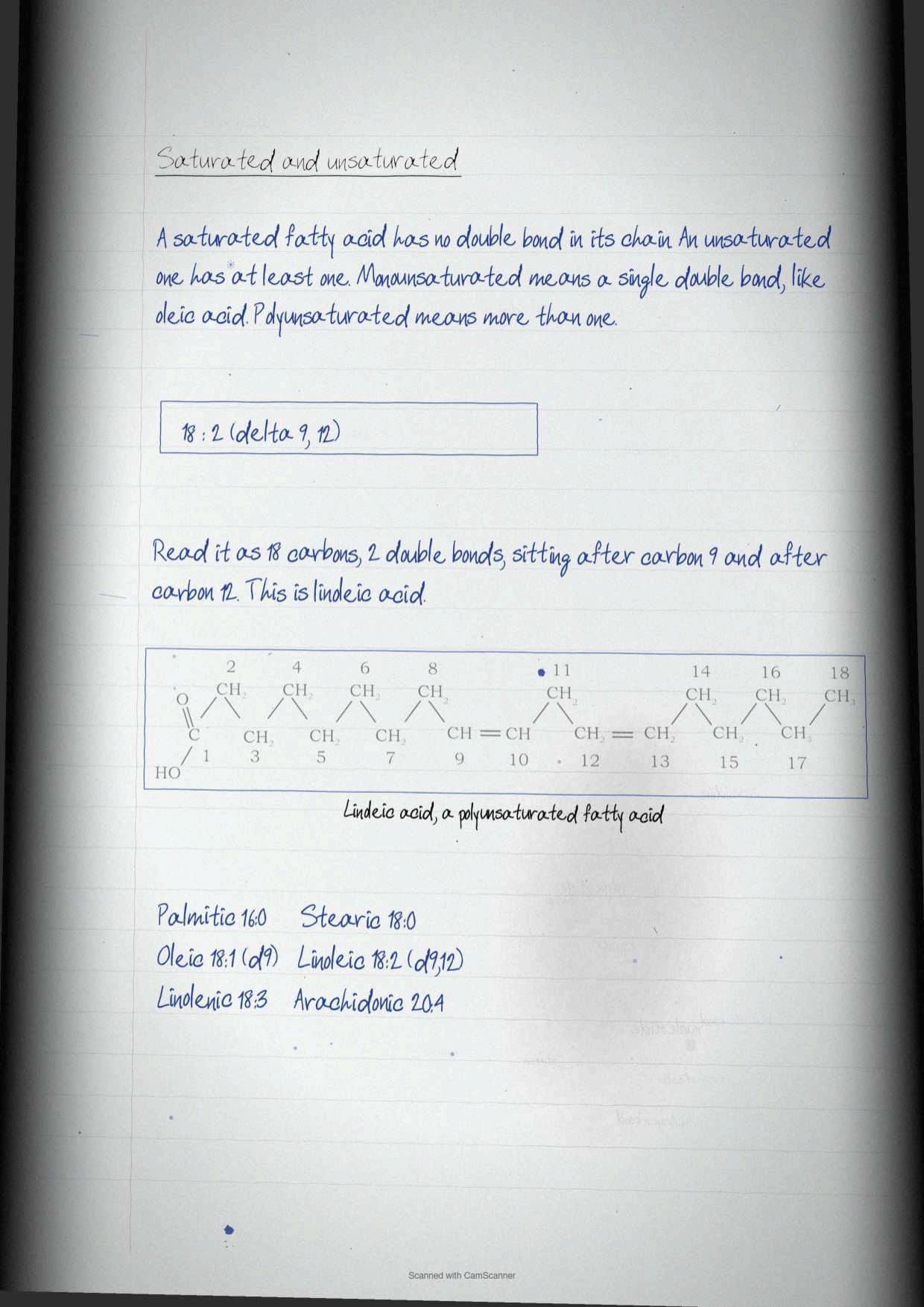
Lipids are a group of organic compounds that vary widely in structure and function but share one property. Because they are hydrophobic and non-polar, lipids are soluble in organic solvents. They are primarily made of hydrocarbon chains connected to glycerol by an ester linkage.
Fatty acids are obtained by hydrolysis of fats. Because naturally occurring fatty acids are synthesised from two carbon units, they almost always contain an even number of carbon atoms. From there the split is simple: saturated fatty acids have no double bonds, and unsaturated fatty acids have one or more.
The naming system is the part students most often get wrong, so learn it as a formula. Number the carbons starting from the carboxyl carbon as C-1 and ending at the methyl carbon. Then write the total number of carbons, a colon, the total number of double bonds, and the positions of those double bonds as superscripts on a delta symbol.
- 18:0 means eighteen carbons and no double bonds, which is stearic acid.
- 18:2 (Δ9,12) means eighteen carbons with double bonds between C-9 and C-10 and between C-12 and C-13, which is linoleic acid.
- Monounsaturated fatty acids carry exactly one double bond, for example oleic acid.
- Polyunsaturated fatty acids carry more than one: linoleic acid has two, linolenic acid three, and arachidonic acid four.
| Common name | Symbol | Number of carbons | Type |
|---|---|---|---|
| Lauric acid | 12:0 | 12 | Saturated |
| Myristic acid | 14:0 | 14 | Saturated |
| Palmitic acid | 16:0 | 16 | Saturated |
| Stearic acid | 18:0 | 18 | Saturated |
| Arachidic acid | 20:0 | 20 | Saturated |
| Palmitoleic acid | 16:1 (Δ9) | 16 | Monounsaturated |
| Oleic acid | 18:1 (Δ9) | 18 | Monounsaturated |
| Linoleic acid | 18:2 (Δ9,12) | 18 | Polyunsaturated |
| Arachidonic acid | 20:4 (Δ5,8,11,14) | 20 | Polyunsaturated |
Classification of Lipids: Simple Lipids and Compound Lipids
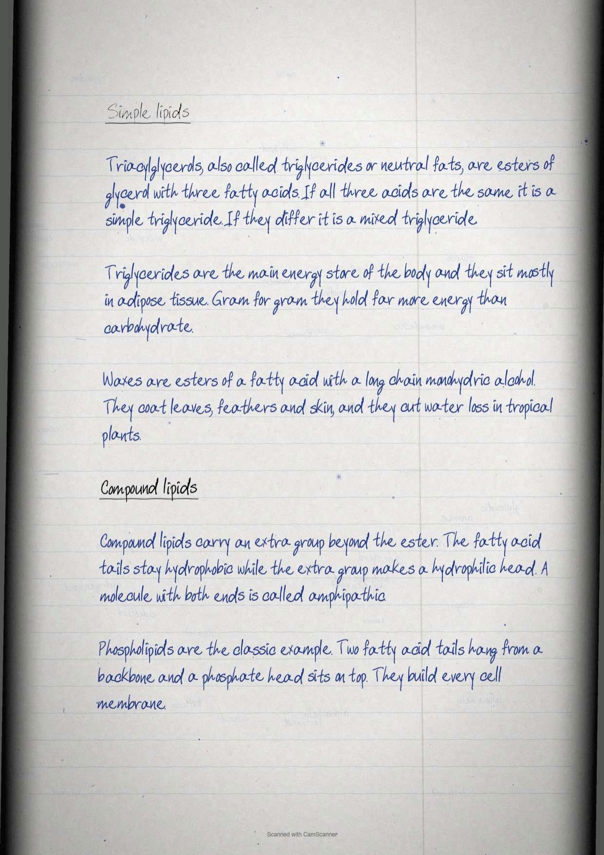
Lipids are broadly classified into two categories, and every named lipid in the chapter belongs to one of them. A simple lipid is a fatty acid ester with an alcohol and no other group. A compound lipid is a fatty acid ester with an alcohol that carries an additional group.
| Category | Member | Structure and role |
|---|---|---|
| Simple lipids | Triacylglycerols | Esters of glycerol and fatty acids, also called triglycerides or neutral fats. Same fatty acid in all three positions gives a simple triglyceride, more than one type gives a mixed triglyceride. They are the vehicle of energy storage, primarily in adipose tissue |
| Waxes | Esterification of fatty acids with a monohydric alcohol of high molecular weight. They form a protective coating on plant and animal surfaces and reduce water loss in tropical plants | |
| Compound lipids | Phospholipids | Two hydrophobic fatty acid tails and a hydrophilic phosphate head, found mainly in cell membranes |
| Steroids | A four-fused ring structure, hydrophobic, acting as receptor ligands to control metabolism |
The word the paper wants for compound lipids is amphipathic. The fatty acid components make the hydrophobic tails and the alcohol together with the additional group makes the hydrophilic head, and a molecule built that way is amphipathic. Exercise question 8 asks exactly why membrane lipids are called amphipathic, and that sentence is the answer.
Exercise question 30 is worth noting here too. In glycerolipids the fatty acids are joined to glycerol through an ester bond, not a phosphodiester, glycosidic or peptide bond, and the same distinction returns in every later chapter that touches membranes.
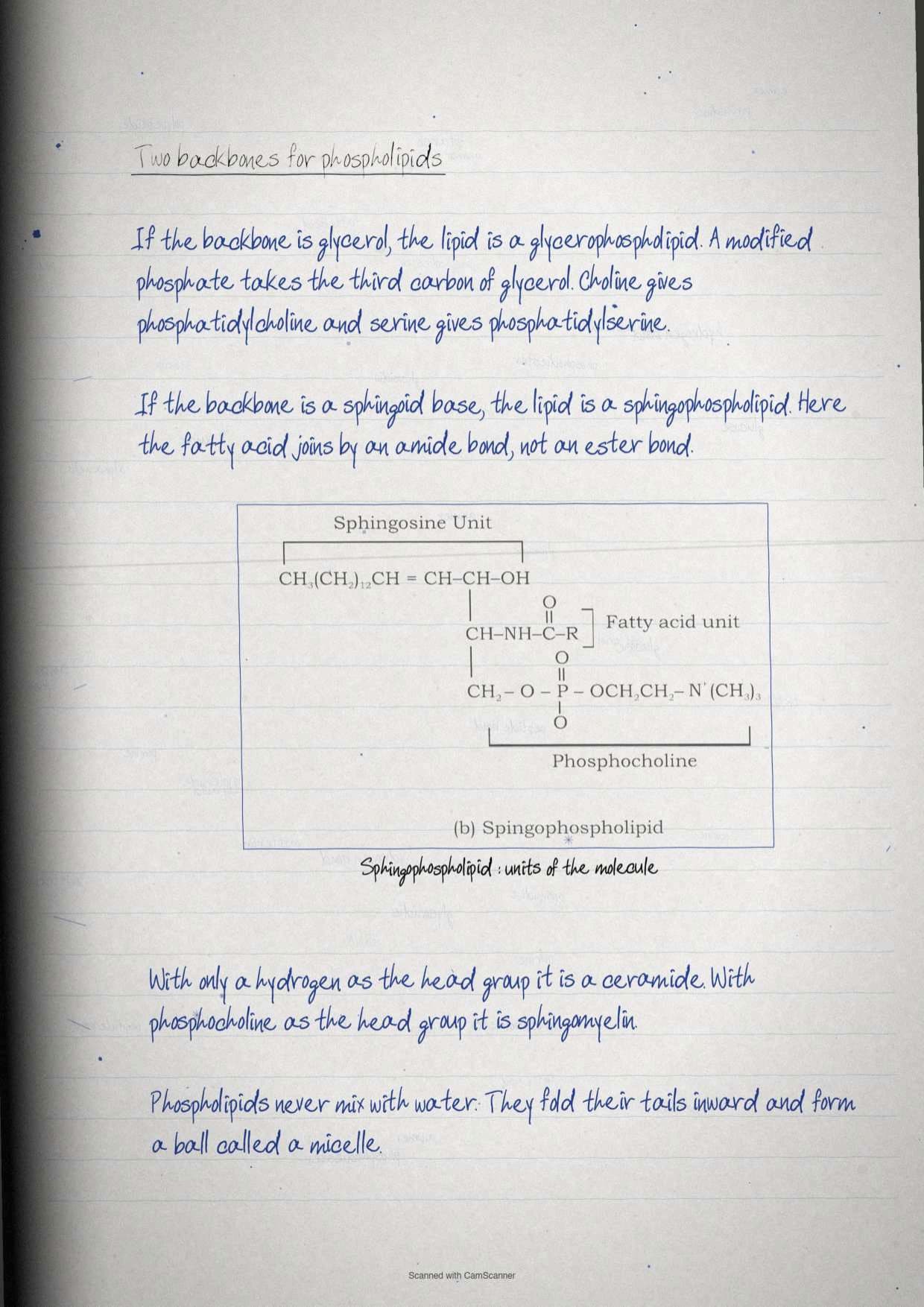
Phospholipids, Micelles and the Two Membrane Backbones
Phospholipids are the reason a cell has an inside and an outside, and the chapter distinguishes them by the backbone they are built on.
- Glycerophospholipids have fatty acid chains attached to a glycerol backbone, with a modified phosphate group occupying the third carbon of the glycerol.
- Sphingophospholipids have their chains attached to a sphingoid base backbone, and the fatty acid is joined by an amide linkage rather than an ester linkage.
A phospholipid is then named by whatever modifier sits on the phosphate group. The two most common are choline, giving phosphatidylcholine, and serine, giving phosphatidylserine. In the sphingolipid family, a head group of just a hydrogen atom gives a ceramide, while a phosphocholine head group gives sphingomyelin.
Phospholipids do not spontaneously mix with water. Instead they acquire a sphere-shaped structure called a micelle, with the tails tucked inside and the heads facing the water. That single behaviour is what a membrane is built on, and it is the answer to exercise question 7 on the difference between sphingolipids and glycerolipids when combined with the backbone point above.
Steroids and Cholesterol: The Four-Fused Ring Family
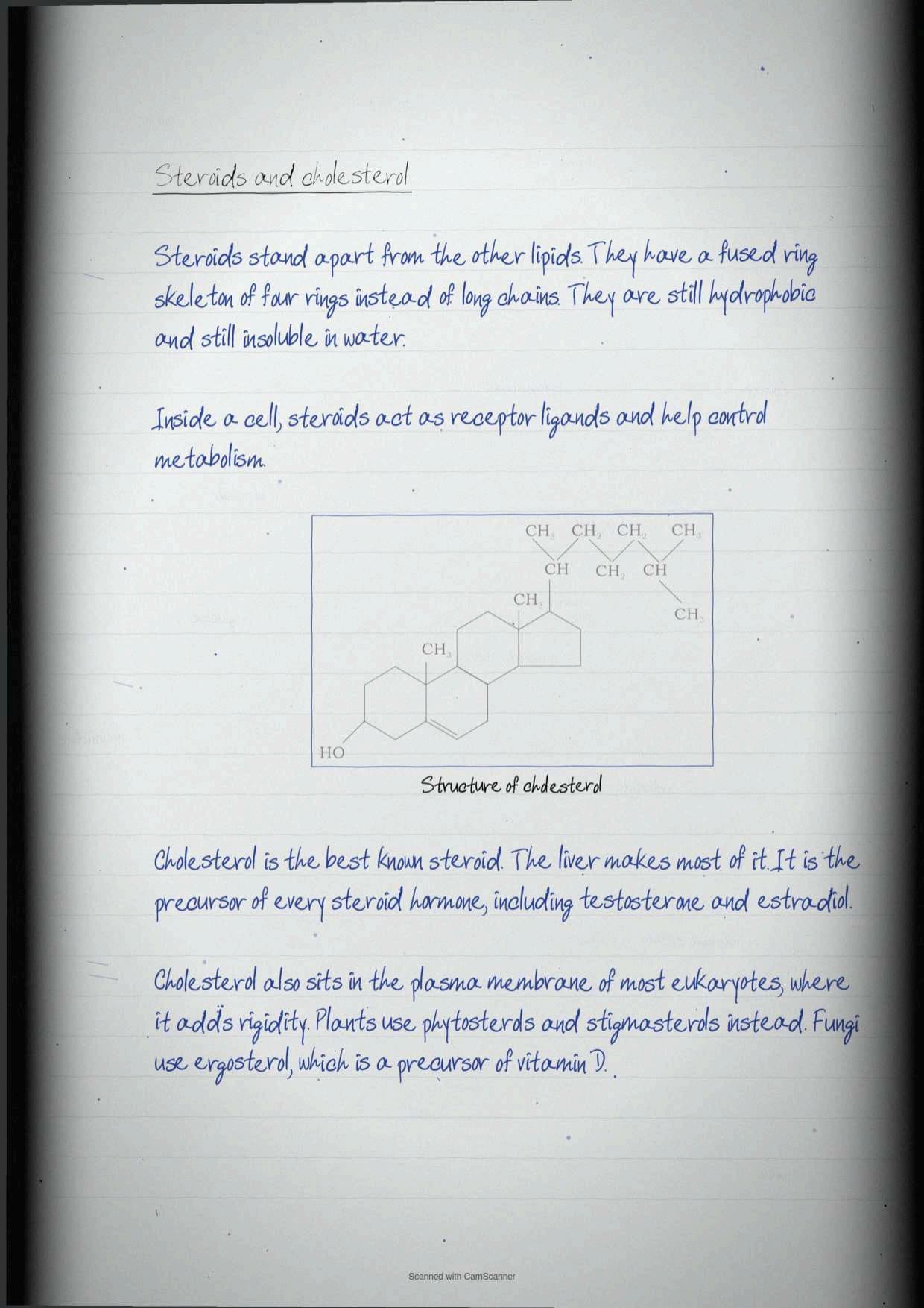
Steroids are distinct from every other lipid in the chapter because of a peculiar four-fused ring structure. Like the rest, they are hydrophobic and insoluble in water, and inside a cell they act as receptor ligands that help control metabolism.
Cholesterol is the most common derivative of steroids and carries the most examinable facts.
| Steroid | Where it is found | What it does |
|---|---|---|
| Cholesterol | Synthesised primarily by the liver, present in the plasma membrane of most eukaryotes | Key precursor of all steroid hormones including testosterone and estradiol, and provides rigidity to the membrane |
| Phytosterols | Plants | Regulate membrane fluidity and permeability |
| Stigmasterols | Plants | Regulate membrane fluidity and permeability |
| Ergosterol | Typically found in fungi | Important precursor of vitamin D |
Tip: cholesterol is asked as a one-mark question far more often than it is asked as a structure. The two facts that score are that it is the precursor of all steroid hormones and vitamin D, and that it stiffens the eukaryotic plasma membrane. Learn those before attempting the ring drawing.
Amino Acids: The General Formula and the Twenty Standard Ones
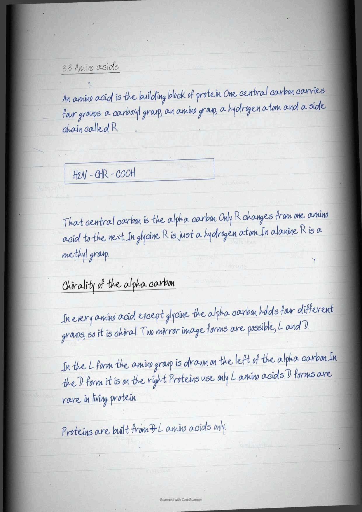
Amino acids are the building blocks of proteins, and their general formula is built around a single carbon atom. The alpha carbon is linked to four different groups: an acidic carboxylic group (-COOH), a basic amino group (-NH2), a hydrogen atom, and an R group called the side chain.
Only the side chain R varies across all 20 amino acids. It can be as simple as a hydrogen atom in glycine or a methyl group in alanine. Because the alpha carbon is tetrahedrally bound to four different groups it is chiral, so amino acids exist in two optically active mirror image forms. In L isomers the amino group is on the left of the alpha carbon and in D isomers it is on the right. Only L isomers are found in proteins, and D isomers are rare in biological protein.
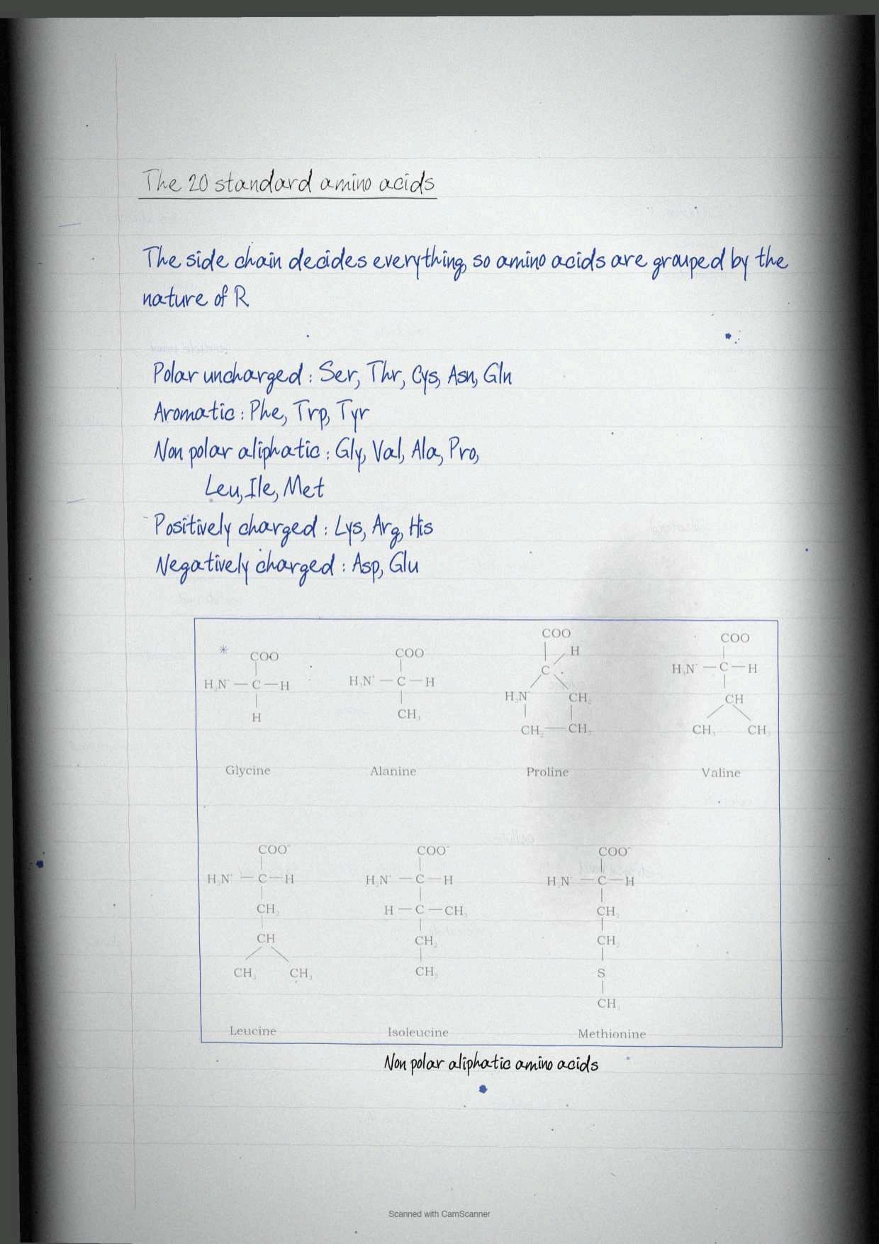
The 20 standard amino acids are grouped into five families by the nature of the R group, and exercise question 10 asks for exactly this classification.
| Category of R group | Amino acids | Single letter symbols |
|---|---|---|
| Polar, uncharged | Serine, Threonine, Cysteine, Asparagine, Glutamine | S, T, C, N, Q |
| Aromatic | Phenylalanine, Tryptophan, Tyrosine | F, W, Y |
| Non-polar aliphatic | Glycine, Valine, Alanine, Proline, Leucine, Isoleucine, Methionine | G, V, A, P, L, I, M |
| Positively charged, basic | Lysine, Arginine, Histidine | K, R, H |
| Negatively charged, acidic | Aspartate, Glutamate | D, E |
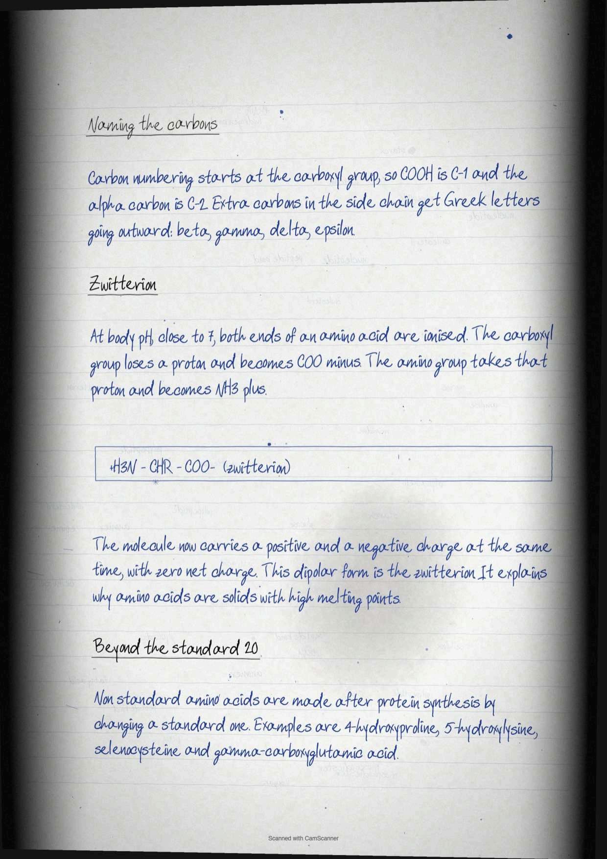
Numbering inside an amino acid uses Greek letters. Carbons added to the alpha carbon within an R group are called beta, gamma, delta and epsilon as they move outward. In ordinary organic numbering the carboxylic carbon is C-1 and the alpha carbon is therefore C-2, and Figure 3.22 shows this on L-lysine.
Zwitterions, Non-standard Amino Acids and Non-protein Amino Acids
At physiological pH, which the chapter fixes at pH 7, both charged groups of an amino acid are ionised at the same time. The alpha carboxyl group loses a proton to become -COO- carrying a negative charge, and the alpha amino group gains it to become -NH3+ carrying a positive charge.
This dipolar state, produced by the migration of a proton from the carboxyl group to the amino group, is called a zwitterion. Exercise question 11 asks for the definition and the structure, and the notebook draws the glycine zwitterion of Figure 3.23 because glycine is the simplest case.
The chapter then adds two categories that students routinely merge into one. They are not the same thing.
| Category | Definition | Examples |
|---|---|---|
| Non-standard amino acids | Occur naturally in cells but take no part in protein synthesis. They are generated after protein synthesis by modification of specific standard amino acids | 4-hydroxyproline, formed by hydroxylation of proline, plus 5-hydroxylysine, selenocysteine and gamma-carboxyglutamic acid |
| Non-protein amino acids | Amino acids that are never part of proteins at all, present widely in plants, animals and microbes | L-ornithine, L-citrulline, beta-alanine, creatine and gamma-aminobutyrate |
The Peptide Bond and the Primary Structure of Proteins
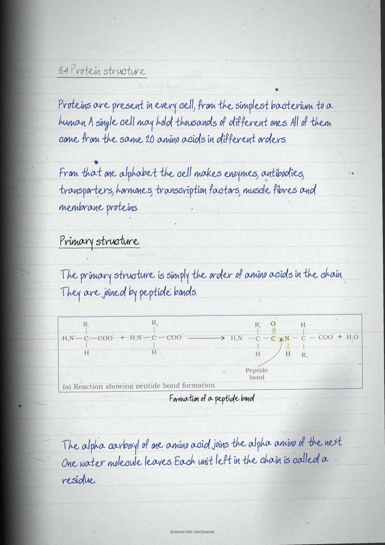
Proteins are present in every cell from the simplest bacterium to human beings and plants, and a single cell may contain thousands of different ones. All of them are built from the same 20 amino acids linked covalently into a linear sequence, and the variety comes purely from the order.
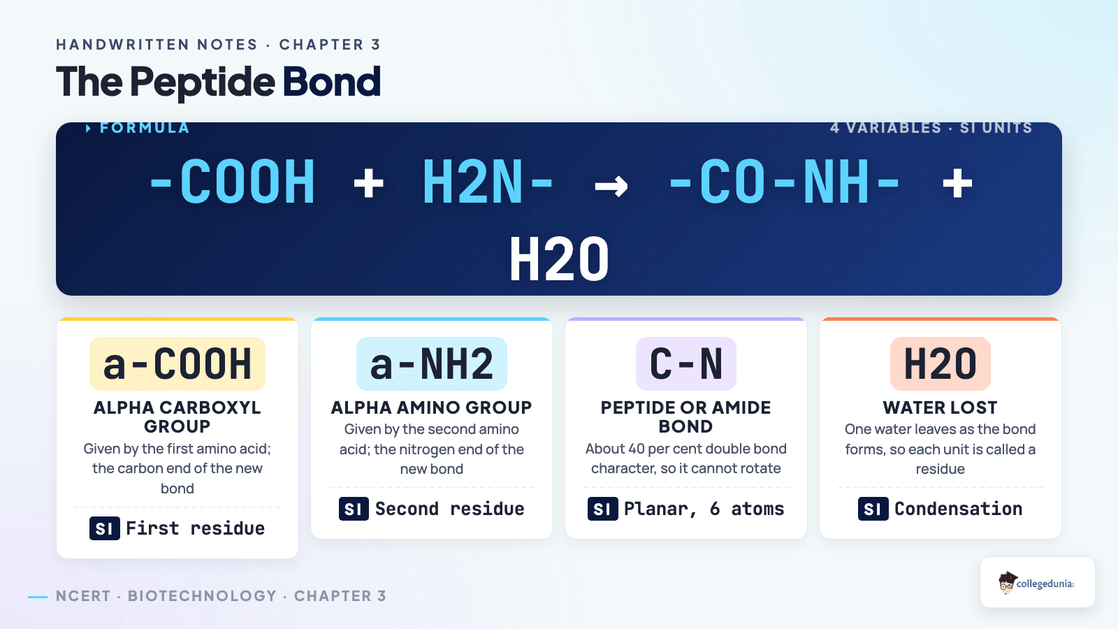
The peptide bond, also called the amide bond, is formed by coupling the alpha carboxyl group of one amino acid to the alpha amino group of another, and the reaction is accompanied by the loss of a water molecule. Because a unit loses water on joining, each amino acid inside a polypeptide is called a residue, not a whole amino acid.
The primary structure of a protein is then simply the linear chain of amino acid sequences linked through peptide bonds. Four rules govern how that chain is written and named.
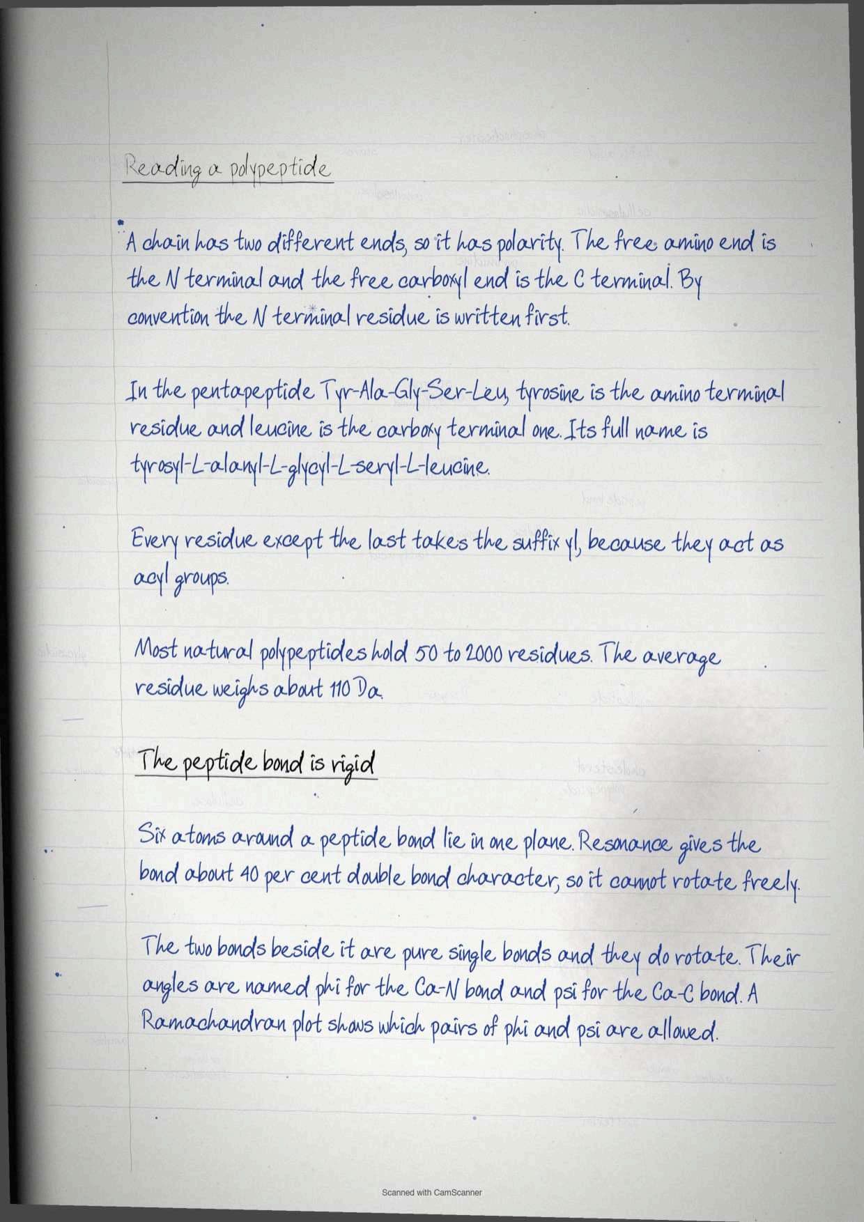
- A chain has polarity because its two ends differ. One is the amino or N-terminal end and the other is the carboxy or C-terminal end.
- By convention the N-terminal residue is written first, at the extreme left, and the C-terminal residue last. In the pentapeptide Tyr-Ala-Gly-Ser-Leu (YAGSL), tyrosine is amino-terminal and leucine is carboxy-terminal.
- In naming, every residue except the last takes the suffix -yl, because all of them are acyl groups. YAGSL is therefore named Tyrosyl-L-alanyl-L-glycyl-L-seryl-L-leucine.
- Chain length and mass follow a rough rule. Most natural polypeptides contain 50 to 2,000 residues, and the mean molecular weight of a residue is about 110 Da.
Box 2 of the chapter adds the geometry, and it is the part that explains why proteins fold at all. Six atoms of a pair of amino acids joined by a peptide bond lie in the same plane: the alpha carbon and CO group of the first amino acid, and the NH group and alpha carbon of the second. This planarity is the result of resonance interactions that give the peptide bond about 40 per cent double bond character, so the bond cannot rotate freely.
The bonds on either side are different. The bonds between the amino group and the alpha carbon, and between the alpha carbon and the carboxyl group, are pure single bonds and rotate freely. Their rotation angles are named phi (φ) for the Cα-N bond and psi (ψ) for the Cα-C bond, and together they decide the path of the whole chain. In 1963, G. N. Ramachandran showed that most pairs of phi and psi values are forbidden by steric clash, and the allowed regions plotted in two dimensions form the Ramachandran plot. For poly-L-serine it shows three allowed regions: one holding the parallel and antiparallel beta sheets and the collagen helix, one holding the right-handed alpha helix, and one holding the left-handed alpha helix, which is sterically allowed but too unstable to occur in real proteins.
Secondary Structure: The Alpha Helix and the Beta Pleated Sheet
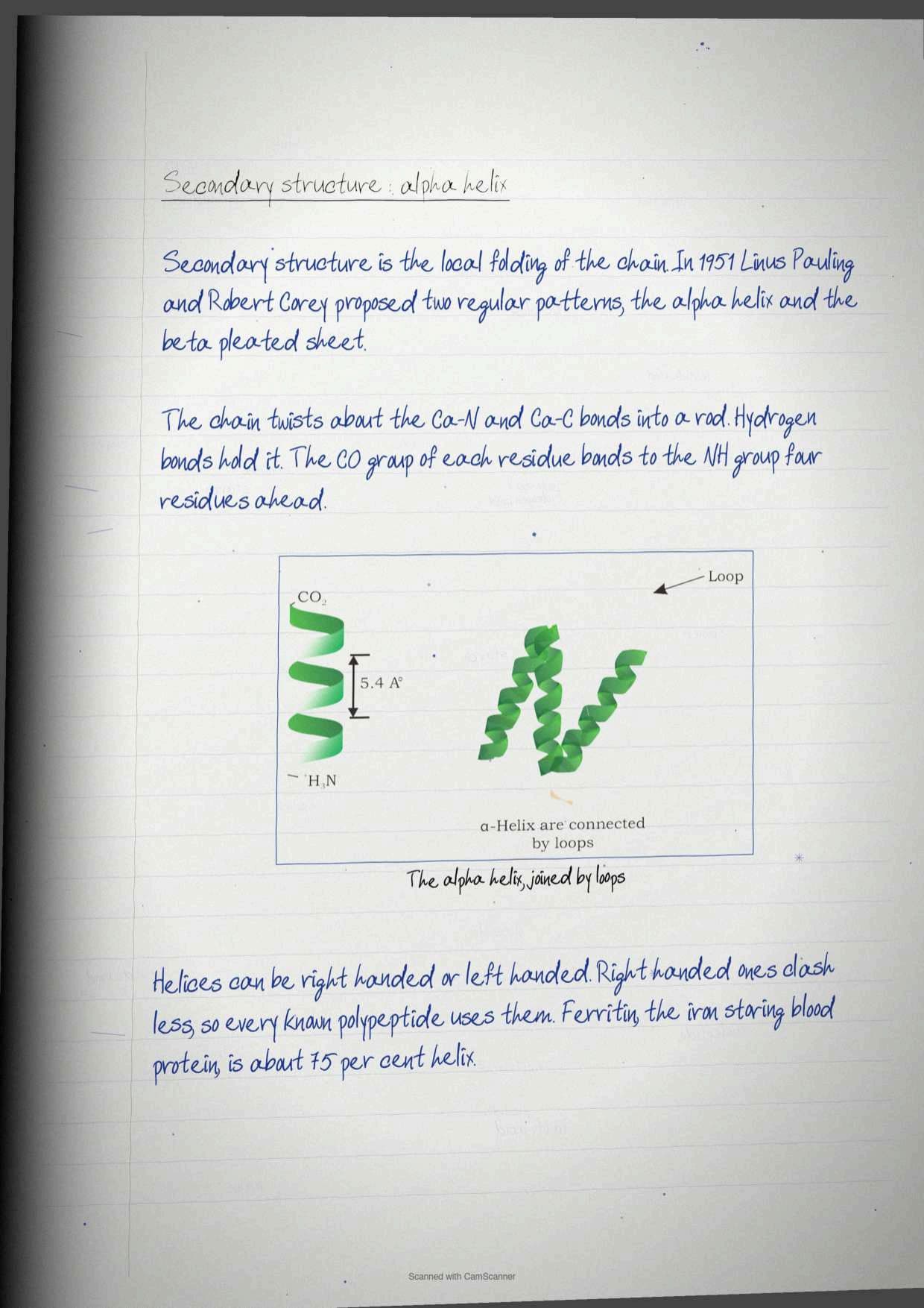
Secondary structure deals with the folding of the polypeptide chain. In 1951, Linus Pauling and Robert Corey proposed two periodic structures, and those two are still the whole of this section.
The alpha helix forms when a chain of planar peptide bonds twists about the Cα-N and Cα-C bonds, producing a rod-like structure. It is held together by hydrogen bonds between the NH and CO groups of the main chain, and the pattern is exact: the CO group of each amino acid hydrogen bonds to the NH group of the amino acid four residues ahead in the sequence. All main chain NH and CO groups are hydrogen bonded except those near the ends, and helices are joined to one another by loops.
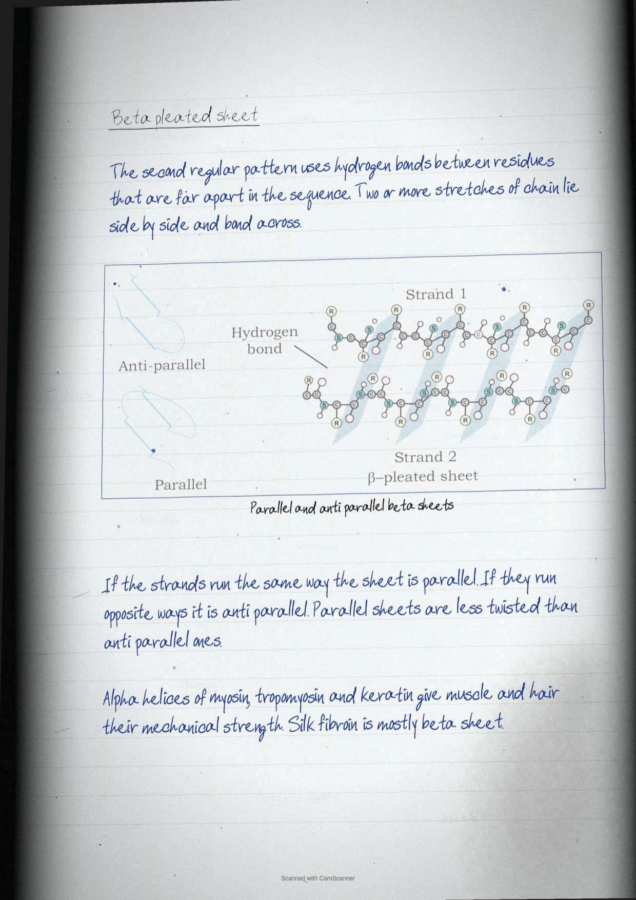
The beta pleated sheet works on the opposite principle. Its hydrogen bonds form between groups on residues that are far apart in the linear sequence. Two or more strands from widely separated parts of the backbone lie side by side and bond across to each other.
| Point of comparison | Alpha helix | Beta pleated sheet |
|---|---|---|
| Proposed by | Pauling and Corey, 1951 | Pauling and Corey, 1951 |
| Shape | Rod-like coil | Extended strands lying side by side |
| Hydrogen bonding | Between residues four apart in the same chain | Between residues distant in the sequence, across strands |
| Handedness or orientation | Right-handed or left-handed, but all known polypeptides use the right-handed form | Parallel if strands run the same way, anti-parallel if they run opposite |
| Stability note | Right-handed helices are sterically more stable, with less clash between side chains and backbone | Parallel sheets are less twisted than anti-parallel sheets |
| Where it is seen | Ferritin has 75 per cent of its residues in alpha helix; myosin, tropomyosin and keratin use it for mechanical strength | Found wherever strands must lie flat against one another in a folded protein |
Tip: exercise question 15 asks students to describe the various secondary structures of protein, and question 26 confirms that the expected answer to which is the most common secondary structure is both the alpha helix and the beta pleated sheet, not one of them alone.
Tertiary and Quaternary Structure of Proteins
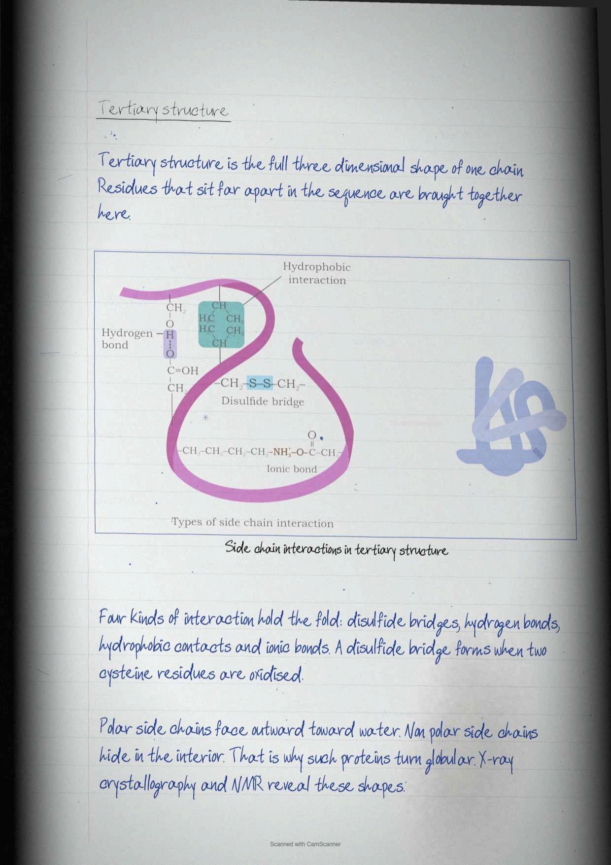
The tertiary structure is the overall three-dimensional arrangement of all residues in a protein. Where secondary structure looks at local folding, tertiary structure looks at long-range interactions: amino acids that are far apart in the chain, sitting in different secondary structures, coming together into one completely folded shape.
Four kinds of additional bond appear at this level, and each one is worth naming separately.
- Disulfide bonds, formed by oxidation of a pair of cysteine residues, the only covalent bond in the list.
- Hydrogen bonds between side chains.
- Hydrophobic interactions between non-polar side chains buried in the interior.
- Ionic bonds between oppositely charged side chains.
These bonds make the protein globular in shape, and some enzymes, transport proteins, peptide hormones and immunoglobulins are globular for this reason. The arrangement inside a globular protein follows one rule worth memorising: polar R groups sit on the outer surface because they are hydrophilic, and non-polar R groups are buried in the interior where they form hydrophobic interactions. These three-dimensional structures are determined experimentally by X-ray crystallography and nuclear magnetic resonance.
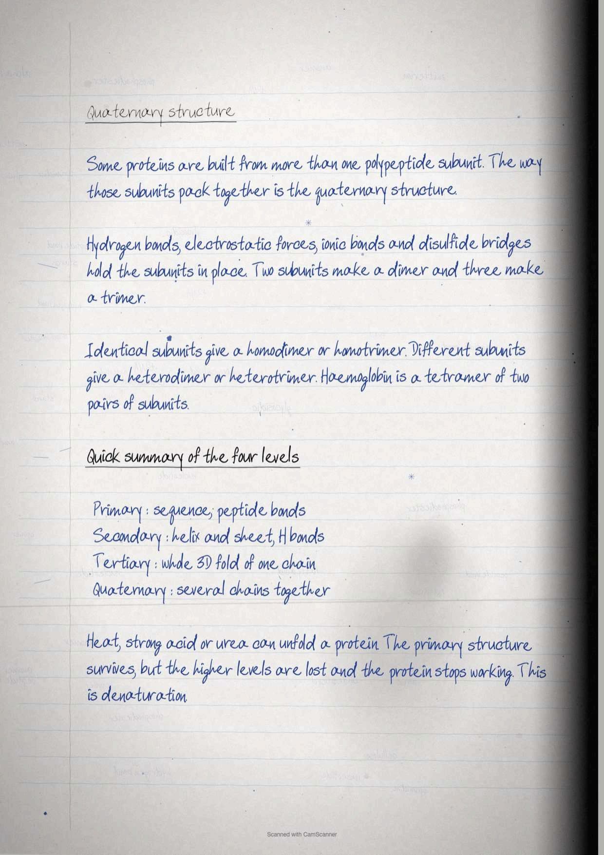
Quaternary structure appears only when a protein consists of more than one polypeptide subunit. It is the spatial arrangement of the protein subunits, which may be identical or different in their primary structure, and it is stabilised by hydrogen bonds, electrostatic interactions, ionic bonds and disulfide bridges.
| Level | What it describes | Bonds involved |
|---|---|---|
| Primary | Linear sequence of amino acids | Peptide bonds only |
| Secondary | Local folding into alpha helix and beta pleated sheet | Hydrogen bonds of the main chain |
| Tertiary | Complete three-dimensional shape of one chain | Disulfide, hydrogen, hydrophobic and ionic |
| Quaternary | Assembly of two or more subunits into one molecule | Hydrogen, electrostatic, ionic and disulfide bridges |
Naming a quaternary assembly follows the subunit count. Two subunits make a dimer, three a trimer and so on. If the subunits are identical the assembly is a homodimer or homotrimer, and if they differ it is a heterodimer or heterotrimer. Figure 3.28 draws a tetrameric protein built from two pairs of identical subunits, which is the diagram exercise question 16 expects.
Nucleic Acids: Sugars, Bases, Nucleosides and Nucleotides
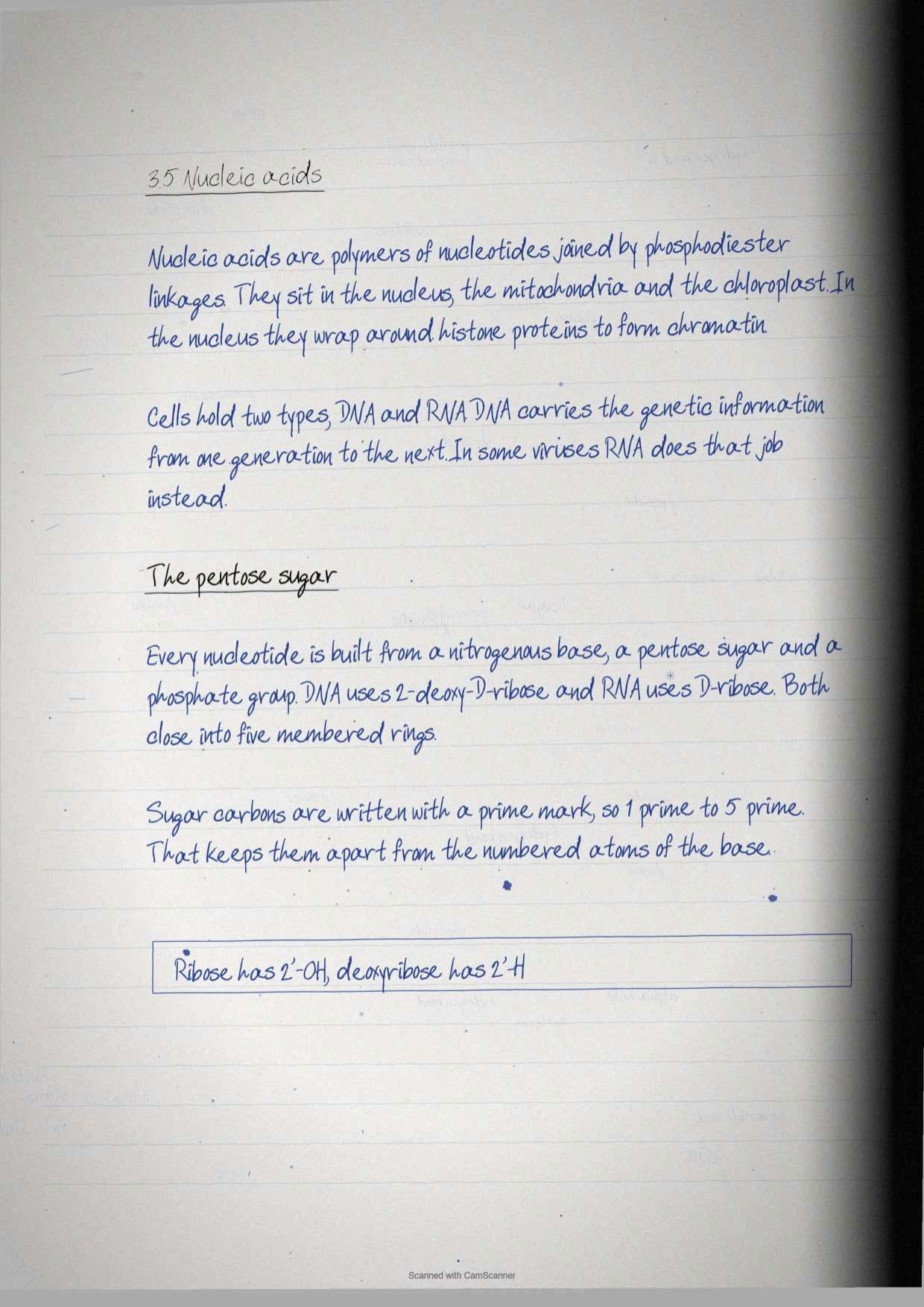
Nucleic acids sit inside the nucleus, the mitochondria and the chloroplast. Within the nucleus they associate with histone proteins to form chromatin. Structurally they are polymers of nucleotides linked through phosphodiester linkages, and there are two of them: deoxyribonucleic acid (DNA) and ribonucleic acid (RNA). DNA acts as the genetic material and passes information between generations, while RNA serves as the genetic material in some viruses.
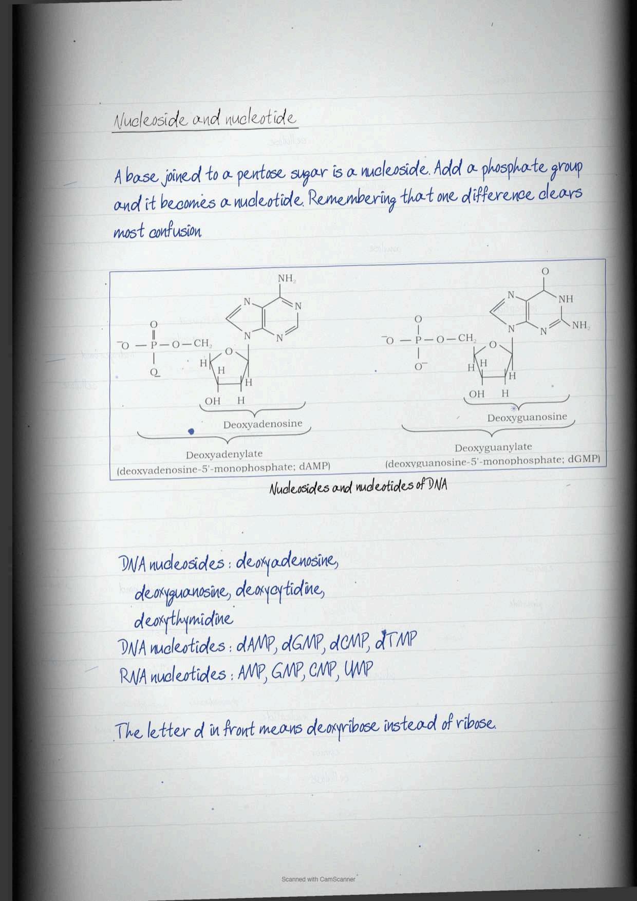
Every nucleotide is assembled from three parts: a nitrogenous base, a sugar and a phosphate. The sugar is always a pentose in a closed five-membered ring, and its carbons are numbered with a prime symbol so that they cannot be confused with the numbered atoms of the base.
| Feature | DNA | RNA |
|---|---|---|
| Pentose sugar | 2'-deoxy-D-ribose | D-ribose |
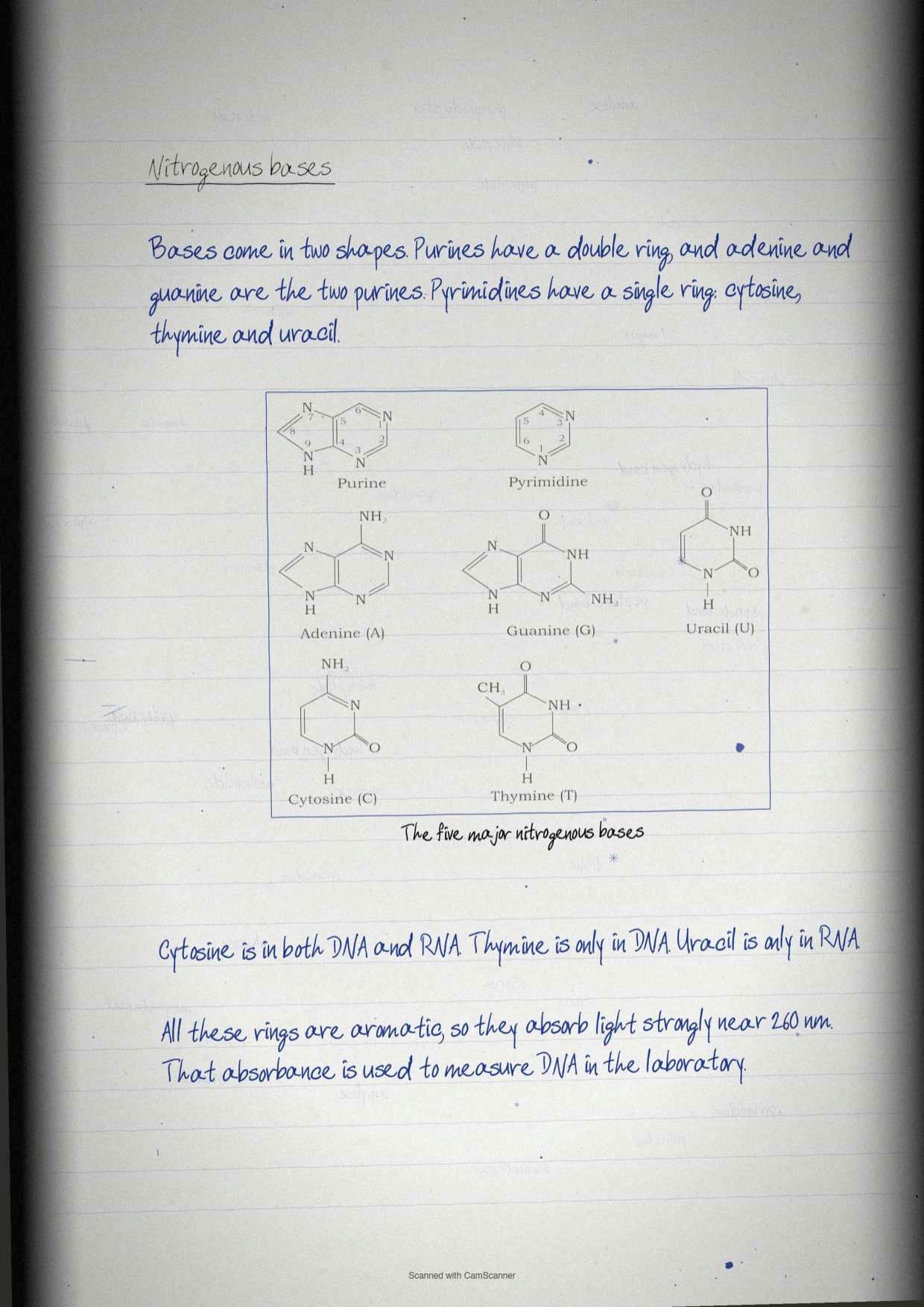
| Purine bases | Adenine (A), Guanine (G) | Adenine (A), Guanine (G) |
| Pyrimidine bases | Cytosine (C), Thymine (T) | Cytosine (C), Uracil (U) |
| Nucleosides | Deoxyadenosine, deoxyguanosine, deoxycytidine, deoxythymidine | Adenosine, guanosine, cytidine, uridine |
| Nucleotides | dAMP, dGMP, dCMP, dTMP | AMP, GMP, CMP, UMP |
The distinction the paper asks for, in exercise question 17, is a one-line one. A base linked to a pentose sugar and a phosphate group is a nucleotide; a base linked only to a pentose sugar, with no phosphate, is a nucleoside. The prefix d in dAMP and its relatives simply marks that the sugar is deoxyribose rather than ribose.
One physical property closes this section and turns up in laboratory questions. Purine and pyrimidine bases contain aromatic ring structures that absorb light at a wavelength near 260 nm, which is how nucleic acid concentration is measured in practice.
The Polynucleotide Chain and the Watson-Crick Double Helix
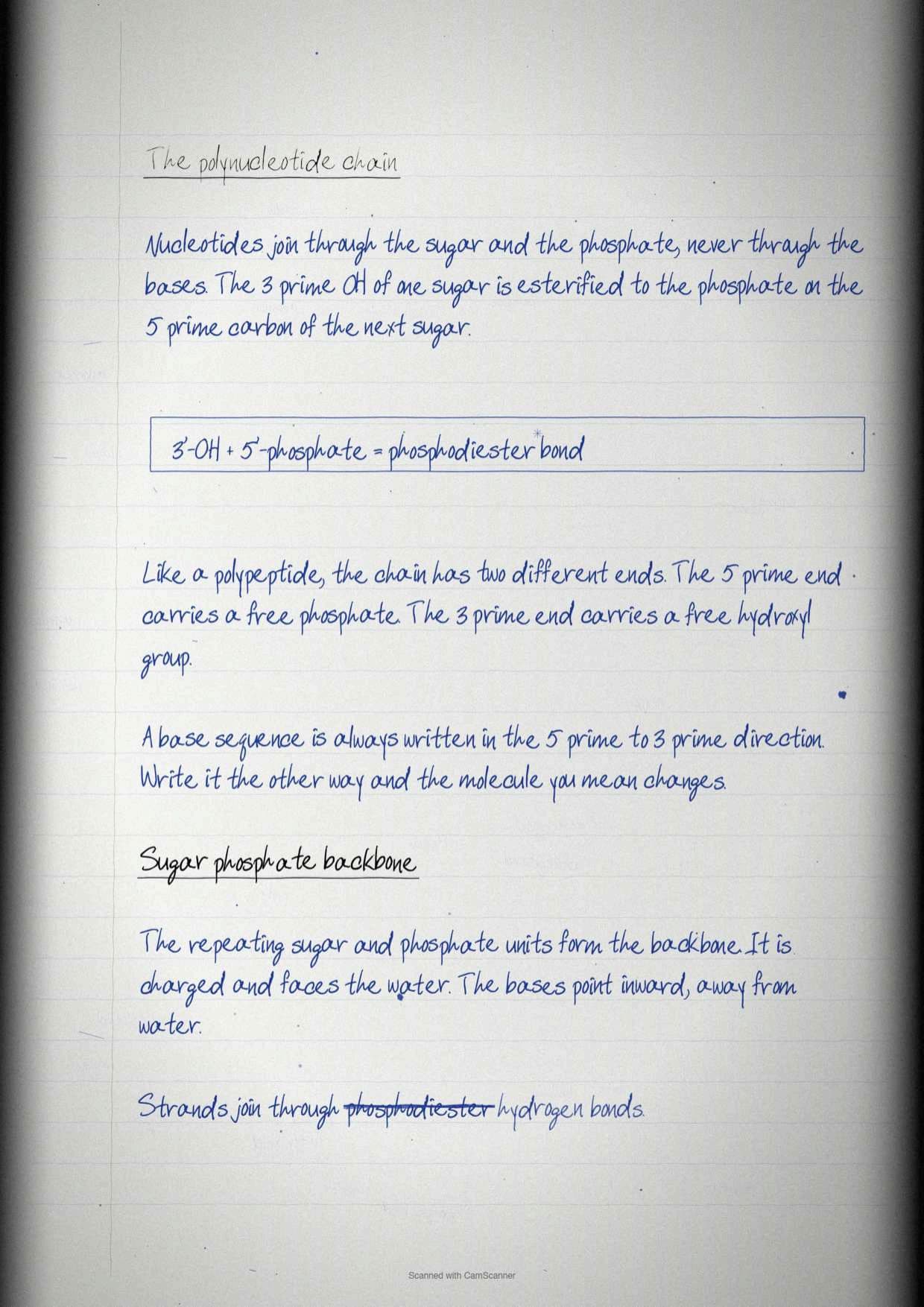
Nucleotides join through a single repeated linkage. The 3' hydroxyl group of the sugar of one nucleotide is esterified to the hydroxyl group of the phosphate attached to the 5' carbon of the next sugar, forming a phosphodiester linkage. Like a polypeptide, a polynucleotide chain therefore has polarity, with a 5' end carrying a phosphate at C-5' and a 3' end carrying a free hydroxyl at C-3'. By convention the base sequence is always written in the 5' to 3' direction.
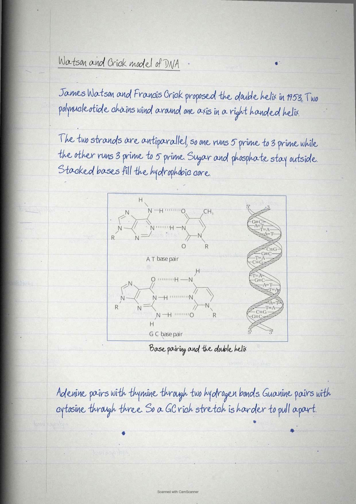
In 1953, James Watson and Francis Crick proposed the double helical three-dimensional structure of DNA, and the chapter lists its features in an order worth copying straight into an answer.
- Two polynucleotide chains wind around the same axis to form a right handed double helix.
- The two strands are antiparallel, so their 3' and 5' phosphodiester bonds run in opposite directions.
- The sugar and phosphate form the backbones, staying exposed to the polar environment outside.
- The bases are stacked inside the core, which makes the interior of the helix hydrophobic.
- Each base of one strand hydrogen bonds in the same plane with a base of the other strand.
Base pairing itself carries a number that must not be swapped. Adenine forms two hydrogen bonds with thymine (A=T), and guanine forms three hydrogen bonds with cytosine (G≡C). A purine always faces a pyrimidine, which is why the helix keeps a constant width.
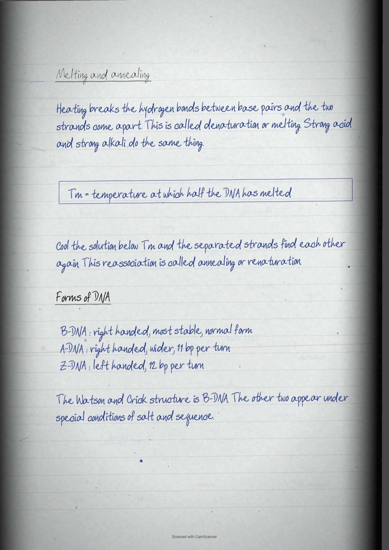
| Form of DNA | Handedness | Base pairs per helix | Note |
|---|---|---|---|
| B-DNA | Right handed | The Watson-Crick structure | The most stable form of DNA |
| A-DNA | Right handed | 11 | Wider than B-DNA |
| Z-DNA | Left handed | 12 | The only left handed form named in the chapter |
Because the two strands are held only by hydrogen bonds, they can be pulled apart. Denaturation, also called melting, is the separation of the two strands when heating disrupts those bonds, and acid or alkali does the same job. The melting temperature (Tm) is the temperature at which half the DNA is denatured. Drop the temperature below Tm and the separated strands spontaneously reassociate, a process called renaturation or annealing, which is the single most useful fact in this chapter for every later chapter on DNA technology.
Types of RNA: mRNA, rRNA and tRNA
Three types of RNA close the chapter, and each one is defined by the job it does during protein synthesis.
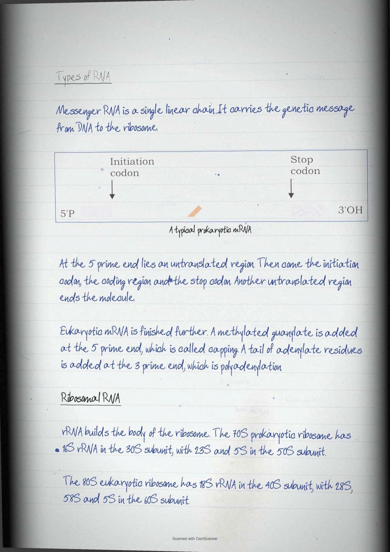
Messenger RNA (mRNA) is a single stranded linear polyribonucleotide chain that carries genetic information from DNA to the ribosome. Reading a prokaryotic mRNA from left to right gives five regions in a fixed order: a 5' untranslated region (UTR) that holds no information for polypeptide synthesis, an initiation codon, the coding region, a stop codon and a second UTR at the 3' end. Eukaryotic mRNA adds two modifications. The 5' end carries a guanylate methylated at its N-7, a process called capping, and the 3' end undergoes polyadenylation, the addition of several adenylate residues.
Ribosomal RNA (rRNA) forms the structural components of ribosomes, and the subunit numbers are pure recall.
| Ribosome | Small subunit | Large subunit |
|---|---|---|
| Prokaryotic, 70S | 30S, containing 16S rRNA | 50S, containing 23S and 5S rRNA |
| Eukaryotic, 80S | 40S, containing 18S rRNA | 60S, containing 28S, 5.8S and 5S rRNA |
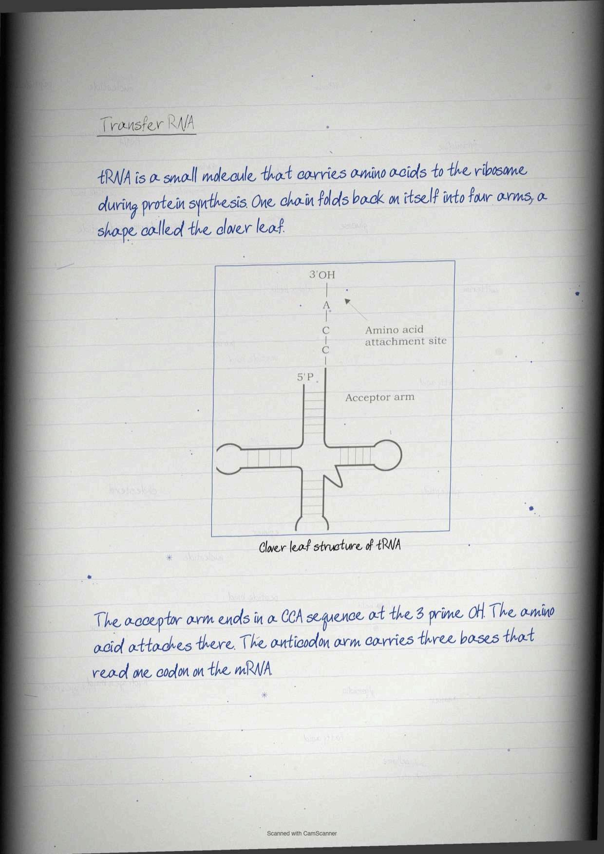
Transfer RNA (tRNA) is a small RNA that carries amino acids to the ribosome during protein synthesis. It is a single polyribonucleotide chain folded into four arms with a clover-leaf secondary structure. Two of those arms carry the marks. The acceptor arm has a CCA sequence at its 3'OH end, which is the amino acid binding site, and the anticodon arm carries the anticodon, a set of three bases that recognises a specific codon on the mRNA during translation. Exercise question 22 asks for the clover leaf model, so the diagram plus those two labels is the complete answer.
Where Enzymes Fit: What This Chapter Does Not Cover
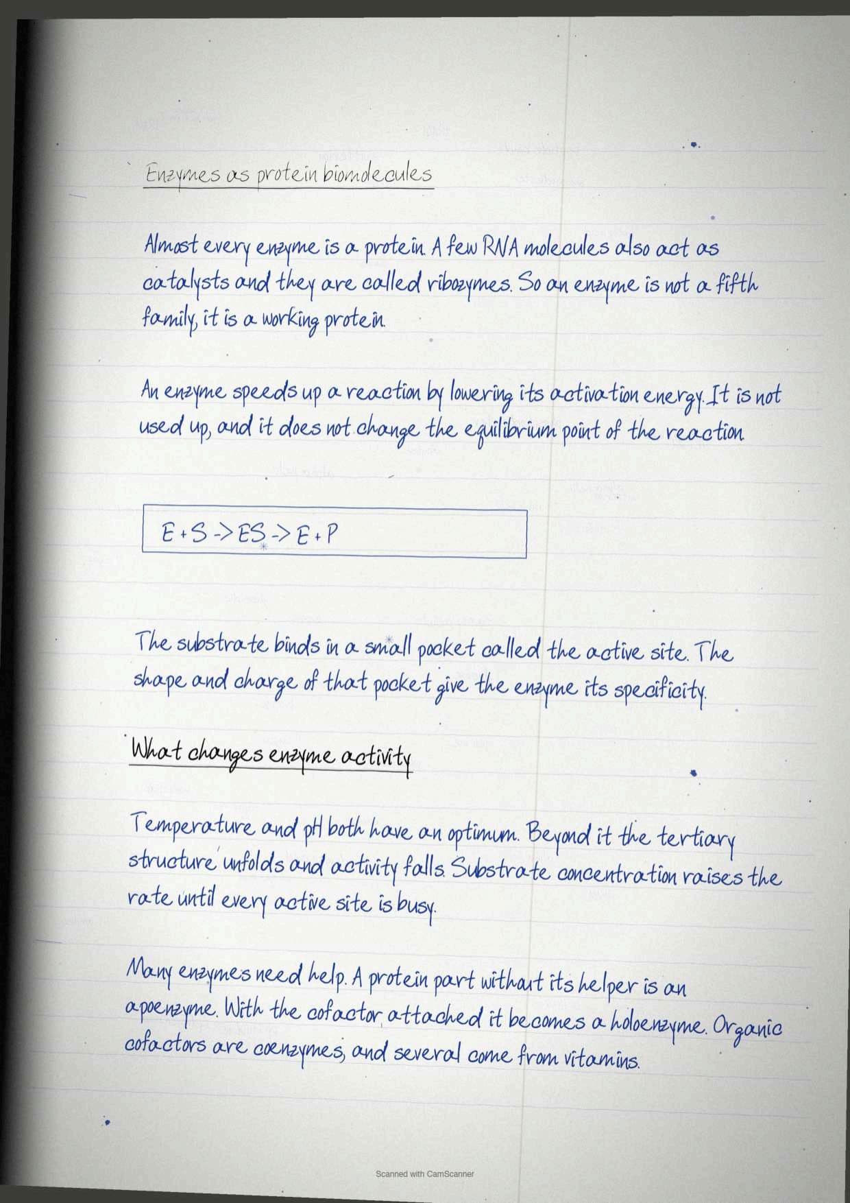
Students often expect a section on enzymes here, because enzymes are proteins and this is the protein chapter. Biomolecules deliberately stops at protein structure. The chapter names enzymes only in passing, as one of the products organisms build from amino acid building blocks, alongside antibodies, transporters, hormones, transcription factors, muscle fibres and membrane proteins.
- What this chapter gives you: the amino acid, the peptide bond, and the four levels of structure that decide a protein's final shape.
- What comes next: enzyme classification, active sites, kinetics and bioenergetics are the subject of Chapter 4, Enzymes and Bioenergetics, in the same Class 11 Biotechnology textbook.
- Why the order matters: an enzyme works because its tertiary structure creates a precisely shaped active site, so the folding rules learnt here are the prerequisite for everything in the next chapter.
The few enzymes that do appear by name in this chapter are there for carbohydrate reasons, not protein reasons: salivary and pancreatic amylase digesting starch, cellulase which humans lack, and lysozyme in tears breaking peptidoglycan. Learn those three as carbohydrate facts and leave enzyme theory for the next chapter.
Common Mistakes Students Make in Biomolecules
Six traps that cost easy marks in the Class 11 Biotechnology paper:
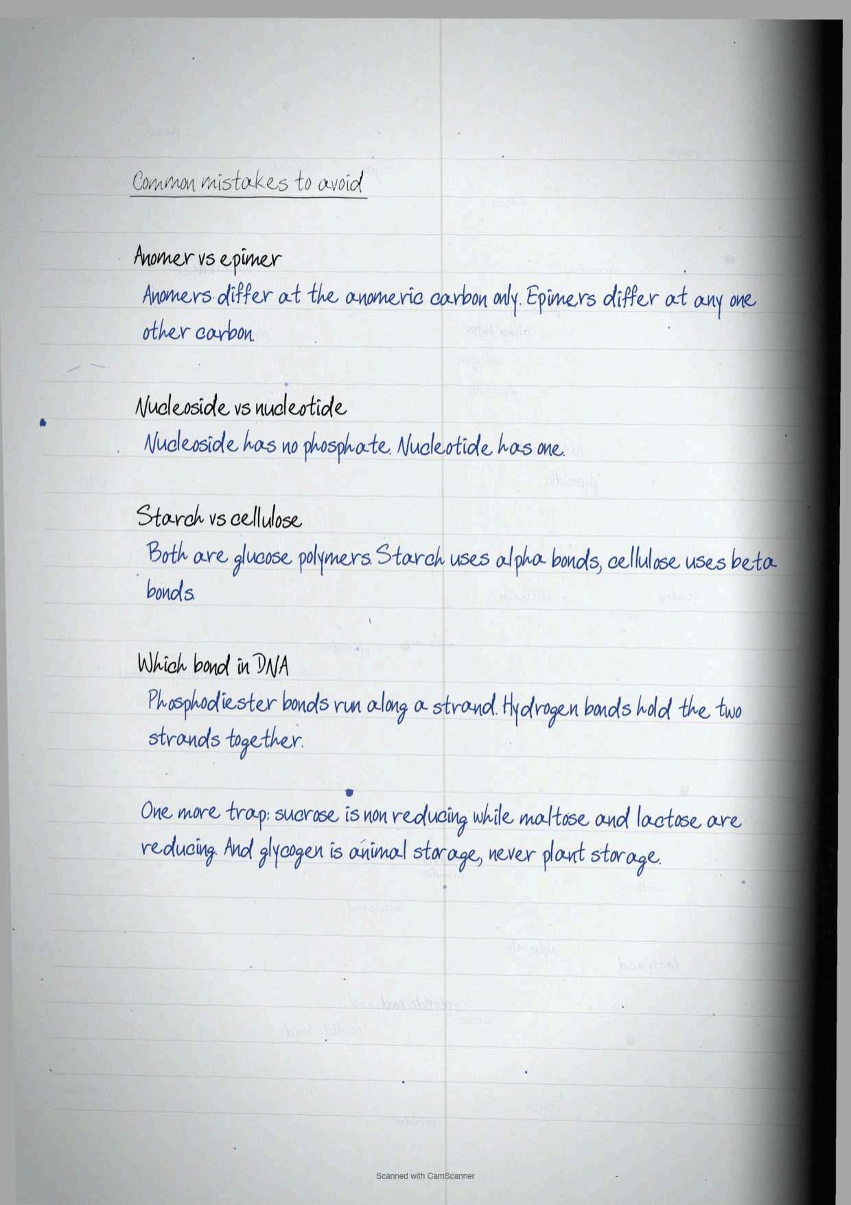
- Swapping the hydrogen bond counts. Adenine and thymine share two bonds, guanine and cytosine share three. The larger pair of letters does not get the larger number, so learn it as GC gets three.
- Confusing anomers with epimers. Anomers differ at the anomeric carbon created when the ring closes. Epimers differ at any one other carbon, which is why mannose and galactose are epimers of glucose but not anomers.
- Calling starch and cellulose the same polymer. Both are glucose chains, but starch uses alpha linkages and cellulose uses beta linkages, and that single letter is why humans digest one and not the other.
- Mixing non-standard with non-protein amino acids. Non-standard ones are made by modifying a standard amino acid after protein synthesis. Non-protein ones never enter a protein at all.
- Writing D isomers into proteins. Only L isomers of amino acids are found in proteins. The D and L rule for sugars runs the other way, since most biological sugars are D.
- Placing the peptide bond rotation wrongly. The peptide bond itself cannot rotate because of its 40 per cent double bond character. The neighbouring single bonds rotate, and those are the phi and psi angles.
Tick each trap off only after you have answered it correctly once in a written attempt. Students who worked this way said the isomer questions stopped costing them marks inside two revision rounds.
Quick Recall Sheet for Biomolecules
This is the page to read the night before and again in the morning. Every term the chapter can ask sits in one place.
| Term | One-line meaning |
|---|---|
| Biomolecules | Carbohydrates, proteins, lipids and nucleic acids, the four macromolecules that build every organelle |
| Monosaccharide | Simple sugar that cannot be hydrolysed further, formula Cn(H2O)n |
| D sugar | Hydroxyl on the carbon most distant from the carbonyl points right |
| Anomer | Isomer differing only at the hemiacetal or hemiketal carbon |
| Epimer | Isomer differing in hydroxyl configuration at exactly one carbon |
| Mutarotation | Interconversion of alpha and beta anomers in aqueous solution |
| Glycosidic bond | Covalent bond between a hydroxyl group and the anomeric carbon of another sugar |
| Cellulose | Beta(1→4) linked glucose, up to 15,000 units, most abundant biomolecule in the biosphere |
| Amphipathic | Molecule with a hydrophobic tail and a hydrophilic head, the definition of a membrane lipid |
| Micelle | Sphere-shaped structure phospholipids adopt instead of mixing with water |
| Cholesterol | Commonest steroid, precursor of all steroid hormones, gives membrane rigidity |
| Zwitterion | Dipolar amino acid at pH 7 with -COO- and -NH3+ together |
| Peptide bond | Amide bond joining an alpha carboxyl to an alpha amino group with loss of water |
| Alpha helix | Rod-like coil, CO hydrogen bonded to NH four residues ahead, right handed in real proteins |
| Beta pleated sheet | Side by side strands, parallel or anti-parallel, bonded across the chain |
| Quaternary structure | Spatial arrangement of two or more polypeptide subunits |
| Nucleoside against nucleotide | Base plus sugar, against base plus sugar plus phosphate |
| Phosphodiester linkage | 3' hydroxyl of one sugar joined to the 5' phosphate of the next |
| Tm | Melting temperature, at which half the DNA is denatured |
| Annealing | Spontaneous reassociation of separated strands below Tm |
The notebook also carries a mnemonic for the running order of the chapter: CLAP N, standing for Carbohydrates, Lipids, Amino acids, Protein structure, Nucleic acids, which is exactly the order of sections 3.1 to 3.5.
What the Biomolecules Handwritten Notes PDF Contains
The file is a scanned notebook written on ruled paper, with the textbook figures placed where the chapter uses them. Nothing is typed, so it reads the way a classmate's notes read.
- Pages 1 to 8: cover, the four classes of biomolecules, carbohydrate roles, the three-way classification and the monosaccharide grid from trioses to hexoses.
- Pages 9 to 15: chirality and the 2n rule, D and L glucose, ring closure, anomers, mutarotation, epimers, and the glycosidic bond with maltose, lactose and sucrose.
- Pages 16 to 22: the homopolysaccharide table, starch against glycogen against cellulose against chitin, peptidoglycan, agar, and the full fatty acid nomenclature with the saturated and unsaturated table.
- Pages 23 to 30: simple and compound lipids, phospholipids and micelles, cholesterol, the twenty amino acids by R group, the zwitterion, and the peptide bond with all four levels of protein structure.
- Pages 31 to 37: nucleic acids, nucleosides against nucleotides, the polynucleotide chain, base pairing, the double helix with A, B and Z forms, the three RNA types, the quick recall sheet and every exercise question worked out.
Textbook Exercise Questions and How These Notes Answer Them
The chapter closes with 30 exercises, of which 22 are written questions and 8 are multiple choice. The notebook writes out a model answer for each, and the table below shows where in the notes the material for each group sits.
| Exercise questions | Where the answer is built |
|---|---|
| Questions 1 to 6 on carbohydrate classification, D and L glucose, disaccharide structures, starch and glycogen diagrams, carbohydrate functions and isomerisation | The carbohydrate classification table, the chirality section and the anomer and epimer table |
| Questions 7 to 9 on sphingolipids against glycerolipids, amphipathic membrane lipids and saturated against unsaturated fatty acids | The lipid classification table, the phospholipid backbone list and the fatty acid symbol table |
| Questions 10 to 14 on amino acid categories, the zwitterion, non-standard amino acids, peptide bond formation and the Lys-Glu-Lys structure | The five-family amino acid table, the zwitterion explanation and the peptide bond section |
| Questions 15 and 16 on secondary structures and tertiary against quaternary structure | The alpha helix against beta sheet comparison and the four-level structure table |
| Questions 17 to 22 on nucleosides against nucleotides, the primary structure of DNA, an A-T-C-G oligonucleotide, the Watson and Crick model, DNA forms and the clover leaf tRNA | The nucleic acid comparison table, the five-point double helix list and the RNA section |
| Questions 23 to 30, the multiple choice set on functional groups, non-reducing disaccharides, protein repeating units, secondary structures, nucleotide composition, DNA strand bonding, storage lipids and glycerolipid bonding | Answered inline in the notebook margin against the relevant section |
How to Use These Handwritten Notes Most Effectively
This chapter is four short chapters wearing one number, so treating it as a single block is what makes it feel heavy. Students who split it by biomolecule class reported finishing it in about three and a half hours.
- Block 1, 55 minutes. Carbohydrates up to the glycosidic bond. Finish by drawing D-glucose and L-glucose side by side from memory.
- Block 2, 40 minutes. Polysaccharides. Cover the biological significance column of the homopolysaccharide table and rebuild it, then cover the monosaccharide column and rebuild that.
- Block 3, 45 minutes. Fatty acids and lipids. Write out the symbol for stearic, oleic and linoleic acid without looking, then define amphipathic in one sentence.
- Block 4, 50 minutes. Amino acids and all four levels of protein structure. Draw the peptide bond reaction, then list the bonds present at each level.
- Block 5, 40 minutes. Nucleic acids. Draw the two base pairs with the correct hydrogen bond counts and label a tRNA clover leaf.
- Night before, 15 minutes. Quick recall table and the six common mistakes, nothing else.
How These Handwritten Notes Pair with Other Biomolecules Resources
Also Check: the same four molecule classes are taught again in Class 11 Biology and in Class 12, and several of those pages are already published on Collegedunia. Read this chapter first for the biotechnology framing, then use the pages below for extra worked practice.
| Resource | Best used for | Open it |
|---|---|---|
| Class 11 Biology Notes | The same four biomolecule classes with more emphasis on metabolic roles and enzyme kinetics | Biomolecules Class 11 Biology Notes |
| Class 11 Biology Handwritten Notes | A second scanned revision pass over sugars, lipids, proteins and nucleic acids | Biomolecules Class 11 Biology Handwritten Notes |
| Class 11 Biology Formula Sheet | One-page recall of formulae, bond types and classification trees | Biomolecules Class 11 Biology Formula Sheet |
| Class 12 Biology Notes | The double helix, base pairing and DNA packaging taken much further | Molecular Basis of Inheritance Class 12 Notes |
| Class 12 Chemistry Notes | Carbohydrate, protein and nucleic acid chemistry from the organic chemistry side | Biomolecules Class 12 Chemistry Notes |
| Class 11 Biotechnology Notes | Typed revision notes for this chapter with every term explained | Biomolecules Class 11 Biotechnology Notes (coming soon) |
| Class 11 Biotechnology Book PDF | The official Class 11 Biotechnology chapter text with all original figures | Biomolecules Class 11 Biotechnology Book PDF (coming soon) |
NCERT Handwritten Notes for Class 11 Biotechnology: All Chapters
Related Links: handwritten revision notes for every chapter of the Class 11 Biotechnology textbook, written to the 2026-27 syllabus.
| Chapter | Class 11 Biotechnology Handwritten Notes |
|---|---|
| Chapter 1 | An Introduction to Biotechnology Class 11 Handwritten Notes |
| Chapter 2 | Cellular Organelles Class 11 Handwritten Notes (coming soon) |
| Chapter 3 | Biomolecules Class 11 Handwritten Notes (this page) |
| Chapter 4 | Enzymes and Bioenergetics Class 11 Handwritten Notes (coming soon) |
| Chapter 5 | Cellular Processes Class 11 Handwritten Notes (coming soon) |
| Chapter 6 | Basic Principles of Inheritance Class 11 Handwritten Notes (coming soon) |
| Chapter 7 | Basic Processes of DNA Class 11 Handwritten Notes (coming soon) |
| Chapter 8 | Genetic Disorder Class 11 Handwritten Notes (coming soon) |
| Chapter 9 | Introduction to Bioinformatics Class 11 Handwritten Notes (coming soon) |
| Chapter 10 | Protein Informatics and Cheminformatics Class 11 Handwritten Notes (coming soon) |
| Chapter 11 | Programming and Systems Biology Class 11 Handwritten Notes (coming soon) |
Biomolecules Class 11 Handwritten Notes FAQs
Questions Students Ask Before Downloading These Notes
Ques. What are the four major types of biomolecules?
Ans. The four major types are carbohydrates, proteins, lipids and nucleic acids. They are also called macromolecules because they are the ingredients of every cellular organelle: the cell membrane is made of lipids and proteins, the cell wall of carbohydrates, chromosomes of proteins and DNA, and ribosomes of proteins and RNA. Besides being structural, each class also carries out functions in cellular processes.
Ques. How do you tell a D sugar from an L sugar?
Ans. Look at the hydroxyl group on the carbon that is most distant from the carbonyl carbon. If that hydroxyl is on the right side of the carbon bearing it, the sugar is the D isomer, and if it is on the left, the sugar is the L isomer. Most sugars present in biological systems are D sugars, and Figure 3.3 of the chapter shows L-glucose and D-glucose drawn side by side.
Ques. What is the difference between anomers and epimers?
Ans. Anomers differ only in the configuration about the hemiacetal or hemiketal carbon, which is the anomeric carbon created when the sugar ring closes, giving the alpha and beta forms of D-glucopyranose. Epimers differ in the configuration of the hydroxyl group at exactly one carbon atom other than that one. Mannose is the epimer of glucose at C-2 and galactose is the epimer at C-4.
Ques. Why can humans digest starch but not cellulose?
Ans. Both are polymers of glucose, but starch is linked by alpha(1 to 4) glycosidic bonds while cellulose is linked by beta(1 to 4) bonds. Salivary amylase and pancreatic amylase hydrolyse alpha(1 to 4) linkages, so starch is digested to glucose. The human gut lacks cellulase, the enzyme that hydrolyses the beta(1 to 4) bond, so cellulose passes through. Cattle and termites digest cellulose only because symbiotic microorganisms in their gut secrete cellulase.
Ques. Why are membrane lipids called amphipathic?
Ans. Because a single molecule carries both a water-hating and a water-loving end. The fatty acid components form hydrophobic tails, while the alcohol moiety together with the additional group forms a hydrophilic head. Phospholipids built this way do not spontaneously mix with water and instead take up a sphere-shaped structure called a micelle, which is the basis of membrane formation.
Ques. How is a peptide bond formed?
Ans. The peptide bond, also called the amide bond, is formed by coupling the alpha carboxyl group of one amino acid to the alpha amino group of another amino acid, and the reaction is accompanied by the loss of a water molecule. Because of this loss, each amino acid unit inside a polypeptide is called a residue. Resonance gives the bond about 40 per cent double bond character, so it is planar and cannot rotate freely.
Ques. What is the difference between the alpha helix and the beta pleated sheet?
Ans. Both were proposed by Linus Pauling and Robert Corey in 1951. The alpha helix is a rod-like coil in which the CO group of each amino acid hydrogen bonds to the NH group of the residue four positions ahead in the same chain, and all known polypeptides use the right-handed form. The beta pleated sheet forms hydrogen bonds between residues that are distant in the linear sequence, with two or more strands lying side by side, either parallel or anti-parallel.
Ques. How many hydrogen bonds are there between the DNA base pairs?
Ans. Adenine forms two hydrogen bonds with thymine, written A=T, and guanine forms three hydrogen bonds with cytosine, written G with a triple bond to C. The two strands run antiparallel around a right handed axis, the sugar and phosphate backbones face the polar environment outside, and the stacked bases make the core of the helix hydrophobic. Heating breaks these hydrogen bonds, which is denaturation or melting.
Ques. Does this chapter cover enzymes?
Ans. No. Biomolecules stops at protein structure and names enzymes only as one of the products organisms build from amino acids. Enzyme classification, active sites, kinetics and bioenergetics are covered in Chapter 4 of the same Class 11 Biotechnology textbook. The four levels of protein folding taught here are the prerequisite, because an enzyme works through the active site shaped by its tertiary structure.
Ques. Where can I download the Class 11 Biotechnology Biomolecules handwritten notes PDF?
Ans. Use the download option at the top of this page. The file is free and printable, runs to 37 handwritten pages, and follows the 2026-27 chapter order from carbohydrates through to the three types of RNA, so it can sit beside the textbook while you revise.



Comments