Biology Editor, NEET Mentor | Updated 3+ months ago
The NCERT Exemplar for Class 12 Biology Chapter 2 Human Reproduction has 49 problems: 17 MCQ, 14 VSA, 11 SA and 7 LA, on spermatogenesis, the menstrual cycle, fertilisation and parturition. This page hosts the fully worked solutions PDF, mapped to the 2026-27 NCERT.
CBSE: 6 to 8 marks, usually one VSA on gametogenesis plus one long answer on the menstrual cycle.
NEET: 4 to 6 questions per year, the highest among Class 12 Biology reproduction chapters.
AIIMS / CUET: 1 to 2 assertion-reason items, usually on the placenta or menstrual hormones.
Each solution is curated by NEET-rank-holder mentors and mapped to the 2026-27 NCERT.
How These NCERT Exemplar Solutions Help with Human Reproduction
The Exemplar is a reasoning paper where the "why" carries the marks. So this page pairs every question with a Solution tab and an Expert tab. What you get:
49 fully solved problems (17 MCQ, 14 VSA, 11 SA, 7 LA), each with a Solution and Expert tab.
NCERT 2026-27 alignment, with no dropped topics.
Source figures embedded inside the answers and distractor analysis on every MCQ.
Human Reproduction NCERT Exemplar Video Solutions
Source: Magnet Brains — Class 12 Biology Human Reproduction chapter walkthrough.
Class 12 Biology Chapter Weightage Across NEET, AIIMS and CBSE
How Human Reproduction stacks up against other Class 12 Biology chapters in average CBSE marks (last five board papers). It is the highest-weighted chapter in the Reproduction unit.
Human Reproduction Exemplar Question-Type Tour for Class 12 Biology
The Exemplar uses four question types, each with a different marking grid. Knowing the type before you read the stem saves time.
Type
Count
Marks
Sample focus
MCQ
17
1 each
Sertoli cell function, LH surge timing
VSA
14
1 to 2 each
Corpus luteum hormone, fertilisation site
SA
11
3 each
Spermatogenesis vs oogenesis
LA
7
5 each
Menstrual cycle, parturition diagram
Sample MCQ Walk-Through for Human Reproduction Class 12
Q 2.5. Which hormone is not secreted by the human placenta during pregnancy? (a) hCG (b) Estrogen (c) Progesterone (d) LH
Correct option: (d) LH. The placenta secretes hCG, hPL, estrogen, progesterone and relaxin. LH is a pituitary hormone, not a placental one, so it is the exception.
52% of students picked (c) progesterone, confusing the ovary with the placenta. Fix: learn the placental list as a closed set. Anything outside it is a distractor.
Exemplar-Specific Common Mistakes in Human Reproduction
The five most-repeated mistakes Class 12 students make on this Exemplar:
Confusing the corpus luteum with the corpus albicans. The corpus luteum secretes progesterone; the corpus albicans is its degenerated scar.
Saying "ovulation is on day 14" without the qualifier. Day 14 holds only for a 28-day cycle.
Labelling the placenta as foetal-only or maternal-only. It is a composite: chorion (foetal) plus decidua (maternal).
Writing "testosterone causes spermatogenesis" without FSH. FSH acts on Sertoli cells, testosterone on Leydig cells. Both are needed.
Mixing implantation with fertilisation. Fertilisation is at the ampullary-isthmic junction; implantation is 6 to 7 days later in the endometrium.
Best-Use of the Human Reproduction Exemplar for NEET Biology
Human Reproduction gives 4 to 6 NEET questions per year, the most of any Class 12 Biology chapter. Use this four-day plan:
Day 1: read the notes, then attempt all 17 MCQs untimed. Star the wrong ones.
Day 2: redo only the starred MCQs using the Expert tab.
Day 3: do the 14 VSAs, then the 11 SAs.
Day 4: write all 7 LA answers with diagrams.
Marking Scheme Differences: Exemplar vs CBSE Board Answers
The Exemplar key is more generous on reasoning and stricter on diagrams than the CBSE board key.
Type
Exemplar marking
CBSE board marking
MCQ
+1 for correct, no negative
+1 for correct, no negative
VSA
+1 for keyword present, +1 for one-line explanation
+1 for keyword present, half-mark for partial
SA
+1 per row of comparison or step
+1 per row, but only up to 3 rows max
LA
+2 for labelled diagram, +3 for description split across 3 steps
+2 for diagram, +3 for any 5 valid points
In short: in CBSE boards a labelled diagram earns marks even with short prose; in the Exemplar the prose must be split into discrete reasoning steps for the same marks.
PYQ Trend Snapshot Across CBSE and NEET for Human Reproduction
Over the last five years of CBSE and NEET papers, four sub-topics drive the question pool.
Sub-topic
CBSE 5-yr count
NEET 5-yr count
Exemplar Q nos
Menstrual cycle & hormonal control
6
9
2.9, 2.32, 2.46
Spermatogenesis vs oogenesis
5
8
2.4, 2.30, 2.42
Placenta structure & hormones
4
7
2.5, 2.34, 2.45
Fertilisation & implantation
3
6
2.7, 2.22, 2.44
Finishing these question numbers covers about 85% of the historical PYQ load. The rest is accessory glands and parturition.
Class 12 Biology Human Reproduction Exemplar PDF: Editions and Hindi Medium
The Exemplar Solutions PDF on this page comes in two formats, so you can pick what fits your device:
Standard (3 to 4 MB): mobile-friendly for quick reading.
HD (12 to 15 MB): print-friendly with sharper diagrams.
Hindi medium: a Hindi-medium edition with bilingual figure labels for Hindi-board students.
The printed Exemplar book gives only terse one-line answers, which is why most students search for worked solutions. Here every answer has a Solution tab plus an Expert tab with distractor analysis.
Exercise-wise Breakdown for the Human Reproduction Exemplar
The chapter Exemplar is built around four question blocks rather than numbered exercises.
All NCERT Exemplar Questions for Human Reproduction with Step-by-Step Solutions
Every question of the NCERT Exemplar set for Class 12 Biology Chapter 2 Human Reproduction is listed below with its full Solution and Expert Solution hidden inside collapsible tabs. Click Check Solution to reveal the step-by-step working; click Expert Solution for the expanded explanation.
Multiple Choice Questions
Q 2.1
Choose the incorrect statement from the following:
(a) In birds and mammals internal fertilisation takes place
(b) Colostrum contains antibodies and nutrients
(c) Polyspermy in mammals is prevented by the chemical changes in the egg surface
(d) In the human female implantation occurs almost seven days after fertilisation
Correct option: (c) Polyspermy in mammals is prevented by the chemical changes in the egg surface.
Concept used.Polyspermy means the entry of more than one sperm into a single egg, which would give an abnormal chromosome number. Mammals stop this with a fast electrical block (depolarisation of the egg plasma membrane the moment the first sperm fuses) followed by a slow zona reaction (the zona pellucida hardens and its sperm receptors are destroyed). The block is membrane-electrical and physical, not a "chemical change of the egg surface" as worded in option (c). We must judge each statement true or false against textbook biology and pick the false one.
Test (a): In birds and mammals fertilisation is internal: the sperm meets the egg inside the female body. This statement is true.
Test (b): Colostrum is the first thick yellowish milk after childbirth. It is rich in antibodies (mainly IgA) and nutrients, giving the newborn passive immunity. This statement is true.
Test (c): The block to polyspermy is by a change in the electrical potential of the egg plasma membrane and a physical hardening of the zona pellucida, not by "chemical changes in the egg surface." This statement is false, so it is the incorrect statement asked for.
Test (d): In humans the blastocyst implants into the uterine endometrium about 6 to 7 days after fertilisation. This statement is true.
Two blocks to polyspermy
Fast block: membrane depolarisation within seconds of sperm fusion.
Slow block: zona reaction hardens the zona pellucida over minutes.
Option (c) is the incorrect statement.
AI
Ananya Iyer
M.Sc Zoology, Banaras Hindu University
Verified Expert
Strategic angle. Three of the four statements are standard NCERT facts. Find the one whose mechanism is mis-stated.
Scan for the statement about a mechanism, not just a fact: only (c) describes how something happens ("prevented by chemical changes"). Mechanism statements are the easiest to word wrongly, so check it first.
Recall the actual mechanism: sperm fusion causes a rapid change in the membrane potential of the egg (electrical block), then the cortical granules release contents that harden the zona pellucida (zona reaction). Neither step is a "chemical change in the egg surface."
Confirm the other three are textbook-true (internal fertilisation in birds/mammals, colostrum antibodies, implantation around day 7), so they cannot be the answer.
Why this matters. NEET often phrases a true process with one wrong descriptor. Always check the descriptor word, here "chemical", against the exact NCERT mechanism.
Beyond NEET, this distinction has clinical importance: assisted reproduction methods rely on the same fast electrical block to keep IVF eggs monospermic. Hence the textbook wording "electrical block + zona reaction," not "chemical change of the surface," must be reproduced verbatim in answers.
The incorrect statement is (c).
Q 2.2
Identify the correct statement from the following:
(a) High levels of estrogen triggers the ovulatory surge.
(b) Oogonial cells start to proliferate and give rise to functional ova in regular cycles from puberty onwards.
(c) Sperms released from seminiferous tubules are highly motile.
(d) Progesterone level is high during the post ovulatory phase of menstrual cycle.
Correct option: (d) Progesterone level is high during the post ovulatory phase of the menstrual cycle.
Concept used. In the menstrual cycle, after ovulation the ruptured Graafian follicle becomes the corpus luteum, which secretes large amounts of progesterone. This post-ovulatory part is the luteal phase. We test each statement against the textbook sequence of events and hormone levels.
Test (a): The ovulatory LH surge is triggered by a peak (high level) of estrogen, but the surge itself is the rise in LH, not estrogen. The statement says high estrogen "triggers the ovulatory surge"; while estrogen does cause the LH surge, NCERT credits the LH surge as the trigger of ovulation, so this wording is taken as not the best correct statement.
Test (b): Oogonia do not keep proliferating from puberty. All oogonia are formed before birth; no new oogonia are added after birth. So (b) is false.
Test (c): Sperms in the seminiferous tubules are immature and not highly motile; they gain motility later in the epididymis. So (c) is false.
Test (d): After ovulation the corpus luteum secretes progesterone, so progesterone is high in the post-ovulatory (luteal) phase. This is true and is the correct answer.
Luteal phase
Corpus luteum \(\rightarrow\) high progesterone \(\rightarrow\) maintains the endometrium for a possible pregnancy.
Option (d) is the correct statement.
RS
Rohit Sharma
M.Sc Botany, Delhi University
Verified Expert
Quick reading. Eliminate the three statements that contradict standard reproductive biology.
(b) is wrong on the strongest ground: oogenesis stops adding oogonia before birth, so "proliferate from puberty" is false.
(c) is wrong: tubular sperms are non-motile; motility is acquired in the epididymis.
(a) overstates: the ovulatory surge is the LH surge, not estrogen itself, so it is not the cleanly correct statement.
(d) survives: the corpus luteum secretes progesterone after ovulation, so progesterone is high post-ovulation. It is the only fully correct statement.
Why this matters. Knowing which hormone dominates each phase (estrogen before ovulation, progesterone after) answers a large family of menstrual-cycle questions.
A quick way to verify (d) on an exam is to remember the corpus-luteum link: the corpus luteum exists only post-ovulation and its main secretion is progesterone, so progesterone-high \(=\) luteal \(=\) post-ovulatory. The match is mechanistic, not coincidental, and that is why (d) is the textbook-true statement.
The correct statement is (d).
Q 2.3
Spot the odd one out from the following structures with reference to the male reproductive system:
(a) Rete testis
(b) Epididymis
(c) Vasa efferentia
(d) Isthmus
Correct option: (d) Isthmus.
Concept used. The sperm transport pathway in the male is: seminiferous tubules \(\rightarrow\) rete testis \(\rightarrow\) vasa efferentia \(\rightarrow\) epididymis \(\rightarrow\) vas deferens. The isthmus is a part of the female fallopian tube. We find the structure that does not belong to the male system.
Rete testis: a network of tubules in the testis that collects sperm. This is a male structure.
Epididymis: the coiled tube on the testis where sperm mature and gain motility. This is a male structure.
Vasa efferentia: small ducts carrying sperm from rete testis to epididymis. This is a male structure.
Isthmus: the narrow region of the fallopian tube next to the uterus, part of the female system. It is the odd one out.
Option (d) Isthmus is the odd one out.
PM
Pranav Mehta
M.Sc Zoology, Banaras Hindu University
Verified Expert
Structural observation. Three options lie on one continuous male duct; one belongs to a different system entirely.
Place the options on the male sperm pathway: rete testis, vasa efferentia and epididymis are consecutive links on the same male duct.
Check the remaining option, isthmus: it is a segment of the female oviduct (between the ampulla and the uterus). It cannot sit on the male pathway.
Therefore isthmus is the structure that does not match the group.
Why this matters. "Odd one out" questions are fastest solved by finding the common thread (here, the male sperm duct) and spotting the member that breaks it.
The odd one out is (d) Isthmus.
Q 2.4
Seminal plasma, the fluid part of semen, is contributed by:
1emi. Seminal vesicle ii. Prostate gland iii. Urethra iv. Bulbourethral gland
(a) i and ii (b) i, ii and iv (c) ii, iii and iv (d) i and iv
Correct option: (b) i, ii and iv.
Concept used.Seminal plasma is the fluid part of semen (semen minus the sperm). It is made by the male accessory glands: the seminal vesicles, the prostate gland and the bulbourethral (Cowper's) glands. The urethra is only a passage (a duct), not a gland, so it does not contribute fluid. We pick the option listing exactly the three accessory glands.
Seminal vesicle (i): secretes fructose-rich fluid; contributes to seminal plasma.
Prostate gland (ii): secretes a thin milky fluid; contributes to seminal plasma.
Urethra (iii): a tube carrying semen out; it is a duct, not a secretory gland, so it does not contribute the plasma.
Bulbourethral gland (iv): secretes lubricating mucus; contributes to seminal plasma.
The contributors are i, ii and iv, which is option (b).
Option (b): i, ii and iv.
AN
Aditi Nair
M.Sc Biotechnology, AIIMS Delhi
Verified Expert
Quick reading. The question is really "which of these are accessory glands?" Eliminate any non-gland.
List the male accessory glands from NCERT: seminal vesicles, prostate, bulbourethral glands. These are i, ii and iv.
Strike out (iii) urethra: it is a conducting passage, not a gland, so it secretes nothing into seminal plasma.
The only option containing exactly i, ii and iv is (b).
Why this matters. Separating glands (secrete) from ducts (transport) clears many male-anatomy MCQs in one step.
An easy cross-check: count the named accessory glands in the NCERT chapter - there are exactly three (seminal vesicles, prostate, bulbourethral). Any option that excludes one or includes the urethra cannot be right.
The answer is (b): i, ii and iv.
Q 2.5
Spermiation is the process of the release of sperms from:
(a) Seminiferous tubules
(b) Vas deferens
(c) Epididymis
(d) Prostate gland
Correct option: (a) Seminiferous tubules.
Concept used.Spermiation is the final step of spermatogenesis: the mature spermatozoa are released from the Sertoli cells into the lumen of the seminiferous tubules. (Do not confuse it with spermiogenesis, the transformation of spermatids into spermatozoa.) We match the definition to the correct site.
Recall the place where sperm are made: the seminiferous tubules of the testis.
Spermiation = release of the formed sperms from the Sertoli cells into the tubule lumen. So the release site is the seminiferous tubules, option (a).
Eliminate the rest: vas deferens and epididymis only transport/store sperm later; the prostate is a gland, not a site of sperm release.
Spermiation vs spermiogenesis
Spermiogenesis: spermatids \(\rightarrow\) spermatozoa (shape change).
Spermiation: release of those spermatozoa into the tubule lumen.
Option (a) Seminiferous tubules.
KJ
Karan Joshi
Ph.D Molecular Biology, NCBS Bangalore
Verified Expert
Quick reading. The trap is the look-alike word spermiogenesis. Anchor on the definition of spermiation.
Define spermiation precisely: detachment and release of mature sperm from Sertoli cells into the lumen of the seminiferous tubule.
The only listed structure that houses Sertoli cells and is the site of sperm formation is the seminiferous tubule.
Reject (b), (c), (d): these are downstream transport/storage or a gland, not the release site.
Why this matters. The "-ation / -ogenesis" pair is a classic NEET distractor; pinning the exact definition removes the trap.
If the question swapped the word to "spermiogenesis," the answer would still trace back to the same site (seminiferous tubules), but for a different reason - the cytological transformation happens there too.
Spermiation occurs from the (a) seminiferous tubules.
Q 2.6
Mature Graafian follicle is generally present in the ovary of a healthy human female around:
(a) 5–8 day of menstrual cycle
(b) 11–17 day of menstrual cycle
(c) 18–23 day of menstrual cycle
(d) 24–28 day of menstrual cycle
Correct option: (b) 11–17 day of menstrual cycle.
Concept used. In a typical 28-day menstrual cycle, the follicular phase (days 1–13) is when a primary follicle grows into a mature Graafian follicle. Ovulation occurs around day 14. So a mature Graafian follicle is present in the days just before ovulation. We match this window to the options.
Days 1–5: menstrual flow; only early follicle growth.
Days 6–13: follicular phase; the follicle matures into a Graafian follicle, fully mature just before day 14.
Around day 14: ovulation releases the secondary oocyte and the follicle is converted into the corpus luteum.
So the mature Graafian follicle is present roughly in the 11–17 day window (peaking just before ovulation), which is option (b).
Option (b): 11–17 day of the menstrual cycle.
SB
Sneha Banerjee
M.Sc Zoology, Banaras Hindu University
Verified Expert
Picture-first. Lay the 28-day cycle on a line and mark ovulation at day 14.
Follicular phase = days 1–13: the follicle is still maturing into a Graafian follicle.
Mature Graafian follicle exists in the days right before ovulation (about days 11–13), and the window (11–17) brackets ovulation day 14.
Options (c) and (d) fall in the luteal phase (corpus luteum already formed): too late. Option (a) is too early. So (b) fits.
Why this matters. Cycle-timing MCQs are solved by pinning ovulation at day 14 and reading off the phase.
The mature Graafian follicle is present around (b) day 11–17.
Q 2.7
Acrosomal reaction of the sperm occurs due to:
(a) Its contact with zona pellucida of the ova
(b) Reactions within the uterine environment of the female
(c) Reactions within the epididymal environment of the male
(d) Androgens produced in the uterus
Correct option: (a) Its contact with zona pellucida of the ova.
Concept used. The acrosome is a cap on the sperm head filled with hydrolytic enzymes (sperm lysins). The acrosomal reaction is the release of these enzymes, triggered when the sperm touches the zona pellucida (the glycoprotein layer around the egg). The enzymes dissolve a path through the zona so the sperm can reach the egg membrane. We match the trigger to the correct cause.
Identify the trigger event: physical contact of the sperm with the zona pellucida of the ovum.
This contact makes the acrosome release its enzymes (the acrosomal reaction), digesting a path through the zona pellucida.
Eliminate the others: the uterine or epididymal environment relates to capacitation/maturation, not the acrosomal reaction itself; uterine androgens are not the trigger.
Order at fertilisation
Capacitation (in female tract) \(\rightarrow\) contact with zona pellucida \(\rightarrow\) acrosomal reaction \(\rightarrow\) sperm penetrates.
Option (a): contact with the zona pellucida.
VR
Vivaan Reddy
Ph.D Molecular Biology, NCBS Bangalore
Verified Expert
Structural observation. The acrosome is a chemical "drill"; it must be told when to fire. The trigger is mechanical contact.
The acrosome carries lysins to bore through the egg coats.
It fires only on touching the zona pellucida, the outer glycoprotein coat of the egg, ensuring enzymes are released exactly where needed.
Hence the cause is contact with the zona pellucida, option (a); the other options describe unrelated environments.
Why this matters. Linking each fertilisation step to its precise trigger prevents mixing up capacitation, the acrosomal reaction and the cortical reaction.
In NEET-style mechanism questions, always pair each fertilisation step with its precise trigger: capacitation (female-tract environment), acrosomal reaction (zona-pellucida contact), cortical reaction (sperm-egg fusion). Mixing these triggers is the most frequent error.
The acrosomal reaction occurs due to (a) contact with the zona pellucida.
Q 2.8
Which one of the following is not a male accessory gland?
(a) Seminal vesicle
(b) Ampulla
(c) Prostate
(d) Bulbourethral gland
Correct option: (b) Ampulla.
Concept used. The male accessory glands are exactly three: the seminal vesicles, the prostate, and the bulbourethral (Cowper's) glands. The ampulla is the wide central part of the female fallopian tube (the usual site of fertilisation); it is not a male gland. We pick the structure that is not in the list of three.
Seminal vesicle: a male accessory gland (fructose-rich secretion).
Prostate: a male accessory gland (thin milky secretion).
Bulbourethral gland: a male accessory gland (lubricating mucus).
Ampulla: a part of the female oviduct, not a gland and not male. So it is the answer.
Option (b) Ampulla is not a male accessory gland.
AK
Aanya Kapoor
M.Sc Microbiology, JNU
Verified Expert
Quick reading. Memorise the closed set of three male accessory glands; anything outside it is the answer.
The set of male accessory glands is fixed: seminal vesicle, prostate, bulbourethral.
Match the options: (a), (c), (d) are all in the set.
(b) Ampulla is not in the set (it is an oviduct region), so it is the structure that is not a male accessory gland.
Why this matters. Keeping a fixed list of the three accessory glands makes this and similar MCQs instant.
(b) Ampulla is not a male accessory gland.
Q 2.9
The spermatogonia undergo division to produce sperms by the process of spermatogenesis. Choose the correct one with reference to above.
(a) Spermatogonia have 46 chromosomes and always undergo meiotic cell division
(b) Primary spermatocytes divide by mitotic cell division
(c) Secondary spermatocytes have 23 chromosomes and undergo second meiotic division
(d) Spermatozoa are transformed into spermatids
Correct option: (c) Secondary spermatocytes have 23 chromosomes and undergo second meiotic division.
Concept used.Spermatogenesis sequence: spermatogonium (\(2n=46\)) divides by mitosis; some become primary spermatocytes (\(2n=46\)); a primary spermatocyte undergoes meiosis I to give two secondary spermatocytes (\(n=23\)); each secondary spermatocyte undergoes meiosis II to give two spermatids (\(n=23\)); spermatids change shape into spermatozoa (spermiogenesis). We test each statement against this order and chromosome count.
Test (a): Spermatogonia have 46 chromosomes (true) but they divide by mitosis, not "always meiotic." So (a) is false.
Test (b): Primary spermatocytes divide by meiosis I, not mitosis. So (b) is false.
Test (c): A secondary spermatocyte is haploid with 23 chromosomes and it undergoes the second meiotic division (meiosis II) to form spermatids. This is true.
Test (d): The direction is reversed: spermatids are transformed into spermatozoa, not the other way round. So (d) is false.
Quick reading. Three statements break the spermatogenesis sequence; one matches it exactly.
(a) fails on division type: spermatogonia divide by mitosis, not "always meiotic."
(b) fails on division type: primary spermatocytes divide by meiosis I, not mitosis.
(d) fails on direction: spermatids \(\rightarrow\) spermatozoa, not spermatozoa \(\rightarrow\) spermatids.
(c) holds: secondary spermatocyte is haploid (\(n=23\)) and undergoes meiosis II. It is correct.
Why this matters. Track two things at every stage: the chromosome number and the type of division. That decides every spermatogenesis MCQ.
A reliable check: at every spermatogenesis stage write the ploidy (\(2n\) or \(n\)) and the division type beside the cell name. Option (c) is the only one where ploidy (\(n=23\)) and division (meiosis II) both match the textbook sequence.
The correct statement is (c).
Q 2.10
Match between the following representing parts of the sperm and their functions and choose the correct option. Column I: A. Head B. Middle piece C. Acrosome D. Tail Column II: i. Enzymes ii. Sperm motility iii. Energy iv. Genetic material
(a) A-ii, B-iv, C-i, D-iii (b) A-iv, B-iii, C-i, D-ii
(c) A-iv, B-i, C-ii, D-iii (d) A-ii, B-i, C-iii, D-iv
Correct option: (b) A-iv, B-iii, C-i, D-ii.
Concept used. A spermatozoon has four parts. The head holds the haploid nucleus (genetic material). The acrosome is a cap over the head storing hydrolytic enzymes. The middle piece is packed with mitochondria that supply energy (ATP). The tail (flagellum) beats to give motility. We match each part to its function.
A. Head \(\rightarrow\) carries the nucleus, i.e. genetic material \(=\) iv.
B. Middle piece \(\rightarrow\) has mitochondria, supplies energy \(=\) iii.
C. Acrosome \(\rightarrow\) stores hydrolytic enzymes \(=\) i.
D. Tail \(\rightarrow\) provides sperm motility \(=\) ii.
So A-iv, B-iii, C-i, D-ii, which is option (b).
!%
[See diagram in the PDF version]
%
Option (b): A-iv, B-iii, C-i, D-ii.
DP
Diya Pillai
M.Sc Biotechnology, AIIMS Delhi
Verified Expert
Picture-first. Draw a sperm from front to back and read off the function of each part.
Front cap (acrosome): enzymes to drill into the egg \(=\) i.
Nucleus region (head): DNA, i.e. genetic material \(=\) iv.
Mitochondrial collar (middle piece): the engine, energy \(=\) iii.
Whip (tail): swimming, motility \(=\) ii.
Reading A,B,C,D in the question's order gives A-iv, B-iii, C-i, D-ii \(=\) option (b).
Why this matters. A labelled sperm sketch converts a 4-way matching question into a one-glance answer.
If you mix up middle piece and tail, remember that the tail is a flagellum (mechanical motion) while the middle piece is a powerhouse stack of mitochondria (chemical energy production). Mechanical action vs energy generation is the key separation.
The match is (b): A-iv, B-iii, C-i, D-ii.
Q 2.11
Which among the following has 23 chromosomes?
(a) Spermatogonia (b) Zygote (c) Secondary oöcyte (d) Oögonia
Correct option: (c) Secondary oöcyte.
Concept used.Diploid cells have \(2n=46\) chromosomes; haploid cells have \(n=23\). The secondary oocyte is formed when the primary oocyte completes meiosis I, so it is haploid (\(n=23\)). We check the chromosome number of each option.
Spermatogonia: diploid germ cells, \(2n=46\). Not 23.
Zygote: formed by fusion of sperm (\(n=23\)) and egg (\(n=23\)), so it is diploid, \(2n=46\). Not 23.
Secondary oocyte: produced after meiosis I of the primary oocyte, so it is haploid, \(n=\mathbf{23}\). This is the answer.
Oogonia: diploid germ cells, \(2n=46\). Not 23.
Option (c) Secondary oöcyte has 23 chromosomes.
TR
Tara Rao
M.Sc Zoology, Banaras Hindu University
Verified Expert
Quick reading. Tag each cell diploid or haploid; pick the haploid one.
Spermatogonia and oogonia are stem germ cells: diploid, \(2n=46\).
Zygote is the product of fertilisation: diploid, \(2n=46\).
Secondary oocyte is after meiosis I: haploid, \(n=23\). It is the only 23-chromosome cell listed.
Why this matters. "How many chromosomes" questions are solved by knowing which stage crosses the meiosis-I line into the haploid state.
(c) Secondary oöcyte (\(n=23\)).
Q 2.12
Match the following and choose the correct options: Column I: A. Trophoblast B. Cleavage C. Inner cell mass D. Implantation Column II: i. Embedding of blastocyst in the endometrium ii. Group of cells that would differentiate as embryo iii. Outer layer of blastocyst attached to the endometrium iv. Mitotic division of zygote
(a) A-ii, B-i, C-iii, D-iv (b) A-iii, B-iv, C-ii, D-i
(c) A-iii, B-i, C-ii, D-iv (d) A-ii, B-iv, C-iii, D-i
Correct option: (b) A-iii, B-iv, C-ii, D-i.
Concept used. After fertilisation the zygote undergoes cleavage (rapid mitotic divisions) to form a morula, then a blastocyst. The blastocyst has an outer trophoblast (attaches to the endometrium) and an inner cell mass (forms the embryo proper). Implantation is the embedding of the blastocyst in the uterine endometrium. We match each term to its definition.
A. Trophoblast \(\rightarrow\) outer layer of blastocyst attached to the endometrium \(=\) iii.
B. Cleavage \(\rightarrow\) mitotic division of the zygote \(=\) iv.
C. Inner cell mass \(\rightarrow\) group of cells that would differentiate as embryo \(=\) ii.
D. Implantation \(\rightarrow\) embedding of blastocyst in the endometrium \(=\) i.
So A-iii, B-iv, C-ii, D-i, which is option (b).
Option (b): A-iii, B-iv, C-ii, D-i.
AV
Aditya Verma
M.Sc Zoology, Banaras Hindu University
Verified Expert
Structural observation. Two terms are blastocyst structures, two are processes; pair like with like.
Structures: Trophoblast is the outer layer (iii); inner cell mass is the future embryo (ii).
Processes: Cleavage is mitotic division of the zygote (iv); implantation is the embedding of the blastocyst (i).
Why this matters. Sorting matching items into "structure" vs "process" groups halves the search space instantly.
A useful framing: the trophoblast becomes the placenta (it must contact maternal tissue), while the inner cell mass becomes the embryo (kept inside, protected). The first cell-fate decision in mammals is therefore an outside-vs-inside split.
The match is (b): A-iii, B-iv, C-ii, D-i.
Q 2.13
Which of the following hormones is not secreted by human placenta?
(a) hCG (b) Estrogens (c) Progesterone (d) LH
Correct option: (d) LH.
Concept used. The placenta acts as an endocrine gland during pregnancy. It secretes hCG (human chorionic gonadotropin), hPL (human placental lactogen), estrogens and progestogens (progesterone). LH (luteinising hormone) is secreted by the anterior pituitary, not the placenta. We pick the hormone the placenta does not make.
hCG: secreted by the placenta (trophoblast); maintains the corpus luteum early in pregnancy. Placental.
Estrogens: secreted by the placenta in increasing amounts during pregnancy. Placental.
Progesterone: secreted by the placenta, maintaining the endometrium. Placental.
LH: a pituitary gonadotropin, not secreted by the placenta. So (d) is the answer.
Option (d) LH is not secreted by the placenta.
MC
Meera Chatterjee
M.Sc Biotechnology, AIIMS Delhi
Verified Expert
Quick reading. Recall the closed placental-hormone list; the option outside it is the answer.
LH (d) is a pituitary hormone, outside the list, so it is the one not secreted by the placenta.
Why this matters. Knowing which gland makes which hormone resolves a whole class of "not secreted by" MCQs.
(d) LH is not a placental hormone.
Q 2.14
The vas deferens receives duct from the seminal vesicle and opens into urethra as:
(a) Epididymis (b) Ejaculatory duct (c) Efferent ductule (d) Ureter
Correct option: (b) Ejaculatory duct.
Concept used. In the male tract, the vas deferens carries sperm upward, then joins the duct of the seminal vesicle. The combined channel is the ejaculatory duct, which opens into the urethra. We identify this combined duct.
The vas deferens ascends and reaches near the urinary bladder.
There it is joined by the duct of the seminal vesicle.
The short channel formed by this union is the ejaculatory duct.
The ejaculatory duct opens into the urethra, so the answer is (b).
Male duct sequence
Vas deferens \(+\) seminal-vesicle duct \(\rightarrow\) ejaculatory duct \(\rightarrow\) urethra.
Option (b) Ejaculatory duct.
YG
Yash Gupta
M.Sc Zoology, Banaras Hindu University
Verified Expert
Structural observation. The question describes a junction; name the duct formed at that junction.
Two inputs meet: vas deferens and seminal-vesicle duct.
Their union forms one short duct that empties into the urethra: by definition the ejaculatory duct.
Reject the rest: epididymis and efferent ductule are upstream of the vas deferens; the ureter belongs to the urinary system.
Why this matters. Reading anatomy questions as "what is formed where A meets B" pinpoints the named structure quickly.
The combined duct is the (b) ejaculatory duct.
Q 2.15
Urethral meatus refers to the:
(a) Urinogenital duct
(b) Opening of vas deferens into urethra
(c) External opening of the urinogenital duct
(d) Muscles surrounding the urinogenial duct
Correct option: (c) External opening of the urinogenital duct.
Concept used. The word meatus means a body opening or passage. The urethral meatus is specifically the external opening through which the urethra (the urinogenital duct in the male) opens to the outside. We match the term to its precise meaning.
"Urethral" refers to the urethra (the urinogenital duct in the male).
"Meatus" means an external opening.
Combining, urethral meatus \(=\) the external opening of the urinogenital duct, option (c).
Eliminate the rest: (a) is the whole duct, (b) is an internal junction, (d) refers to muscles, none is an external opening.
Option (c): the external opening of the urinogenital duct.
SB
Sanya Bhat
M.Sc Microbiology, JNU
Verified Expert
Quick reading. Decode the term word by word.
Meatus \(=\) an opening (not a duct, not a muscle).
Urethral \(=\) of the urethra (the urinogenital duct).
So the urethral meatus is the external opening of that duct: option (c). The other options are either the whole duct, an internal junction, or muscle.
Why this matters. Anatomical terms are often self-defining once you split the root words; "meatus" always means an opening.
(c) External opening of the urinogenital duct.
Q 2.16
Morula is a developmental stage:
(a) Between the zygote and blastocyst
(b) Between the blastocyst and gastrula
(c) After the implantation
(d) Between implantation and parturition
Correct option: (a) Between the zygote and blastocyst.
Concept used. After fertilisation the zygote divides by cleavage into 2, 4, 8, 16 cells. The compact 8 to 16-celled solid ball is the morula. The morula then develops a cavity to become the blastocyst. So the morula sits between the zygote and the blastocyst. We place the morula in the developmental sequence.
The morula comes after the zygote and before the blastocyst.
Therefore it is the stage between the zygote and the blastocyst, option (a). Implantation happens later, ruling out (c) and (d); gastrula is much later, ruling out (b).
!%
[See diagram in the PDF version]
%
Option (a): between the zygote and the blastocyst.
KS
Krishna Singh
Ph.D Molecular Biology, NCBS Bangalore
Verified Expert
Picture-first. Lay the early-development timeline on a line and find where the morula sits.
Order the early stages: zygote, then morula (solid ball), then blastocyst (hollow), then implantation.
The morula is sandwiched between zygote and blastocyst.
Hence option (a); all other options place it too late in development.
Why this matters. Memorising the early-embryo order zygote \(\rightarrow\) morula \(\rightarrow\) blastocyst answers many development questions at once.
Putting an approximate timing on each early stage helps too: zygote at hour 0, morula by day 3–4, blastocyst by day 5, implantation by day 7. The morula sits in the middle of this 7-day window, far before any gastrulation.
The morula lies (a) between the zygote and the blastocyst.
Q 2.17
The membranous cover of the ovum at ovulation is:
(a) Corona radiata (b) Zona radiata (c) Zona pellucida (d) Chorion
Correct option: (a) Corona radiata.
Concept used. At ovulation the secondary oocyte is released surrounded by coats. From inside out: the plasma membrane, then the zona pellucida (a glycoprotein layer), then the corona radiata, the outermost layer of follicular (granulosa) cells radiating around the egg. The outermost membranous cover at ovulation is therefore the corona radiata. We identify the outermost cover present at ovulation.
List the coats of the released oocyte from inside out: oocyte membrane \(\rightarrow\) zona pellucida \(\rightarrow\) corona radiata.
"Zona radiata" is not a standard human term here; "chorion" is an extra-embryonic membrane formed much later, not a cover at ovulation.
The outermost cover present at ovulation is the corona radiata, option (a).
Egg coats at ovulation
Inside \(\rightarrow\) out: plasma membrane, zona pellucida, corona radiata (outermost).
Option (a) Corona radiata.
ID
Ishita Desai
M.Sc Zoology, Banaras Hindu University
Verified Expert
Picture-first. Draw the released oocyte as concentric rings and read the outermost.
Centre: the oocyte with its plasma membrane.
Next ring out: the zona pellucida (glycoprotein).
Outermost ring: the corona radiata (follicular cells). This is the cover at ovulation, option (a).
Why this matters. Keeping the egg coats in inside-to-out order distinguishes corona radiata from zona pellucida in any fertilisation question.
The corona radiata cells continue to nourish the ovulated oocyte for some hours and also help the fimbriae grip the oocyte for transport into the oviduct. So this outermost cover is both a structural cover and a functional carrier.
(a) Corona radiata is the cover at ovulation.
Q 2.18
Identify the odd one from the following:
(a) Labia minora (b) Fimbriae (c) Infundibulum (d) Isthmus
Correct option: (a) Labia minora.
Concept used. The fallopian tube (oviduct) has, from the ovary towards the uterus: fimbriae (finger-like projections), infundibulum (funnel), ampulla, and isthmus. The labia minora are folds of the external genitalia (vulva), not part of the fallopian tube. We find the structure that is not part of the oviduct.
Fimbriae: finger-like ends of the oviduct that collect the ovum. Oviduct part.
Infundibulum: the funnel-shaped opening of the oviduct near the ovary. Oviduct part.
Isthmus: the narrow part of the oviduct near the uterus. Oviduct part.
Labia minora: skin folds of the external genitalia, not part of the oviduct. So it is the odd one out.
Option (a) Labia minora is the odd one out.
NJ
Neha Joshi
M.Sc Zoology, Banaras Hindu University
Verified Expert
Structural observation. Find the common group, then the member that breaks it.
Fimbriae, infundibulum and isthmus are all consecutive regions of the fallopian tube.
Labia minora belongs to the external genitalia, a different region of the female system.
Hence labia minora is the odd one out, option (a).
Why this matters. "Odd one out" is solved by spotting the unifying anatomical region (here, the oviduct) and the lone outsider.
Group the rest by location: fimbriae, infundibulum and isthmus are all consecutive parts of the fallopian tube. The labia minora belong to the external genitalia (vulva), an entirely different anatomical region, which is why it is the outsider.
The odd one out is (a) Labia minora.
Very Short Answer Type Questions
Q 2.19
Given below are the events in human reproduction. Write them in correct sequential order.
Insemination, gametogenesis, fertilisation, parturition, gestation, implantation
Concept used.Human reproduction proceeds in a fixed order: gametes are first formed (gametogenesis), sperm are deposited in the female tract (insemination), the sperm fuses with the egg (fertilisation), the embryo embeds in the uterus (implantation), the embryo develops over the pregnancy period (gestation), and finally the baby is delivered (parturition). We arrange the six terms in this natural cause-to-effect order.
Gametes must exist before anything else: gametogenesis comes first.
Sperm must be delivered into the female tract next: insemination.
Sperm and egg then fuse: fertilisation.
The resulting blastocyst embeds in the uterine wall: implantation.
The embryo grows for about nine months: gestation.
The fully developed baby is delivered: parturition.
Quick reading. Anchor on the obvious endpoints: it must start with making gametes and end with birth.
Start: gametogenesis (no reproduction without gametes).
End: parturition (birth is the last event).
Fill the middle by cause and effect: insemination \(\rightarrow\) fertilisation \(\rightarrow\) implantation \(\rightarrow\) gestation.
Why this matters. Sequencing questions are reliably solved by fixing the first and last steps, then chaining the middle by what must happen before what.
A useful classroom check: write each event with the body location where it happens - gametogenesis (gonads), insemination (vagina), fertilisation (oviduct), implantation (uterus), gestation (uterus), parturition (birth canal). Mismatched locations flag a misordered sequence.
The path of sperm transport is given below. Provide the missing steps in blank boxes.
Seminiferous tubules \(\rightarrow\) 2.2cm \(\rightarrow\) 2.2cm \(\rightarrow\) 2.2cm \(\rightarrow\) Epididymis (and onward).
Source flow chart, NCERT Exemplar Class 12 Biology, Chapter 3 Human Reproduction (VSA Q2).
Concept used. The sperm transport pathway in the male begins where sperm are made and ends where semen leaves the body: seminiferous tubules \(\rightarrow\) rete testis \(\rightarrow\) vasa efferentia \(\rightarrow\) epididymis \(\rightarrow\) vas deferens \(\rightarrow\) ejaculatory duct \(\rightarrow\) urethra. We fill the missing links between the seminiferous tubules and the epididymis (and beyond).
!%
[See diagram in the PDF version]
%
Sperm leave the seminiferous tubules and collect in the rete testis (a network of tubules).
From the rete testis they pass through the vasa efferentia (small ducts).
The vasa efferentia open into the epididymis, where sperm mature and gain motility.
From the epididymis sperm move on through the vas deferens, then the ejaculatory duct, and finally the urethra.
Picture-first. Trace one sperm from its birthplace to the exit.
Birthplace: seminiferous tubule. First collecting station: rete testis.
Connecting ducts: vasa efferentia carry sperm to the epididymis.
Onward: epididymis \(\rightarrow\) vas deferens \(\rightarrow\) ejaculatory duct \(\rightarrow\) urethra to the exterior.
Why this matters. A single mental "journey of a sperm" fixes the whole male duct order for every related question.
A handy sanity check is the order of "male duct sizes": tubules in the testis are microscopic (rete testis, vasa efferentia), the epididymis is a long highly-coiled tube on the testis, then the vas deferens becomes a thicker muscular tube, and finally the ejaculatory duct opens into the urethra. Diameter and structure increase along the path.
Missing links: rete testis, vasa efferentia (then vas deferens, ejaculatory duct, urethra after the epididymis).
Q 2.21
What is the role of cervix in the human female reproductive system?
Concept used. The cervix is the narrow lower part of the uterus that opens into the vagina through the cervical canal. Its functions follow from this position between the uterus and the vagina.
It forms the passage that connects the uterus to the vagina, so sperm deposited in the vagina can enter the uterus through the cervical canal.
During childbirth the cervix dilates widely to form part of the birth canal through which the baby is delivered.
The cervix connects the uterus to the vagina; its canal is the route for sperm entry, and it forms part of the birth canal during parturition.
RB
Riya Banerjee
M.Sc Zoology, Banaras Hindu University
Verified Expert
Quick reading. Read its role straight off its location: a gateway between uterus and vagina.
Position: lower neck of the uterus opening into the vagina.
Therefore inward: a passage for sperm into the uterus.
And outward: it dilates to become part of the birth canal at delivery.
Why this matters. For a structure connecting two organs, its functions are simply "what passes each way through it."
Cervix: passage for sperm into the uterus and part of the birth canal at childbirth.
Q 2.22
Why are menstrual cycles absent during pregnancy?
Concept used.Menstruation happens only when the endometrium is shed because no pregnancy has occurred. During pregnancy, high progesterone (from the corpus luteum, then the placenta) maintains the endometrium and suppresses the gonadotropins (FSH, LH) that would otherwise start a new follicular cycle. We explain why no cycle runs during pregnancy.
After fertilisation and implantation, the corpus luteum (later the placenta) secretes large amounts of progesterone.
High progesterone maintains the endometrium (it is not shed) and, by negative feedback, suppresses FSH and LH from the pituitary.
Without the FSH/LH-driven follicular development and ovulation, no new menstrual cycle can occur, so menstruation stops throughout pregnancy.
High progesterone during pregnancy maintains the endometrium and suppresses FSH/LH, so no follicular cycle or menstruation occurs.
AV
Ankit Verma
M.Sc Biotechnology, AIIMS Delhi
Verified Expert
Strategic angle. Menstruation \(=\) shedding the endometrium. Pregnancy needs the endometrium kept, so shedding is switched off.
Pregnancy hormone: progesterone is sustained at high levels (corpus luteum, then placenta).
High progesterone keeps the endometrium intact (no shedding \(=\) no menstruation) and inhibits FSH/LH.
No FSH/LH means no new follicle or ovulation, so the cycle cannot restart until after delivery.
Why this matters. The endometrium is built for the embryo; the body will not demolish it while a pregnancy is being supported.
Sustained high progesterone maintains the endometrium and blocks FSH/LH, halting the menstrual cycle during pregnancy.
Q 2.23
Female reproductive organs and associated functions are given below in column A and B. Fill the blank boxes.
Column A: Ovaries, Oviduct, 1.5cm (b), Vagina. Column B: 1.5cm (a), Pregnancy, 1.5cm, Birth.
Source figure, NCERT Exemplar Class 12 Biology, Chapter 3 Human Reproduction (VSA Q5).
Concept used. Each major female reproductive organ has a defining function: ovaries (ovulation, i.e. release of the ovum), oviduct (site of fertilisation), uterus (pregnancy/implantation and development), vagina (birth canal). We fill the blanks by pairing each organ with its function.
!%
[See diagram in the PDF version]
%
Ovaries \(\rightarrow\) Ovulation (blank a): the ovary releases the secondary oocyte.
Oviduct \(\rightarrow\) Fertilisation: the third blank in column B; fusion of sperm and egg occurs at the ampullary-isthmic junction.
Column A blank b \(\rightarrow\) Uterus, whose function is Pregnancy (implantation and development).
Vagina \(\rightarrow\) Birth: it serves as the birth canal.
(a) Ovulation; (b) Uterus; missing column-B function \(=\) Fertilisation (Oviduct).
PI
Priya Iyer
M.Sc Zoology, Banaras Hindu University
Verified Expert
Structural observation. Pair each organ with the one job it is famous for.
Ovary is famous for releasing the egg \(\rightarrow\) ovulation (blank a).
Oviduct is famous as the fertilisation site \(\rightarrow\) fertilisation (missing B).
Uterus is famous for housing pregnancy \(\rightarrow\) so the organ blank (b) is the uterus.
Vagina is the birth canal \(\rightarrow\) birth.
Why this matters. A one-organ-one-signature-function map clears every female-anatomy matching question.
An easy mnemonic for the function map is "OOUV \(\to\) OFPB": Ovary \(\to\) Ovulation, Oviduct \(\to\) Fertilisation, Uterus \(\to\) Pregnancy, Vagina \(\to\) Birth. The matching letter pairs make the table easy to recall during exams.
(a) Ovulation, (b) Uterus, and Oviduct \(\rightarrow\) Fertilisation.
Q 2.24
From where do the parturition signals arise, mother or foetus? Mention the main hormone involved in parturition.
Concept used.Parturition (childbirth) is triggered by signals that originate from the fully developed foetus and the placenta. These set off a neuroendocrine foetal ejection reflex, in which oxytocin from the maternal pituitary is the main hormone driving uterine contractions.
The signals for parturition arise from the foetus (and placenta), not the mother. They build up only when the foetus is fully developed.
These signals trigger the foetal ejection reflex; in response the maternal posterior pituitary releases oxytocin.
Oxytocin causes strong contractions of the uterine muscles, leading to delivery of the baby.
Parturition signals arise from the foetus (and placenta); the main hormone is oxytocin.
RN
Rahul Nair
M.Sc Biotechnology, AIIMS Delhi
Verified Expert
Quick reading. Two facts: who signals (foetus) and which hormone delivers (oxytocin).
Source of signal: the mature foetus and placenta (mild uterine contractions begin here).
Reflex: the foetal ejection reflex triggers oxytocin release from the mother's pituitary.
Effect: oxytocin strengthens uterine contractions until the baby is born.
Why this matters. The baby effectively signals its own readiness for birth; oxytocin is the contraction driver throughout labour.
Signals: from the foetus (and placenta). Main hormone: oxytocin.
Q 2.25
What is the significance of epididymis in male fertility?
Concept used. The epididymis is a long coiled tube on the back of each testis that receives sperm from the vasa efferentia. Sperm leaving the testis are immature and non-motile; the epididymis is where they finish maturing.
Sperm from the seminiferous tubules are immature and immotile.
In the epididymis they undergo maturation and acquire motility and the ability to fertilise.
The epididymis also stores sperm temporarily until ejaculation.
The epididymis matures and stores sperm, giving them motility and fertilising ability, which is essential for male fertility.
AS
Aarav Sharma
M.Sc Zoology, Banaras Hindu University
Verified Expert
Strategic angle. Sperm leave the testis "unfinished"; the epididymis is the finishing line.
Input: immature, non-motile sperm from the testis.
Process in epididymis: maturation, gain of motility and fertilising capacity.
Output and storage: functional sperm held until ejaculation.
Why this matters. Without epididymal maturation, sperm cannot swim to or fertilise the egg, so it is central to fertility.
Epididymis matures, activates and stores sperm, making them fertile and motile.
Q 2.26
Give the names and functions of the hormones involved in the process of spermatogenesis. Write the names of the endocrine glands from where they are released.
Concept used.Spermatogenesis is controlled by the hypothalamo-hypophyseal axis. GnRH from the hypothalamus stimulates the anterior pituitary to release FSH and LH; LH acts on Leydig cells to make androgens (testosterone), and FSH acts on Sertoli cells. We name each hormone, its gland and its role.
GnRH (gonadotropin releasing hormone), from the hypothalamus: stimulates the anterior pituitary to secrete FSH and LH.
LH (luteinising hormone), from the anterior pituitary: acts on Leydig (interstitial) cells to secrete androgens (testosterone).
Testosterone/androgens, from the Leydig cells of the testis: stimulate the process of spermatogenesis.
FSH (follicle stimulating hormone), from the anterior pituitary: acts on Sertoli cells, which release factors needed for spermiogenesis (sperm maturation).
Why this matters. Every gonad-hormone question becomes easy once you draw the hypothalamus \(\rightarrow\) pituitary \(\rightarrow\) gonad axis.
Note that testosterone itself feeds back negatively to both the hypothalamus (less GnRH) and the pituitary (less LH), keeping levels stable. This negative-feedback loop is why exogenous testosterone use can suppress natural spermatogenesis.
The mother germ cells are transformed into a mature follicle through a series of steps. Provide the missing steps in the blank boxes.
Oogonia \(\rightarrow\) 2cm \(\rightarrow\) 2cm \(\rightarrow\) Primary follicle \(\rightarrow\) 2cm \(\rightarrow\) Mature (Graafian) follicle.
Source flow chart, NCERT Exemplar Class 12 Biology, Chapter 3 Human Reproduction (VSA Q9).
Concept used. In oogenesis, the oogonia (mother germ cells) multiply and enter meiosis I to become primary oocytes. A primary oocyte gets surrounded by granulosa cells to form a primary follicle, which grows into a secondary follicle and then a tertiary follicle with a fluid-filled antrum, finally becoming the Graafian follicle. We supply the missing stages.
!%
[See diagram in the PDF version]
%
Oogonia divide and enlarge into primary oocytes (which enter and pause in meiosis I).
The primary oocyte completes meiosis I (around ovulation) to give the secondary oocyte.
The oocyte surrounded by a granulosa layer is the primary follicle; this grows into a secondary follicle, then a tertiary follicle (with antrum), and finally the Graafian follicle.
The missing boxes are primary oocyte, secondary oocyte, and the secondary/tertiary follicle stages.
Why this matters. Tracking the egg cell and its follicle wrapper separately avoids mixing the two oogenesis sequences.
It is important to remember that the human oocyte already pauses at meiosis I prophase during fetal life and can stay arrested for decades. So the "oogonia \(\to\) primary oocyte" transition listed here happens before birth, not at puberty.
During reproduction, the chromosome number (\(2n\)) reduces to half (\(n\)) in the gametes and again the original number (\(2n\)) is restored in the offspring. What are the processes through which these events take place?
Concept used.Meiosis is a reduction division that halves the chromosome number, producing haploid (\(n\)) gametes from diploid (\(2n\)) germ cells. Fertilisation is the fusion of two haploid gametes, which restores the diploid (\(2n\)) number in the zygote. We name the process for each event.
Halving (\(2n \rightarrow n\)): occurs during gametogenesis by meiosis, which separates homologous chromosomes so each gamete gets only \(n=23\) chromosomes.
Restoring (\(n + n \rightarrow 2n\)): occurs at fertilisation, when a haploid sperm (\(n\)) fuses with a haploid egg (\(n\)) to form a diploid zygote (\(2n=46\)).
Reduction to \(n\): meiosis (during gametogenesis). Restoration to \(2n\): fertilisation (fusion of two haploid gametes).
PD
Pooja Desai
M.Sc Zoology, Banaras Hindu University
Verified Expert
Strategic angle. Two opposite operations: one divides the number, the other adds it back.
Divide by two: meiosis in the germ cells gives \(n\) gametes.
Add back: fertilisation fuses two \(n\) gametes to give \(2n\).
Net effect: the species chromosome number stays constant generation after generation.
Why this matters. Meiosis and fertilisation together keep the chromosome number stable across generations.
Without meiosis, every generation would double the chromosome number; without fertilisation, the species would lose the diploid state. The two processes are inverses of each other and together hold the karyotype constant.
Meiosis halves it; fertilisation restores it.
Q 2.29
What is the difference between a primary oöcyte and a secondary oöcyte?
Concept used. In oogenesis, the primary oocyte is the cell that enters meiosis I; the secondary oocyte is formed after meiosis I is completed. They differ in ploidy and the meiotic stage they are at.
Primary oocyte: diploid (\(2n=46\)); it is arrested in prophase of meiosis I until just before ovulation.
Secondary oocyte: haploid (\(n=23\)); it is formed when the primary oocyte completes meiosis I (with an unequal split giving one large secondary oocyte and a small first polar body). It then begins meiosis II and is arrested at metaphase II until fertilisation.
Primary oocyte: diploid (\(2n\)), in meiosis I. Secondary oocyte: haploid (\(n\)), formed after meiosis I, arrested in meiosis II until fertilisation.
VJ
Vivaan Joshi
Ph.D Molecular Biology, NCBS Bangalore
Verified Expert
Structural observation. The dividing line is meiosis I: before it vs after it.
Before meiosis I: primary oocyte, diploid (\(2n\)).
After meiosis I: secondary oocyte, haploid (\(n\)), plus a tiny first polar body.
The secondary oocyte then waits at metaphase II for a sperm.
Why this matters. Every oogenesis comparison reduces to "which side of meiosis I" the cell is on.
Also note the cytoplasmic difference: the secondary oocyte keeps almost all the cytoplasm from the primary oocyte; the leftover cytoplasm goes into the tiny first polar body. This unequal split is unique to oogenesis.
Primary \(=\) \(2n\), pre-meiosis-I; Secondary \(=\) \(n\), post-meiosis-I, paused in meiosis II.
Q 2.30
What is the significance of ampullary–isthmic junction in the female reproductive tract?
Concept used. The fallopian tube has, near the ovary, a wide ampulla and, near the uterus, a narrow isthmus. Where these meet is the ampullary-isthmic junction, the normal site of fertilisation.
The ovum released at ovulation is picked up by the fimbriae and moves into the ampulla.
Sperm swim up the female tract to the same region.
Fertilisation (fusion of sperm and ovum) normally occurs at the ampullary-isthmic junction.
The ampullary-isthmic junction is the usual site of fertilisation in the female reproductive tract.
AM
Aanya Mehta
M.Sc Zoology, Banaras Hindu University
Verified Expert
Quick reading. One fact to remember: it is where sperm meets egg.
Ovum travels into the ampulla after ovulation.
Sperm ascend to the junction of ampulla and isthmus.
They fuse here: the junction is the fertilisation site.
Why this matters. Knowing the exact fertilisation site explains why this junction is clinically important (e.g. ectopic tubal pregnancy).
It is the site where fertilisation normally takes place.
Q 2.31
How does zona pellucida of ovum help in preventing polyspermy?
Concept used. The zona pellucida is the glycoprotein coat around the ovum. When the first sperm contacts and fuses with the egg, the egg's cortical reaction releases enzymes that change the zona pellucida (the zona reaction), blocking further sperm entry. This prevents polyspermy (entry of more than one sperm).
The first sperm binds the zona pellucida and induces the acrosomal reaction, then fuses with the egg membrane.
Fusion triggers the cortical reaction: cortical granules release enzymes into the zona pellucida.
These enzymes harden the zona pellucida and inactivate its sperm receptors (the zona reaction), so no other sperm can bind or penetrate. This blocks polyspermy.
Sperm fusion triggers the cortical/zona reaction, hardening the zona pellucida and inactivating its sperm receptors, so additional sperm cannot enter, preventing polyspermy.
SR
Sneha Rao
M.Sc Biotechnology, AIIMS Delhi
Verified Expert
Strategic angle. The zona changes the instant the first sperm arrives, locking the door behind it.
First sperm fuses: this is the trigger.
Cortical granules dump enzymes into the zona pellucida.
Zona hardens and its receptors are destroyed: no second sperm can attach or pass.
Why this matters. A single, self-triggered chemical lock guarantees exactly one sperm fertilises the egg.
If polyspermy were not blocked, the zygote would form with \(3n\) or \(4n\) chromosomes - a state called triploidy or tetraploidy - and would be developmentally non-viable. So the zona reaction is essentially a chromosomal-quality control mechanism.
The zona reaction hardens the zona pellucida and removes sperm receptors after the first sperm enters, preventing polyspermy.
Q 2.32
Mention the importance of LH surge during the menstrual cycle.
Concept used.LH (luteinising hormone) rises sharply around the middle of the menstrual cycle; this rapid rise is the LH surge. Its main job is to cause ovulation and to form the corpus luteum.
The LH surge peaks around day 14 of a 28-day cycle.
It causes the mature Graafian follicle to rupture and release the secondary oocyte: this is ovulation.
After ovulation, LH converts the empty follicle into the corpus luteum, which secretes progesterone to prepare the endometrium.
The LH surge triggers ovulation (rupture of the Graafian follicle) and the formation of the corpus luteum.
KP
Karan Pillai
Ph.D Molecular Biology, NCBS Bangalore
Verified Expert
Quick reading. The LH surge is the cycle's "release trigger."
Timing: a sharp mid-cycle peak (about day 14).
Immediate effect: ruptures the Graafian follicle, releasing the oocyte (ovulation).
After-effect: forms the corpus luteum for progesterone secretion.
Why this matters. The LH surge is the single hormonal event that defines mid-cycle and links the follicular and luteal phases.
LH surge: causes ovulation and corpus-luteum formation.
Q 2.33
Which type of cell division forms spermatids from the secondary spermatocytes?
Concept used. A secondary spermatocyte is haploid (\(n=23\)), formed after meiosis I. It next undergoes the second meiotic division (meiosis II), which is like a mitotic division (separates sister chromatids without changing chromosome number), to form two spermatids.
The secondary spermatocyte is already haploid (\(n=23\)) after meiosis I.
It divides by meiosis II (the second meiotic division), separating sister chromatids.
This produces two haploid spermatids (\(n=23\)) from each secondary spermatocyte.
Spermatids are formed from secondary spermatocytes by the second meiotic division (meiosis II).
AS
Aditi Singh
M.Sc Zoology, Banaras Hindu University
Verified Expert
Quick reading. Identify the stage, then the division that follows it.
Meiosis II splits it into two spermatids (still \(n=23\)).
Why this matters. Pinning whether a cell is pre- or post-meiosis-I tells you exactly which division comes next.
Meiosis II (the second meiotic division).
Short Answer Type Questions
Q 2.34
A human female experiences two major changes, menarche and menopause during her life. Mention the significance of both the events.
Concept used.Menarche is the first menstruation, marking the start of the reproductive (fertile) period. Menopause is the permanent stopping of menstruation, marking the end of the reproductive period. Their significance lies in the boundaries they set on a woman's fertile years.
Menarche: occurs at puberty (about 10–14 years). It signals that the ovaries have begun cyclic activity and the female has become capable of reproduction (the start of the fertile phase).
Menopause: occurs around 45–50 years. It signals the end of ovarian cycles and ovulation, so natural reproduction is no longer possible after it.
Menarche marks the onset of the reproductive (fertile) phase; menopause marks its end. Together they define a woman's reproductive lifespan.
TC
Tara Chatterjee
M.Sc Zoology, Banaras Hindu University
Verified Expert
Strategic angle. Treat the two events as the opening and closing brackets of fertility.
Opening bracket, menarche: ovaries start cycling; fertility begins at puberty.
Between the two lies the entire reproductive lifespan.
Why this matters. These two milestones frame every menstrual-cycle and fertility question on the female timeline.
Between the two boundaries lies roughly 30–40 years of cyclic fertility. Tracking this window helps clinicians counsel on ideal reproductive age and on age-related reproductive risks at either end.
Menarche \(=\) start of fertile life; Menopause \(=\) end of fertile life.
Q 2.35
(a) How many spermatozoa are formed from one secondary spermatocyte?
(b) Where does the first cleavage division of zygote take place?
Concept used. (a) In spermatogenesis, one secondary spermatocyte undergoes meiosis II to give spermatids, which mature into spermatozoa. (b) After fertilisation in the oviduct, the zygote begins cleavage while still moving through the fallopian tube towards the uterus.
(a) One secondary spermatocyte (\(n=23\)) undergoes meiosis II \(\rightarrow\) 2 spermatids \(\rightarrow\) each matures into 1 spermatozoon. So 2 spermatozoa are formed from one secondary spermatocyte.
(b) Fertilisation occurs in the ampulla of the fallopian tube. The zygote then starts cleavage as it travels down the tube, so the first cleavage division takes place in the fallopian tube (oviduct).
(a) 2 spermatozoa. (b) In the fallopian tube (oviduct), while the zygote moves towards the uterus.
YB
Yash Banerjee
Ph.D Molecular Biology, NCBS Bangalore
Verified Expert
Quick reading. Two short factual sub-parts; anchor each on its definition.
(a) Meiosis II of one secondary spermatocyte \(=\) 2 cells \(\rightarrow\) 2 spermatozoa.
(b) Fertilisation site is the oviduct; cleavage starts immediately there, before reaching the uterus.
Why this matters. The "\(\times 2\) per secondary spermatocyte" and "cleavage starts in the oviduct" facts recur across development questions.
The first cleavage happens within roughly 30 hours of fertilisation, while the embryo is still travelling through the oviduct. The morula stage is typically reached only as the embryo enters the uterus, around days 3–4.
(a) Two. (b) Fallopian tube (oviduct).
Q 2.36
Corpus luteum in pregnancy has a long life. However, if fertilisation does not take place, it remains active only for 10–12 days. Explain.
Concept used. The corpus luteum secretes progesterone. Its survival depends on hCG (human chorionic gonadotropin): hCG is secreted only when an embryo has implanted, so the corpus luteum is maintained only during pregnancy.
If fertilisation occurs and the embryo implants, the trophoblast secretes hCG, which keeps the corpus luteum active and secreting progesterone for several months (a long life).
If fertilisation does not occur, there is no embryo and so no hCG.
Without hCG support, the corpus luteum degenerates after about 10–12 days, progesterone falls, the endometrium is shed, and menstruation begins.
hCG from the implanted embryo maintains the corpus luteum during pregnancy; without fertilisation there is no hCG, so it degenerates in 10–12 days.
RV
Riya Verma
M.Sc Biotechnology, AIIMS Delhi
Verified Expert
Strategic angle. The corpus luteum lives only as long as it gets the hCG "keep-alive" signal.
Pregnant: embryo makes hCG \(\rightarrow\) corpus luteum kept alive (long life, sustained progesterone).
Not pregnant: no embryo, no hCG \(\rightarrow\) corpus luteum degenerates in about 10–12 days.
Falling progesterone then triggers menstruation.
Why this matters. The hCG dependency explains both the long luteal life in pregnancy and the regular monthly degeneration otherwise.
hCG (from the embryo) sustains the corpus luteum; no fertilisation \(=\) no hCG \(=\) 10–12 day lifespan.
Q 2.37
What is foetal ejection reflex? Explain how it leads to parturition.
Concept used. The foetal ejection reflex is the mild signal from the fully developed foetus and placenta that begins uterine contractions. It sets up a positive feedback loop with oxytocin that drives parturition (childbirth).
When the foetus is fully developed, signals from the foetus and placenta cause mild contractions of the uterus: this is the foetal ejection reflex.
These contractions stimulate the maternal posterior pituitary to release oxytocin.
Oxytocin causes stronger uterine contractions, which in turn trigger more oxytocin release: a positive feedback loop.
This loop intensifies contractions until the cervix dilates and the baby is expelled, i.e. parturition.
!%
[See diagram in the PDF version]
%
The foetal ejection reflex is the foetus/placenta signal that starts uterine contractions; via an oxytocin positive feedback loop it intensifies contractions until childbirth (parturition).
DI
Dev Iyer
M.Sc Zoology, Banaras Hindu University
Verified Expert
Strategic angle. Picture a self-reinforcing loop that ends only when the baby is delivered.
Outcome: the escalating contractions deliver the baby (parturition).
Why this matters. It is a textbook example of a positive feedback loop, contrasted with the negative feedback that governs most hormones.
The positive-feedback nature is the key feature: each contraction triggers more oxytocin, which triggers stronger contractions, with no negative brake. The loop ends only when the baby is delivered and the uterus empties, removing the stimulus.
Except endocrine function, what are the other functions of placenta?
Concept used. The placenta is the structural and functional connection between the foetus and the mother's uterine wall. Apart from secreting hormones (its endocrine role), it serves exchange and protective roles.
Nutrition: it supplies the foetus with nutrients (glucose, amino acids) from the mother's blood.
Respiration (gas exchange): oxygen passes from mother to foetus and carbon dioxide from foetus to mother across the placenta.
Excretion: foetal nitrogenous wastes are removed into the mother's blood through the placenta.
Barrier/protection: it acts as a selective barrier that lets some materials (including some antibodies) pass while restricting others.
Besides endocrine function, the placenta provides nutrition, gas exchange (respiration), excretion of foetal wastes, and acts as a protective selective barrier.
AJ
Ananya Joshi
M.Sc Biotechnology, AIIMS Delhi
Verified Expert
Strategic angle. The placenta is the foetus's combined "lung, gut and kidney" plus a filter.
Gut role: delivers nutrients to the foetus.
Lung role: exchanges \(O_2\) and \(CO_2\).
Kidney role: removes foetal wastes.
Filter role: selective barrier giving some protection.
Why this matters. Grouping placental jobs by the organ they replace makes the list easy to recall in an exam.
The placenta is therefore the foetus's substitute for almost every adult organ - lung, gut, kidney, liver and immune barrier - all packaged into a single transient structure. After birth, each of these jobs reverts to the newborn's own organs.
Nutrition, respiration (gas exchange), excretion, and protective barrier functions.
Q 2.39
Why do doctors recommend breast feeding during the initial period of infant growth?
Concept used. The first milk after childbirth is colostrum, followed by mature breast milk. Both are tailored to the newborn; colostrum in particular gives passive immunity.
Colostrum (the first few days' milk) is rich in antibodies, especially IgA, which give the newborn passive immunity against infections.
Breast milk provides balanced and easily digestible nutrition ideal for the infant's growth.
It is clean, at the right temperature, and strengthens the mother-infant bond.
Breast milk (especially colostrum) supplies antibodies for passive immunity plus complete, easily digested nutrition, so doctors recommend it in early infancy.
AP
Aarav Pillai
M.Sc Microbiology, JNU
Verified Expert
Quick reading. Two reasons: immunity and ideal nutrition.
Immunity: colostrum's IgA antibodies protect the newborn before its own immune system matures.
Nutrition: breast milk is the complete, digestible food for early growth.
Why this matters. Passive immunity from colostrum is the key reason "first milk" must not be discarded.
Discarding the first milk (a common cultural practice in some regions) deprives the newborn of IgA antibodies just when its own immune system is least mature. Modern paediatric guidelines explicitly recommend feeding colostrum within the first hour after birth.
Colostrum gives antibody-based passive immunity, and breast milk gives ideal nutrition for the infant.
Q 2.40
What are the events that take place in the ovary and uterus during the follicular phase of the menstrual cycle?
Concept used. The follicular phase is the first half of the menstrual cycle (about days 1–13). FSH drives follicle growth in the ovary, the growing follicle secretes estrogen, and estrogen rebuilds the endometrium in the uterus.
In the ovary: FSH from the pituitary stimulates a primary follicle to grow into a mature Graafian follicle; the follicle secretes increasing estrogen.
In the uterus: the rising estrogen makes the endometrium proliferate (regrow and thicken) after the previous menstruation, preparing it for a possible pregnancy.
The phase ends with the LH surge that causes ovulation around day 14.
Ovary: FSH-driven growth of the Graafian follicle with rising estrogen. Uterus: estrogen-driven proliferation (thickening) of the endometrium.
PR
Priya Rao
M.Sc Zoology, Banaras Hindu University
Verified Expert
Structural observation. Track two organs in parallel during days 1–13.
Uterus track: estrogen \(\rightarrow\) endometrium proliferates and thickens.
Linkage: ovarian estrogen is the messenger that rebuilds the uterine lining.
Why this matters. Seeing the ovary and uterus as linked by estrogen explains the whole follicular phase in one line.
The phase is also called the proliferative phase from the uterine viewpoint, and the follicular phase from the ovarian viewpoint - two names for the same days 1–13 window. Estrogen is the linking signal that synchronises both organs.
Follicle matures (FSH, rising estrogen) in the ovary; the endometrium proliferates in the uterus.
Q 2.41
Given below is a flow chart showing ovarian changes during menstrual cycle. Fill in the spaces giving the name of the hormones responsible for the events shown.
Primary follicle \(\xrightarrow{\;(a)\;}\) Graafian follicle \(\xrightarrow{\;(b)\;}\) Ovum (ovulation) \(\xrightarrow{\;(c)\;}\) Corpus luteum.
Source flow chart, NCERT Exemplar Class 12 Biology, Chapter 3 Human Reproduction (SA Q8).
Concept used. Ovarian changes are driven by pituitary gonadotropins. FSH matures the follicle, the LH surge causes ovulation, and LH maintains the corpus luteum. We name the hormone for each transition.
!%
[See diagram in the PDF version]
%
(a) Primary follicle \(\rightarrow\) Graafian follicle: driven by FSH (follicle stimulating hormone), which promotes follicular growth and maturation.
(b) Graafian follicle \(\rightarrow\) ovulation: driven by the LH surge, which ruptures the mature follicle and releases the ovum.
(c) After ovulation \(\rightarrow\) corpus luteum: maintained by LH, which converts the empty follicle into the corpus luteum.
(a) FSH; (b) LH surge; (c) LH.
SR
Siddharth Reddy
M.Sc Biotechnology, AIIMS Delhi
Verified Expert
Quick reading. Two hormones do all the work: FSH builds, LH releases and maintains.
Build the follicle: FSH (transition a).
Release the ovum: LH surge (transition b).
Maintain the corpus luteum: LH (transition c).
Why this matters. The FSH-then-LH pattern is the backbone of every ovarian-cycle diagram.
FSH leads and LH closes: think of the cycle as a relay race. FSH grows the follicle in the first half; the LH surge fires the gun at ovulation; steady LH then maintains the corpus luteum and the luteal-phase progesterone supply. Three hormone events drive the entire ovarian cycle.
(a) FSH, (b) LH surge, (c) LH.
Q 2.42
Give a schematic labelled diagram to represent oögenesis (without descriptions).
Concept used.Oogenesis is the formation of the ovum from an oogonium through multiplication, growth and maturation, producing one ovum and three polar bodies from one primary oocyte. We present it as a labelled schematic.
!%
[See diagram in the PDF version]
%
Oogonium (\(2n\)) grows into a primary oocyte (\(2n\)).
Meiosis I gives an unequal split: a large secondary oocyte (\(n\)) and a small first polar body (\(n\)).
Meiosis II of the secondary oocyte (completed at fertilisation) gives the ovum (\(n\)) and a second polar body (\(n\)).
Picture-first. The key visual is the unequal split that conserves cytoplasm for the ovum.
One primary oocyte enters meiosis.
Meiosis I: most cytoplasm goes to the secondary oocyte; the leftover is the tiny first polar body.
Meiosis II: again unequal, yielding one large ovum and a second small polar body.
Why this matters. The unequal divisions explain why one primary oocyte makes only one functional egg (unlike four sperms in spermatogenesis).
The biological pay-off is large: by sacrificing three polar bodies, the female system ensures that the single surviving ovum is loaded with all the cytoplasmic reserves needed to support the early embryo before implantation and placental nutrition take over.
One primary oocyte \(\rightarrow\) one ovum \(+\) three polar bodies (unequal meiotic divisions).
Q 2.43
What are the changes in the oogonia during the transition of a primary follicle to Graafian follicle?
Concept used. As a primary follicle develops into a Graafian follicle, the oocyte inside and the surrounding follicular cells both change. We list the changes.
The oocyte enlarges and a thick glycoprotein layer, the zona pellucida, forms around it.
The single layer of follicular (granulosa) cells multiplies into many layers, and a connective tissue theca forms around the follicle.
A fluid-filled cavity, the antrum, develops among the granulosa cells (secondary \(\rightarrow\) tertiary follicle).
The follicle enlarges greatly to become the mature Graafian follicle, with the oocyte surrounded by the corona radiata and a stalk of granulosa cells, ready for ovulation.
The oocyte enlarges with a zona pellucida; granulosa cells multiply into many layers with a theca; an antrum forms; the follicle enlarges into the Graafian follicle ready for ovulation.
AD
Ankit Desai
Ph.D Molecular Biology, NCBS Bangalore
Verified Expert
Structural observation. Track three things: the egg, its coat, and the surrounding cells.
Egg: enlarges; gains a zona pellucida.
Cells: granulosa layer multiplies; a theca forms; an antrum (fluid cavity) appears.
Whole follicle: swells into the large Graafian follicle ready to ovulate.
Why this matters. Watching the egg, coat and cell layers separately makes the follicular-maturation sequence easy to recall.
Notice three structural milestones during the primary \(\to\) Graafian transition: appearance of the zona pellucida, formation of the theca layer, and development of the antral cavity. Any of these three features in a histology slide indicates an advanced follicular stage.
Oocyte enlarges with zona pellucida; granulosa multiplies, theca and antrum form; follicle matures into Graafian follicle.
Long Answer Questions
Q 2.44
What role do pituitary gonadotropins play during follicular and ovulatory phases of the menstrual cycle? Explain the shifts in steroidal secretions.
Concept used. The pituitary gonadotropins are FSH (follicle stimulating hormone) and LH (luteinising hormone). In the follicular phase FSH dominates and drives follicle growth; the LH surge causes ovulation. The steroids estrogen and progesterone shift in step with these gonadotropins. We trace gonadotropin and steroid changes across the first half of the cycle.
Follicular phase (days 1–13): role of FSH. FSH from the anterior pituitary stimulates a primary follicle to grow into a Graafian follicle. The growing follicle secretes increasing amounts of the steroid estrogen.
Effect of rising estrogen. Estrogen rebuilds and thickens the endometrium (proliferative phase). Estrogen rises steadily through the follicular phase and peaks just before mid-cycle.
Ovulatory phase (around day 14): role of LH. The high estrogen level triggers a sharp surge of LH (and a smaller FSH rise). This LH surge causes the mature Graafian follicle to rupture and release the secondary oocyte: ovulation.
Steroid shift after ovulation. LH converts the ruptured follicle into the corpus luteum, which now secretes large amounts of the steroid progesterone (with some estrogen). So the dominant steroid shifts from estrogen (follicular) to progesterone (post-ovulatory).
!%
[See diagram in the PDF version]
%
FSH drives follicular growth and rising estrogen in the follicular phase; high estrogen triggers the LH surge that causes ovulation. The dominant steroid shifts from estrogen (follicular phase) to progesterone (post-ovulatory, from the corpus luteum).
PC
Pranav Chatterjee
M.Sc Zoology, Banaras Hindu University
Verified Expert
Strategic angle. Follow two gonadotropins (FSH, LH) and watch the steroid baton pass from estrogen to progesterone.
Follicular phase: FSH is the lead gonadotropin; it grows the follicle, which pours out estrogen. Estrogen climbs and rebuilds the endometrium.
Mid-cycle: estrogen peaks and, by positive feedback, triggers a sharp LH surge (with a small FSH rise).
Ovulation: the LH surge ruptures the Graafian follicle, releasing the oocyte around day 14.
Steroid baton pass: LH then forms the corpus luteum, which switches the dominant steroid from estrogen to progesterone for the luteal phase.
Why this matters. Reading the cycle as "FSH then LH" gonadotropins and "estrogen then progesterone" steroids answers nearly every menstrual-cycle question.
Notice the elegant feedback switch: estrogen mostly exerts negative feedback at low/medium levels, but at a sustained high peak it flips to positive feedback and triggers the LH surge. This switch is the single most important regulatory event in the menstrual cycle. The same FSH \(\to\) estrogen \(\to\) LH surge \(\to\) progesterone cascade explains why hormonal contraceptives work - they suppress the surge by keeping estrogen and progesterone artificially steady.
FSH \(\rightarrow\) follicle growth and estrogen rise (follicular); estrogen peak \(\rightarrow\) LH surge \(\rightarrow\) ovulation; then LH \(\rightarrow\) corpus luteum \(\rightarrow\) progesterone dominance.
Q 2.45
Meiotic division during oogenesis is different from that in spermatogenesis. Explain how and why.
Concept used. Both oogenesis and spermatogenesis use meiosis to make haploid gametes, but the cytoplasmic division and timing differ. The difference exists to give the egg a large cytoplasmic store for the early embryo. We compare them point by point.
Equal vs unequal division. In spermatogenesis the meiotic divisions are equal: one primary spermatocyte \(\rightarrow\) 4 equal-sized functional spermatids \(\rightarrow\) 4 sperms. In oogenesis the divisions are unequal: one primary oocyte \(\rightarrow\) 1 large functional ovum \(+\) 3 tiny non-functional polar bodies.
Number of functional gametes. Spermatogenesis yields 4 functional gametes per primary cell; oogenesis yields only 1 functional gamete (the ovum) per primary cell.
Timing/continuity. Spermatogenesis is continuous and completed within the testis. Oogenesis is discontinuous: it begins before birth, the primary oocyte is arrested in meiosis I for years, meiosis I completes only at ovulation, and meiosis II completes only if fertilisation occurs.
Why the difference. The unequal division conserves almost all the cytoplasm and nutrients in one cell, so the ovum carries enough food and organelles to support the zygote and early embryo before implantation. Sperms only need to deliver a nucleus, so equal division giving many small motile cells is efficient.
!%
[See diagram in the PDF version]
%
Spermatogenesis: equal divisions \(\rightarrow\) 4 functional sperms, continuous. Oogenesis: unequal divisions \(\rightarrow\) 1 ovum \(+\) 3 polar bodies, discontinuous (arrested in meiosis I, finishing at ovulation/fertilisation). The unequal split conserves cytoplasm so the ovum can nourish the early embryo.
IB
Ishaan Banerjee
Ph.D Molecular Biology, NCBS Bangalore
Verified Expert
Strategic angle. Compare on three axes: how the cytoplasm splits, how many gametes result, and when meiosis happens; then give the single reason behind all of it.
Cytoplasm: sperm meiosis is symmetric (4 equal cells); egg meiosis is asymmetric (1 big ovum, 3 minute polar bodies).
Yield: 4 functional sperms vs 1 functional ovum per primary cell.
Timing: sperm meiosis is continuous; egg meiosis is paused for years (arrest in meiosis I) and finishes only at ovulation and fertilisation.
The single reason: the ovum must hoard cytoplasm and nutrients for the zygote, so its divisions are unequal; sperm only deliver DNA, so equal divisions maximise numbers and motility.
Why this matters. The "egg keeps the cytoplasm" idea explains the unequal divisions, the single ovum, and even the arrest points in one stroke.
A final consequence worth remembering: because the secondary oocyte only finishes meiosis II at fertilisation, a sperm-egg fusion event triggers both the completion of female meiosis and the start of zygotic life. So in a strict sense, the egg becomes an ovum only at the moment of fertilisation. This single fact answers many "when is meiosis completed?" questions on NEET.
Egg meiosis is unequal, slow and arrested (1 ovum + 3 polar bodies) to conserve cytoplasm for the embryo; sperm meiosis is equal and continuous (4 sperms).
Q 2.46
The zygote passes through several developmental stages till implantation. Describe each stage briefly with suitable diagrams.
Concept used. After fertilisation in the oviduct, the zygote undergoes cleavage (rapid mitotic divisions without growth) to form a morula, then a blastocyst, which finally undergoes implantation in the uterine endometrium. We describe each stage in order.
Zygote: the single diploid cell (\(2n\)) formed by fusion of sperm and egg, in the ampulla of the oviduct.
Cleavage: the zygote divides mitotically into 2, 4, 8, 16 cells called blastomeres, while moving down the oviduct. No growth occurs, so cells get smaller.
Morula: a compact ball of 8–16 blastomeres, still surrounded by the zona pellucida, reaching the uterus.
Blastocyst: the morula develops a fluid-filled cavity; cells arrange into an outer trophoblast and an inner cell mass (the future embryo).
Implantation: the blastocyst loses its zona pellucida and the trophoblast attaches to and embeds in the uterine endometrium, about 7 days after fertilisation.
!%
[See diagram in the PDF version]
%
Zygote \(\rightarrow\) cleavage (blastomeres) \(\rightarrow\) morula \(\rightarrow\) blastocyst (trophoblast + inner cell mass) \(\rightarrow\) implantation in the endometrium (about day 7).
AP
Aditi Pillai
M.Sc Zoology, Banaras Hindu University
Verified Expert
Picture-first. Follow the embryo as a moving ball that subdivides, hollows out, then sticks.
Start: one cell (zygote) in the oviduct.
Subdivide: cleavage makes a cluster of blastomeres (2\(\rightarrow\)16), then a solid morula.
Hollow out: a cavity forms, giving the blastocyst with trophoblast (outside) and inner cell mass (inside).
Stick: the blastocyst implants into the endometrium around day 7.
Why this matters. The "subdivide \(\rightarrow\) hollow \(\rightarrow\) stick" storyline fixes the entire pre-implantation sequence.
An interesting feature is that the embryo gets smaller (per blastomere) during cleavage because the zona pellucida prevents growth: cells divide but the total volume stays constant. Only after the embryo hatches from the zona and implants can it grow by drawing nutrients from the endometrium and, later, the placenta.
Draw a neat diagram of the female reproductive system and label the parts associated with the following: (a) production of gamete, (b) site of fertilisation, (c) site of implantation, and (d) birth canal.
Concept used. In the female reproductive system, the ovary produces the gamete (ovum), the fallopian tube (ampullary-isthmic junction) is the fertilisation site, the uterus (endometrium) is the implantation site, and the vagina (with cervix) is the birth canal. We present a labelled schematic identifying each part.
!%
[See diagram in the PDF version]
%
(a) Ovary produces the gamete (ovum) by oogenesis.
(b) Fallopian tube (ampullary-isthmic junction) is the site of fertilisation.
(c) Uterus (its endometrium) is the site of implantation of the blastocyst.
(d) Vagina (with the cervix) forms the birth canal during parturition.
Picture-first. Tag the four functional zones onto one outline, ovary outward to vagina downward.
Side organs (ovaries): make the egg (a).
Tubes (fallopian tubes): catch the egg and host fertilisation (b).
Central organ (uterus): receives the blastocyst for implantation (c).
Exit channel (vagina + cervix): the birth canal (d).
Why this matters. Mapping function onto position turns an anatomy-labelling question into a quick four-tag exercise.
When you label a diagram in an exam, write the structure and its function together (e.g. "ovary \(=\) gamete production"). The function tag justifies why the structure is being labelled and earns the full mark; the structure name alone usually earns only half. This four-tag function map is also the easiest way to remember the entire female reproductive anatomy.
With a suitable diagram, describe the organisation of mammary gland.
Concept used. The mammary gland is a modified sweat gland present in pairs in females. It is made of glandular tissue (mammary lobes \(\rightarrow\) lobules \(\rightarrow\) alveoli) and a duct system that carries milk to the nipple. We describe its organisation with a labelled schematic.
Each mammary gland has 15–20 mammary lobes, each containing clusters of lobules.
Each lobule is made of grape-like alveoli; the cells of the alveoli secrete milk, which is stored in the alveolar lumen.
Milk passes from the alveoli into mammary tubules; tubules of a lobe join to form a mammary duct.
Several mammary ducts join to form a wider mammary ampulla, which leads through a lactiferous duct to the nipple, from where milk is released.
!%
[See diagram in the PDF version]
%
Alveoli (milk-secreting) \(\rightarrow\) mammary tubules \(\rightarrow\) mammary ducts \(\rightarrow\) mammary ampulla \(\rightarrow\) lactiferous duct \(\rightarrow\) nipple. Lobules of alveoli form lobes (15–20 per gland).
RB
Rahul Bhat
M.Sc Biotechnology, AIIMS Delhi
Verified Expert
Structural observation. Read it as a branching tree: leaves (alveoli) make milk, branches (ducts) carry it to the trunk (nipple).
Leaves: alveoli secrete and store milk; clusters of alveoli \(=\) lobules; lobules \(=\) lobes (15–20).
Twigs to branches: alveoli \(\rightarrow\) mammary tubules \(\rightarrow\) mammary ducts.
Why this matters. The tree analogy makes the lobe\(\rightarrow\)lobule\(\rightarrow\)alveolus\(\rightarrow\)duct\(\rightarrow\)nipple hierarchy easy to reproduce in an exam.
A useful comparison: a mammary gland is essentially a modified sweat gland that secretes milk instead of sweat. The hierarchy (alveoli \(\to\) ducts) mirrors that of any exocrine gland, but the secretion (milk) and the trigger (prolactin + oxytocin) are unique to lactation.
Ques. Where can I download the Human Reproduction Class 12 Biology NCERT Exemplar Solutions PDF?
Ans. You can download the human reproduction class 12 ncert pdf for the Exemplar Solutions directly from this page. Both the Standard and HD editions of the human reproduction class 12 ncert pdf download are available free of cost.
Ques. How many problems does the NCERT Exemplar for Class 12 Biology Chapter 2 Human Reproduction have?
Ans. The Exemplar carries 49 problems — 17 MCQ, 14 Very Short Answer, 11 Short Answer and 7 Long Answer items. Every one is fully solved in the human reproduction class 12 ncert solutions PDF on this page.
Ques. Is this human reproduction class 12 ncert solutions PDF aligned with the 2026-27 NCERT?
Ans. Yes. The solutions follow the 2026-27 syllabus for class 12 biology human reproduction; this chapter remains numbered Chapter 2 Human Reproduction in the latest edition. No answer relies on a topic dropped from the NCERT.
Ques. Are these notes of human reproduction class 12 good for NEET?
Ans. Yes — Human Reproduction supplies 4 to 6 NEET questions every year, the highest count among class 12 biology chapters. The human reproduction class 12 notes for NEET (paired with the human reproduction class 12 notes pdf download on this page) are mapped to the last five NEET answer keys, and pair tightly with this Exemplar PDF for the human reproduction class 12 mcq pool.
Ques. How long are the Human Reproduction Class 12 NCERT Exemplar Solutions?
Ans. The class 12 human reproduction pdf for Exemplar Solutions runs roughly 16 to 20 pages and covers all 49 problems on gametogenesis, menstrual cycle, fertilisation, pregnancy and parturition. Diagrams are included for every figure-quoted item.
Ques. What is the difference between the human reproduction class 12 mcq and the human reproduction class 12 mcq with answers?
Ans. The MCQ-only file lists the questions for self-test, while the human reproduction class 12 mcq with answers file (this page) carries both the correct option and a step-by-step reasoning. For first revision attempt the question-only file; for second revision use the full solutions.
Ques. Where do I find a human reproduction class 12 mind map or human reproduction class 12 ppt?
Ans. Both the mind map and the ppt downloads are linked from the companion Human Reproduction Class 12 Notes page. They make a good 30-minute revision before the board exam or NEET.
Ques. What is spermatogenesis?
Ans. Spermatogenesis is the process by which immature male germ cells (spermatogonia) inside the seminiferous tubules divide and differentiate to form mature haploid spermatozoa. It is regulated by FSH (acting on Sertoli cells) and testosterone (from Leydig cells), and is the basis of class 12 biology human reproduction ncert solutions MCQ Q 2.4.
Ques. How is the menstrual cycle defined?
Ans. The menstrual cycle is the cyclic, hormone-controlled series of changes in the ovary and the uterine endometrium of human females, repeating roughly every 28 days from menarche to menopause. The four phases — menstrual, follicular, ovulatory and luteal — are the focus of Exemplar LA Q 2.46 and the human reproduction class 12 ncert ppt revision deck.
Ques. What are the placental hormones in human reproduction?
Ans. The human placenta secretes five major hormones during pregnancy — hCG (human chorionic gonadotropin), hPL (human placental lactogen), estrogen, progesterone and relaxin. The set is closed; LH and FSH are pituitary, not placental, hormones. This is the single most-tested item in the human reproduction class 12 ncert pdf MCQ block (Q 2.5).





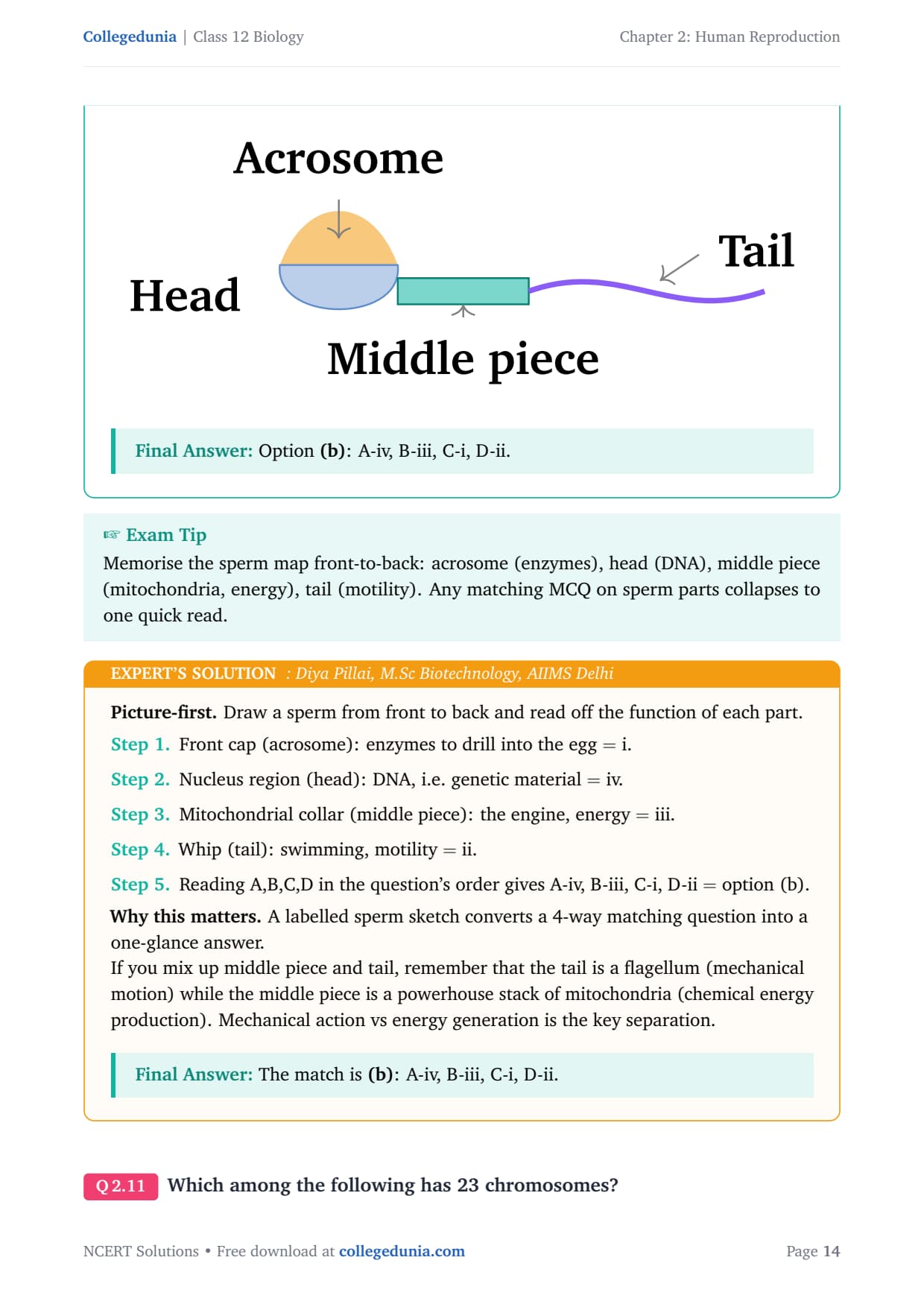
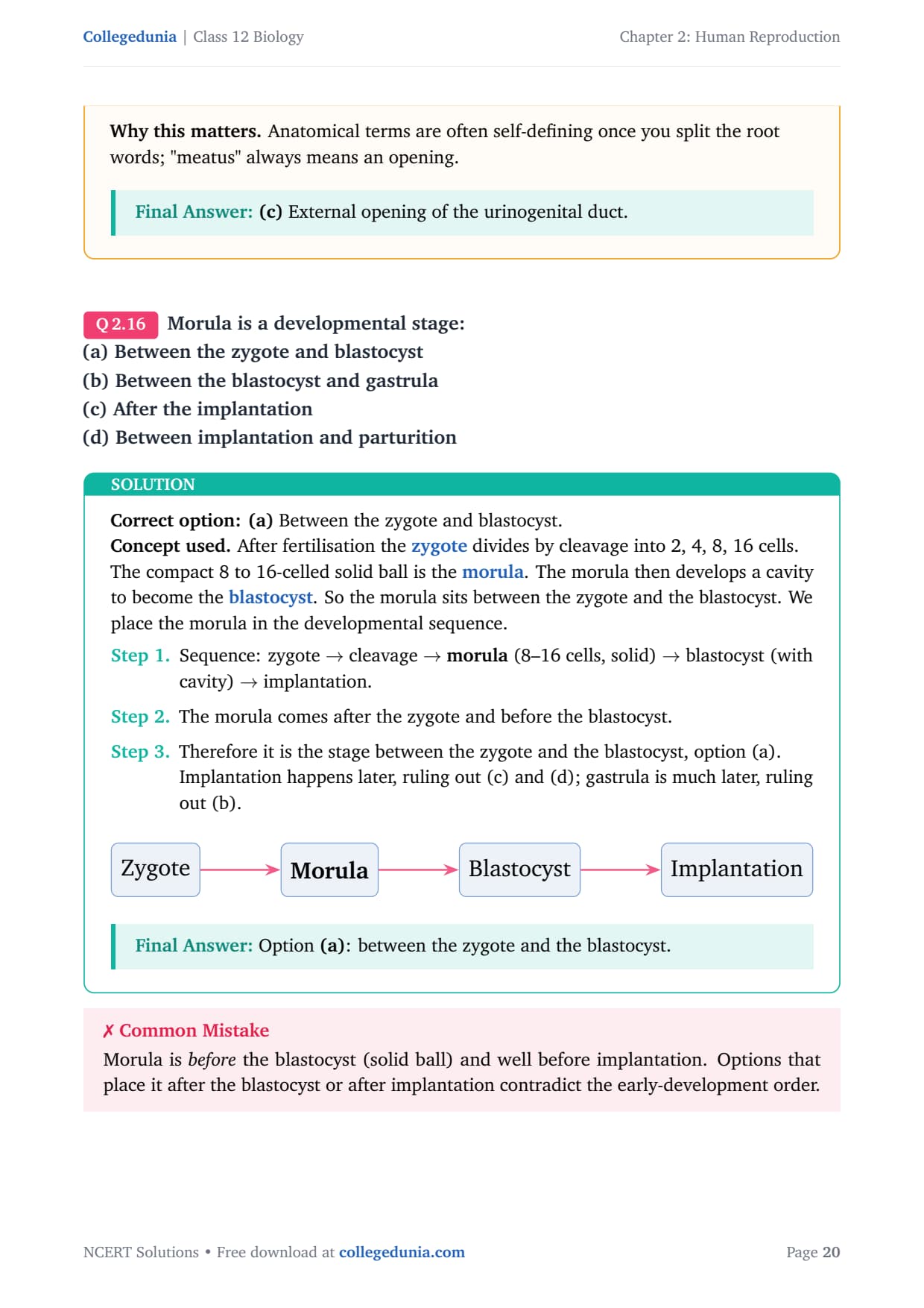
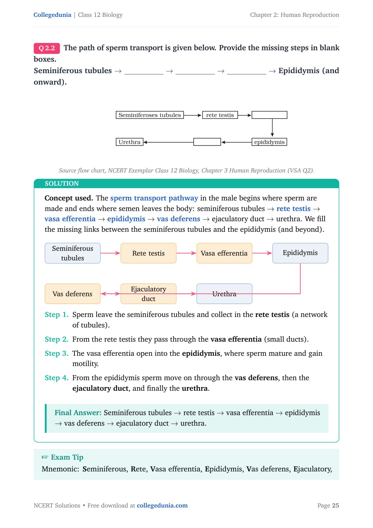
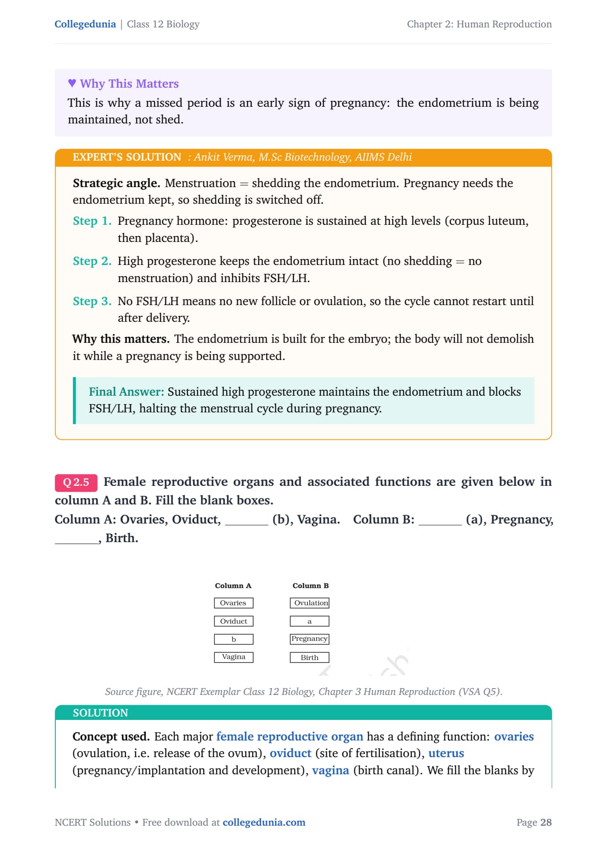
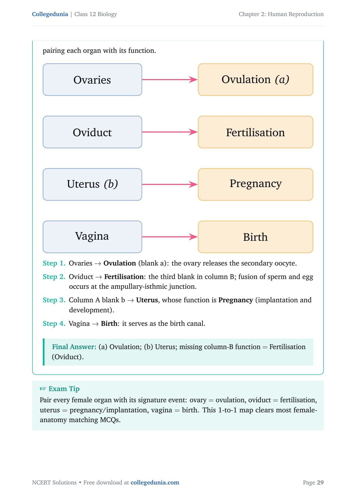
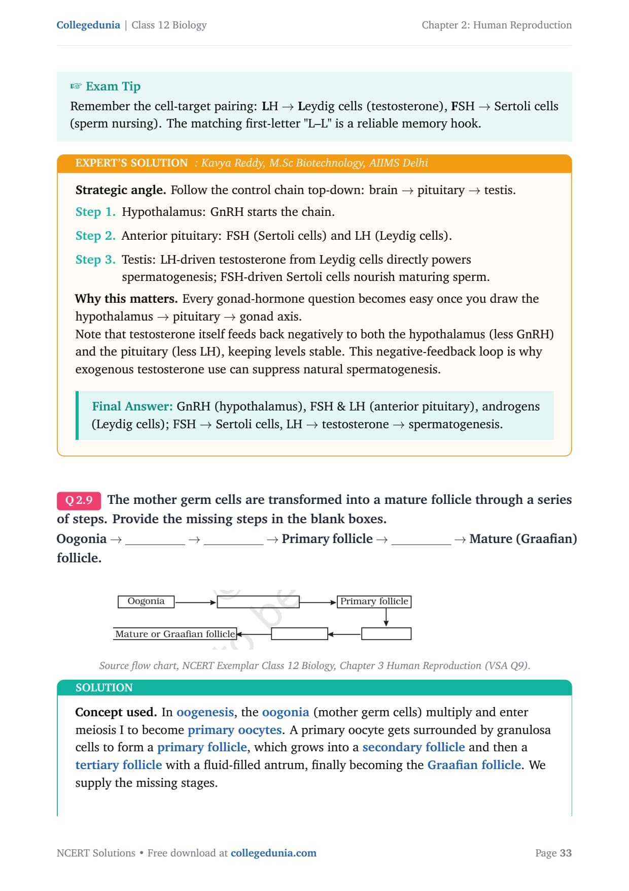
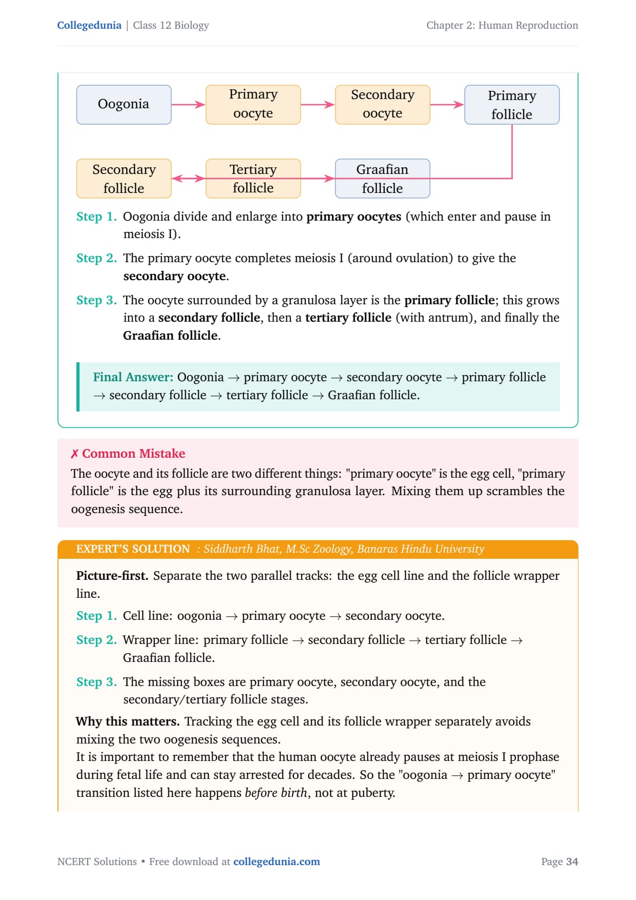

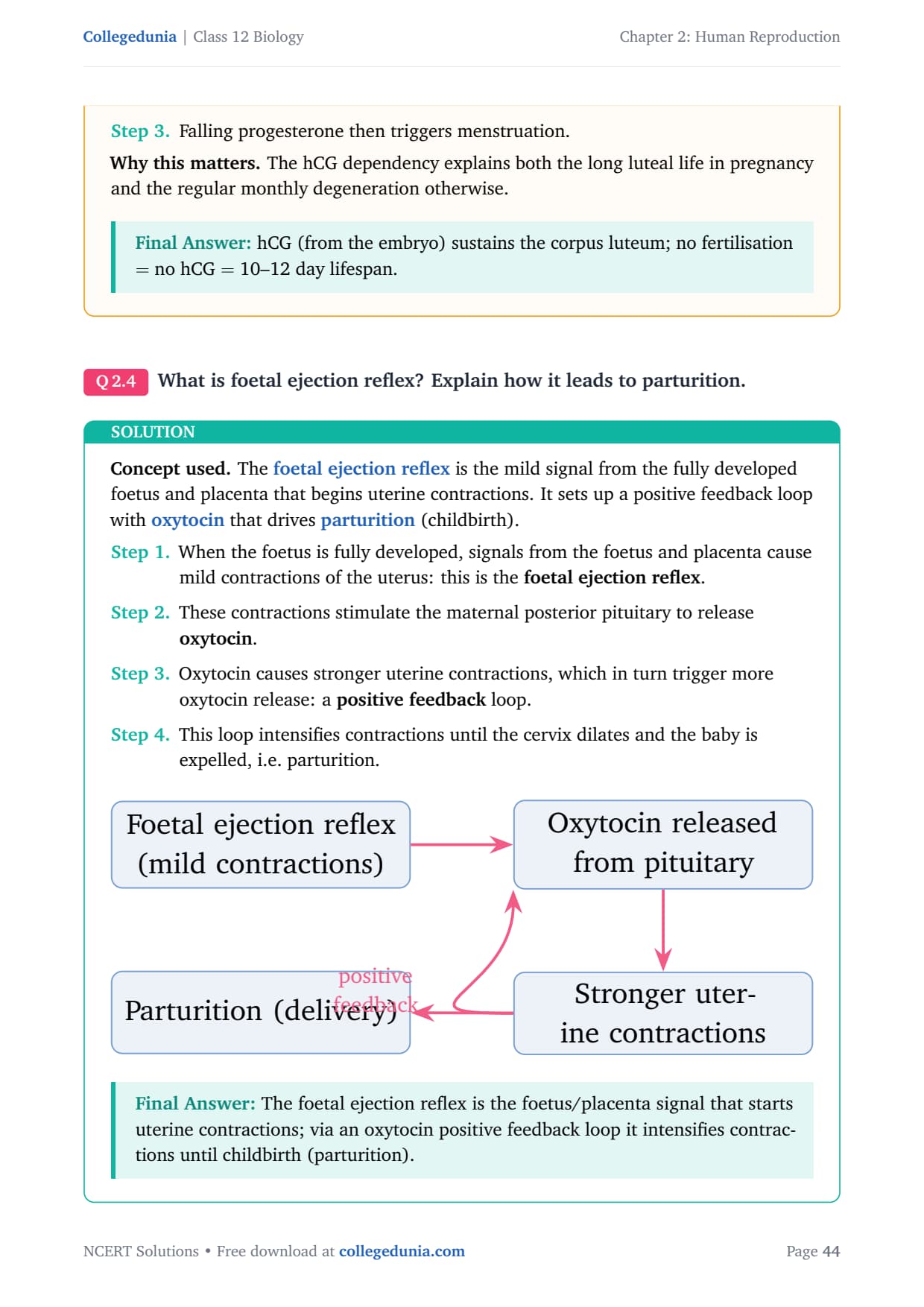
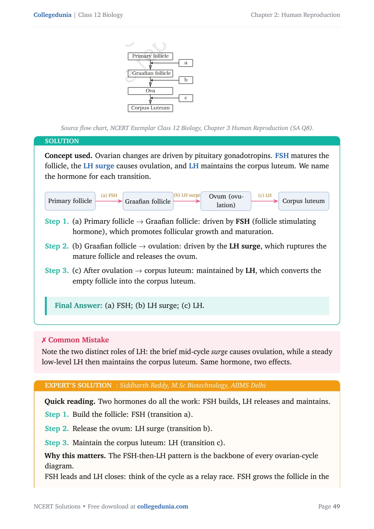
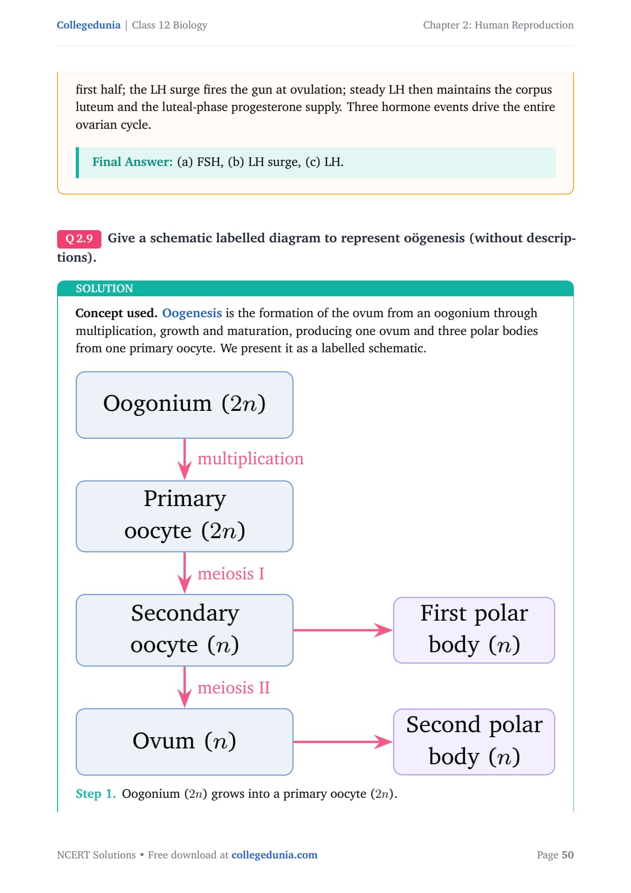
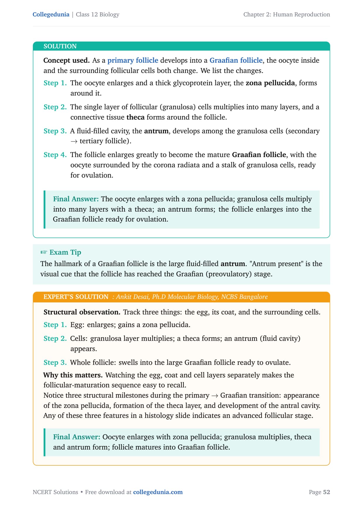
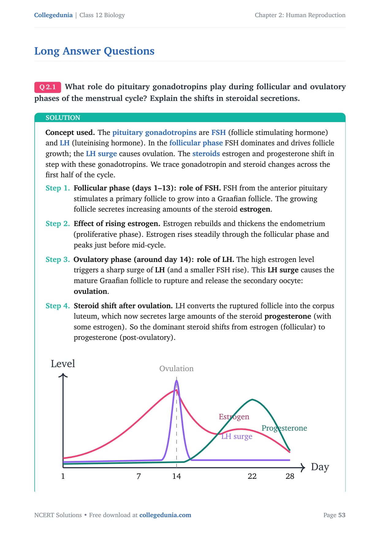
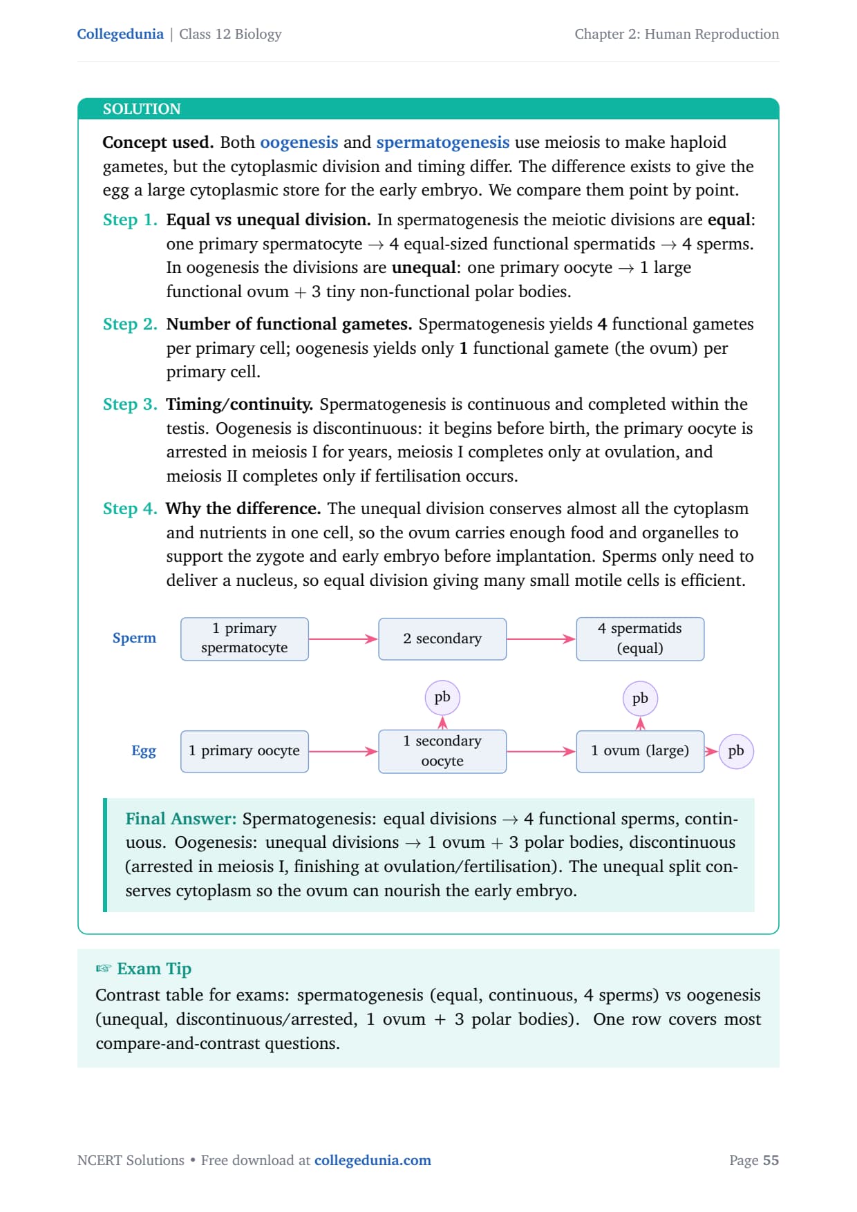
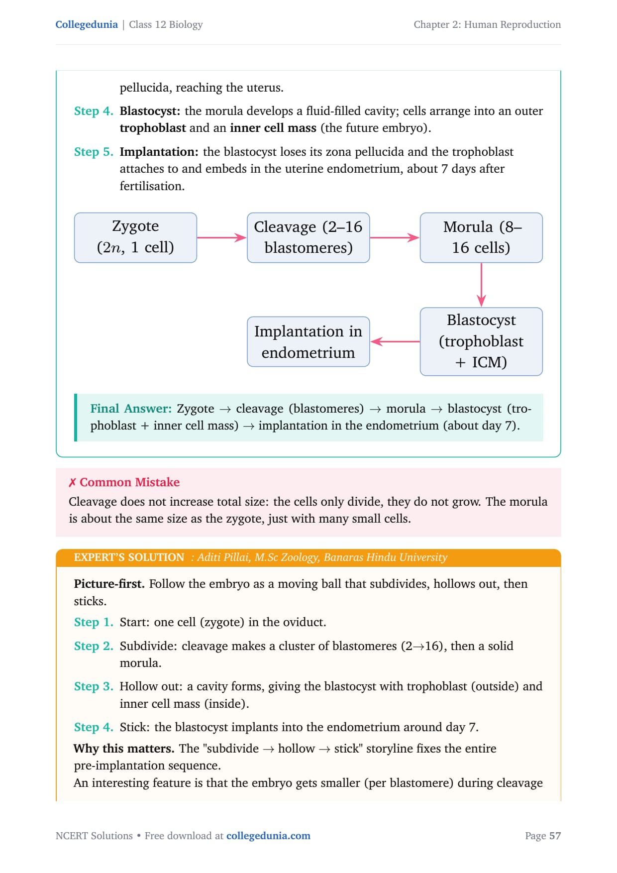
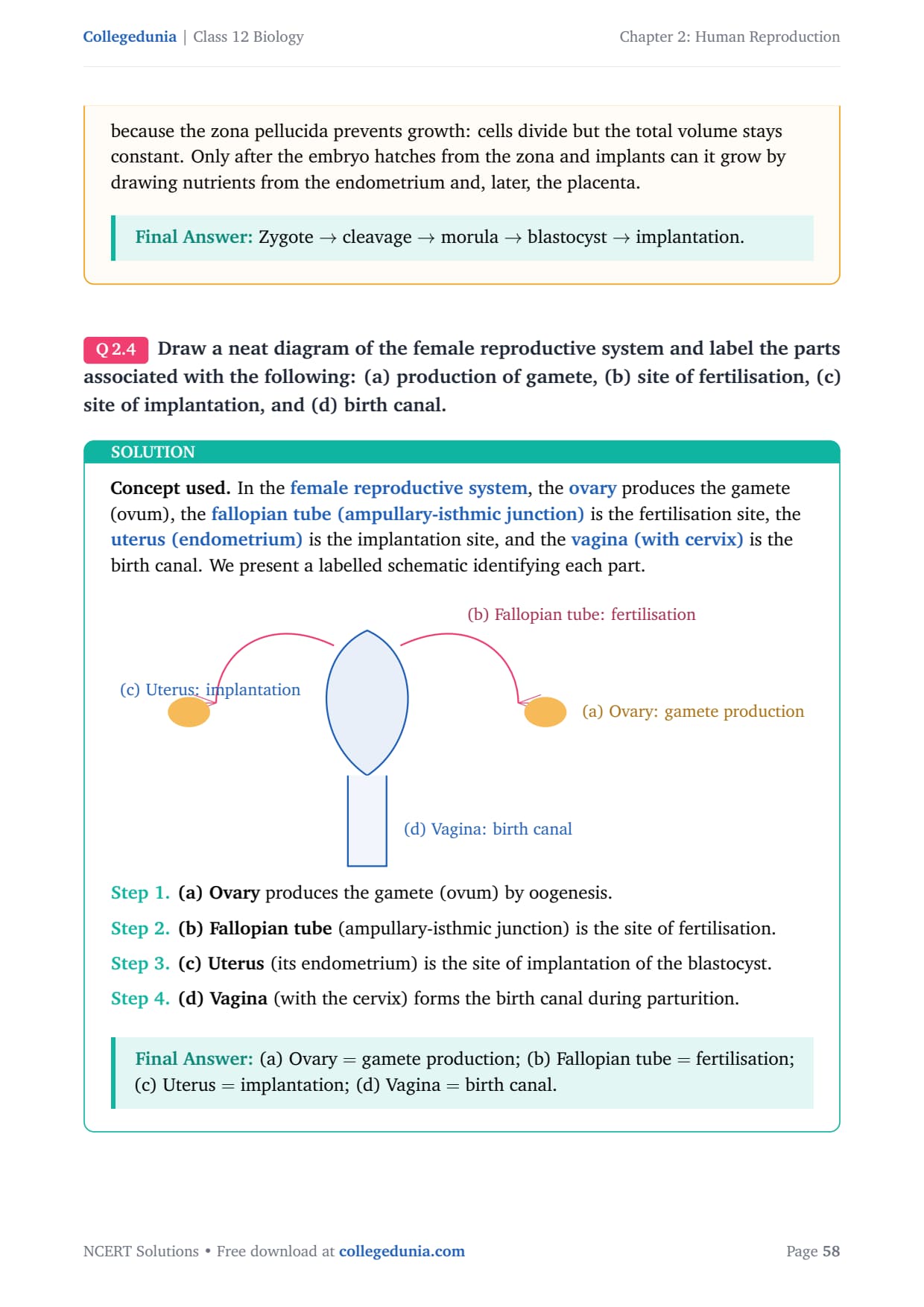
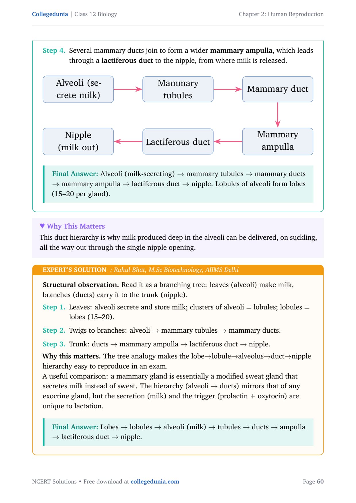
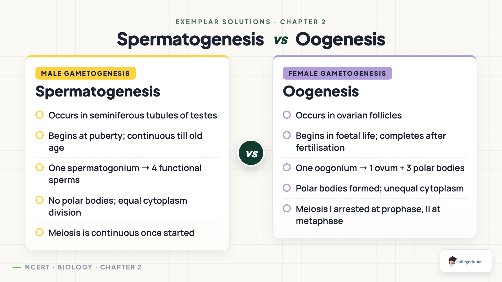
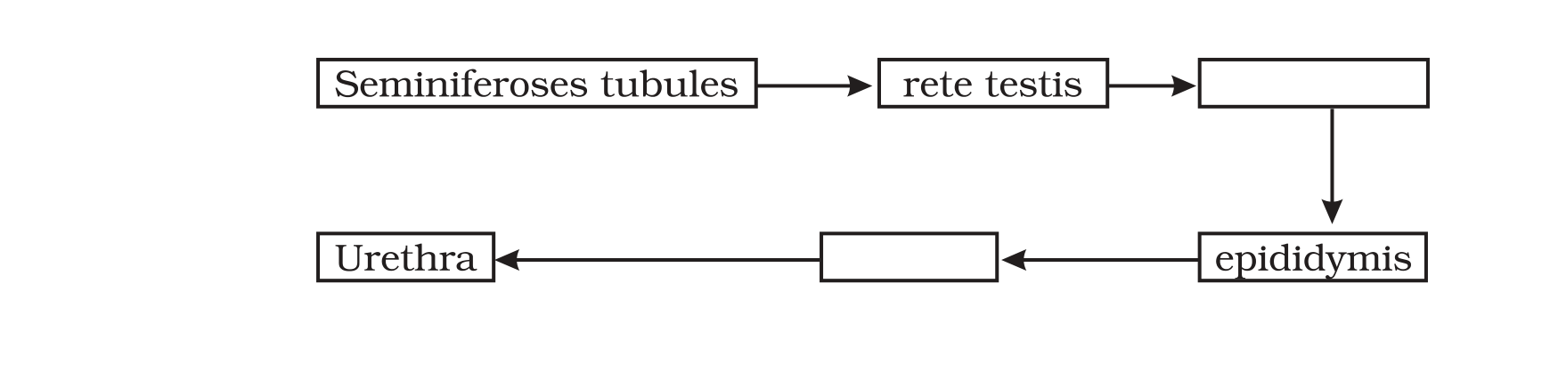
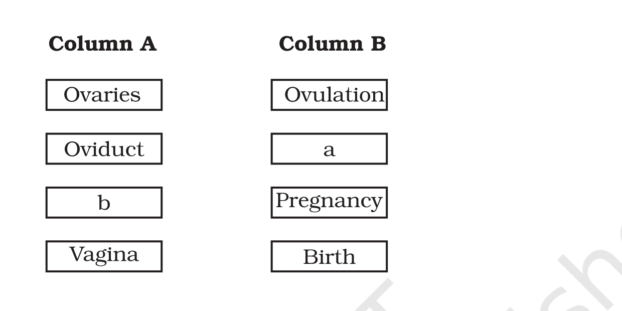

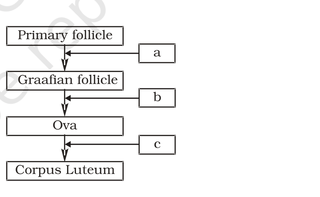



Comments