Physics Mentor, SRM Institute | Updated on - Jul 23, 2026
Chapter 5 Life Processes is the biggest scoring biology chapter of Class 10 Science for 2026-27. The Class 10 Science Chapter 5 NCERT Exemplar Solutions on this page solve every Exemplar problem step by step, in plain language.
CBSE Board weightage: Life Processes carries heavy marks, and at least one long-answer almost always comes from it.
What you get: all MCQ, Short Answer and Long Answer problems solved, with a free PDF.
Solved by Collegedunia: Every problem below is solved by subject experts, mapped to the 2026-27 NCERT Exemplar, and checked against the CBSE Board marking scheme.
Why the NCERT Exemplar Matters for Class 10 Board Preparation
Life Processes is one of the highest-weightage biology chapters, and it is where students slip on reasoning questions. The NCERT Exemplar turns the basics into exam-style questions: multi-statement MCQs, label-the-diagram problems, and reasoning on photosynthesis, respiration and circulation.
Quick Tip: Solve the NCERT textbook exercises first, then the Exemplar. The Exemplar assumes you already know the four life processes and the order of the human alimentary canal.
How Collegedunia's NCERT Exemplar Solutions Help You with Life Processes
Each problem is solved the way a CBSE examiner expects: process named, reason given, every step shown.
Every question type solved: all MCQ, Short Answer and Long Answer problems are worked out, not just the easy ones.
2026-27 alignment: problem numbers and answers match the current edition.
Step-by-step reasoning: each process is explained one stage at a time so you can copy the method.
Trap flags: red boxes mark where students mix up aerobic with anaerobic respiration or arteries with veins.
Best Way to Use the Life Processes Exemplar for Board Revision
Treat the Exemplar as a practice paper, not a re-read of the textbook.
Phase
Exemplar Use
Time
First read
All MCQs
1 hour
Concept practice
Nutrition and respiration Short Answers
1.5 hours
Answer writing
All Long Answers, full working with diagrams
2 hours
Pre-board revision
Re-solve the wrong ones
1 hour
Spend the most time on respiration and the double circulation of blood, which carry the most marks.
Life Processes Exemplar Question Types with One Solved Sample Each
The Exemplar mixes several question formats. The table below previews the shape of each.
Type
Sample Question
Answer Shape
MCQ
Which is the correct sequence of parts in the human alimentary canal?
Single option, with reason
MCQ (statement-based)
Which statements about respiration are true?
Pick the correct set of statements
Short Answer
Why is the rate of breathing in aquatic organisms faster than in land animals?
Two to three line reason
Reasoning
Why do herbivores need a longer small intestine?
Short explanation with logic
Long Answer
Explain the process of double circulation in humans
Several linked parts, diagram
Every one of these is solved in full in the question bank below.
The Four Life Processes Quick Reference
Most Exemplar MCQs test whether you can match a process to its organ and purpose. This table covers the four core jobs.
Life Process
What it does
Key organs
Nutrition
Makes or takes in food and breaks it down
Leaves (plants); mouth, stomach, small intestine (humans)
Respiration
Releases energy from food as ATP
Mitochondria; lungs and alveoli
Transportation
Moves materials around the body
Heart, blood vessels; xylem and phloem in plants
Excretion
Removes waste from the body
Kidneys, nephrons; stomata and old xylem in plants
Energy from respiration is stored as ATP. Photosynthesis is summarised as 6CO2 + 12H2O → C6H12O6 + 6O2 + 6H2O.
Difficulty Step-Up from NCERT Textbook to Exemplar
The Exemplar reuses textbook ideas inside harder wrappers, as the contrast below shows.
Concept
NCERT Textbook
NCERT Exemplar
Photosynthesis
Write the overall equation
Identify the false statement about autotrophs
Digestion
Name the parts of the alimentary canal
Order the parts or match each enzyme to its food
Respiration
Define aerobic and anaerobic respiration
Compare energy yield and end products of both
Circulation
Draw the human heart
Explain why double circulation is needed in mammals
Excretion
Label a nephron
Reason out how urine is concentrated and water saved
The Exemplar gives a situation and asks you to apply the rule and justify it.
Topics Covered in Class 10 Science Chapter 5 Life Processes Exemplar
The Chapter 5 Exemplar spans autotrophic and heterotrophic nutrition, the iodine test for starch, the human digestive system, aerobic and anaerobic respiration, photosynthesis, the role of stomata, haemoglobin, the double circulation of blood, transport through xylem and phloem, transpiration, and excretion by the kidneys and nephrons.
Life Processes Exemplar Common Mistakes That Cost Marks
The Exemplar twists trigger the same wrong reflexes every year. Watch these four.
Mixing up aerobic and anaerobic respiration. Aerobic uses oxygen and gives much energy; anaerobic needs no oxygen and gives little.
Confusing arteries with veins. Arteries carry blood away from the heart; veins carry it back to the heart.
Wrong respiration product for the organism. Yeast gives ethanol and carbon dioxide; human muscle gives lactic acid.
Reversing the inhalation direction. During inhalation the ribs move up and out and the diaphragm moves down, not the other way round.
Watch Out: In a statement-based MCQ, test every statement to the end. Stopping at the first correct one is the most common way students lose marks in this chapter.
Human Digestive System Step by Step
Food moves through the human alimentary canal in a fixed order, and each station does one job.
Step 1 (Mouth): teeth chew the food and saliva adds salivary amylase, which starts breaking starch into sugar.
Step 2 (Oesophagus): the food pipe pushes the food down to the stomach by peristalsis.
Step 3 (Stomach): hydrochloric acid and pepsin begin protein digestion, while mucus protects the stomach wall.
Step 4 (Small intestine): bile, pancreatic juice and intestinal juice finish digestion, and the simple food is absorbed through the villi.
Step 5 (Large intestine): water is absorbed from the leftover, and the waste is removed.
Most Repeated Board Topics from Life Processes
The topics that show up most often in CBSE Board and sample papers.
Topic
How it is asked
Photosynthesis
Write the equation or identify the false autotroph statement
Human digestion
Order the alimentary canal or match enzymes to food
Respiration
Compare aerobic and anaerobic respiration with end products
Double circulation
Explain why blood passes through the heart twice in one cycle
Transport in plants
Distinguish the roles of xylem and phloem
Excretion
Describe urine formation and how water is conserved
All NCERT Exemplar Questions for Life Processes with Step-by-Step Solutions
Every Exemplar question for Chapter 5 Life Processes is listed below with its full Solution and Expert Solution in collapsible tabs.
I. Multiple Choice Questions
Q 5.1
Which of the following statements about the autotrophs is incorrect?
(a) They synthesise carbohydrates from carbon dioxide and water in the presence of sunlight and chlorophyll
(b) They store carbohydrates in the form of starch
(c) They convert carbon dioxide and water into carbohydrates in the absence of sunlight
(d) They constitute the first trophic level in food chains
Correct option: (c) They convert carbon dioxide and water into carbohydrates in the absence of sunlight.
Concept used.Autotrophs are organisms (mostly green plants) that make their own food by photosynthesis. Photosynthesis is the process where chlorophyll traps sunlight and uses it to turn carbon dioxide and water into glucose. Sunlight is the energy source, so the process simply cannot run in the dark.
Check option (a). Autotrophs do make carbohydrates from carbon dioxide and water using sunlight and chlorophyll. This is the textbook definition, so (a) is correct.
Check option (b). The glucose made is stored as starch in plants. So (b) is correct.
Check option (c). It says food is made ``in the absence of sunlight''. Without light there is no energy to drive photosynthesis, so this statement is wrong. This is the answer we want, because the question asks for the incorrect statement.
Check option (d). Green plants are eaten by herbivores, so they form the first trophic level of a food chain. So (d) is correct.
Option (c) is the incorrect statement: autotrophs cannot make food in the absence of sunlight.
AV
Ananya Verma
M.Sc Botany, University of Delhi
Verified Expert
Quick reading (energy-first). Photosynthesis is a light-driven reaction. Any option that claims an autotroph builds food without light is automatically false, so I scan for the word ``absence of sunlight'' and stop there.
Concept used. An autotroph fixes carbon only when chlorophyll has absorbed light energy. No light means no ATP and no NADPH, so carbon dioxide cannot be reduced to carbohydrate. The other three claims (light synthesis, starch storage, first trophic level) are all standard features of green plants.
Spot the energy requirement. Photosynthesis needs light to split water and release electrons. Strike out any option that drops the light condition.
Confirm storage form. Plants keep their spare glucose as starch, not as fat, so the storage option stands.
Confirm the food-chain position. Producers are eaten first, so they sit at trophic level one.
Land on (c) as the false claim, since carbohydrate cannot form in the dark.
Why this matters. Students often confuse autotrophs (which need light) with chemoautotrophic bacteria (which use chemical energy instead of light). For the Class 10 board, ``autotroph'' means ``light-using green plant'', so the dark-synthesis option is always the trap.
Option (c) is incorrect because autotrophs need sunlight to convert carbon dioxide and water into carbohydrates.
Q 5.2
In which of the following groups of organisms, food material is broken down outside the body and absorbed?
(a) Mushroom, green plants, Amoeba
(b) Yeast, mushroom, bread mould
(c) Paramecium, Amoeba, Cuscuta
(d) Cuscuta, lice, tapeworm
Correct option: (b) Yeast, mushroom, bread mould.
Concept used.Saprophytic nutrition is the mode where an organism secretes digestive juices onto dead organic matter, digests the food outside its body, and then absorbs the simple soluble products. Fungi and many moulds feed this way.
Define the group we need: only saprophytes break food down outside the body and then absorb it.
Test (a). Green plants are autotrophs and Amoeba ingests food inside a vacuole, so this group is mixed and wrong.
Test (c) and (d). Paramecium and Amoeba digest food inside the cell; Cuscuta, lice and tapeworm are parasites that take ready-made nutrients from a host. None match.
Test (b). Yeast, mushroom and bread mould are all fungi. They pour enzymes onto the food, digest it externally, and soak up the soluble products. This fits saprophytic nutrition exactly.
Option (b): yeast, mushroom and bread mould are saprophytes that digest food outside the body.
RN
Rahul Nair
M.Sc Microbiology, University of Hyderabad
Verified Expert
Structural observation (group purity). A correct option must have all three members feeding the same way. So I reject any group that mixes an autotroph or an inside-digester with a saprophyte.
Concept used. Saprophytes are decomposers. They release extracellular enzymes onto dead organic matter, break complex molecules into simple ones in the surroundings, and absorb the dissolved nutrients through their body surface. Fungi such as yeast, mushroom and bread mould are the textbook examples.
Scan each group for an odd member. Group (a) has green plants (autotroph) and Amoeba (intracellular) mixed in, so it fails the all-same test.
Groups (c) and (d) contain parasites and intracellular feeders, not external digesters.
Group (b) is pure fungi, all of which feed saprophytically. Select it.
Why this matters. The same external-digestion logic explains why mushrooms grow on rotting logs and bread mould spreads on stale bread: they are recycling dead matter, a role decomposers play in every ecosystem.
Option (b) because yeast, mushroom and bread mould all digest food externally as saprophytes.
Q 5.3
Select the correct statement
(a) Heterotrophs do not synthesise their own food
(b) Heterotrophs use solar energy for photosynthesis
(c) Heterotrophs synthesise their own food
(d) Heterotrophs are capable of converting carbon dioxide and water into carbohydrates
Correct option: (a) Heterotrophs do not synthesise their own food.
Concept used. A heterotroph is an organism that cannot make its own food and depends on other organisms (plants or animals) for nutrition. Animals, fungi and most bacteria are heterotrophs.
State the defining feature: heterotrophs lack chlorophyll and cannot fix carbon, so they cannot prepare food themselves.
Option (a) says exactly this, so it is correct.
Options (b), (c) and (d) all describe photosynthesis, which is the job of autotrophs, not heterotrophs. They are therefore wrong.
Option (a): heterotrophs do not synthesise their own food.
MK
Meera Krishnan
M.Sc Zoology, University of Madras
Verified Expert
Quick reading (definition match). The Greek root says it all: ``hetero'' (other) ``troph'' (feeding) means feeding on others. So the only statement that survives is the one denying self-made food.
Concept used. Heterotrophs do not have chlorophyll and cannot carry out photosynthesis. They obtain pre-formed organic food, then break it down for energy and building blocks. Three of the four options wrongly hand them the autotroph's job of fixing carbon dioxide.
Translate the term: heterotroph = feeds on other organisms.
Eliminate every option that gives heterotrophs photosynthesis or carbon fixing, which removes (b), (c) and (d).
Keep (a), the statement that they cannot make their own food.
Why this matters. The autotroph versus heterotroph split is the foundation of every food chain. Producers (autotrophs) capture energy; consumers (heterotrophs) pass it along. Mixing the two up costs easy marks in ecology questions later.
Option (a) is correct: heterotrophs cannot prepare their own food.
Q 5.4
Which is the correct sequence of parts in human alimentary canal?
(a) Mouth → stomach → small intestine → oesophagus → large intestine
(b) Mouth → oesophagus → stomach → large intestine → small intestine
(c) Mouth → stomach → oesophagus → small intestine → large intestine
(d) Mouth → oesophagus → stomach → small intestine → large intestine
Correct option: (d) Mouth → oesophagus → stomach → small intestine → large intestine.
Concept used. The alimentary canal is the continuous food tube from mouth to anus. Food travels in a fixed order: mouth, food pipe (oesophagus), stomach, small intestine, large intestine.
Start at the mouth, where food is chewed and mixed with saliva.
The oesophagus carries the food down to the stomach by peristalsis.
In the stomach the food is churned with acid and pepsin.
It then passes to the small intestine for final digestion and absorption.
Finally the leftover passes to the large intestine, where water is absorbed. Only option (d) keeps this order.
Option (d): mouth, oesophagus, stomach, small intestine, large intestine.
VI
Vikram Iyer
MBBS, Kasturba Medical College
Verified Expert
Picture-first (trace the food). I imagine swallowing a bite and follow where it goes. That mental movie locks the order without rote memory.
Concept used. The gut is a one-way tube. The oesophagus must come before the stomach because the food pipe delivers food to the stomach. The small intestine must come before the large intestine because digestion finishes before water is reabsorbed from the residue.
Rule out (a): it puts oesophagus after the small intestine, which reverses the food pipe. Wrong.
Rule out (b): it places the large intestine before the small intestine, so absorption would come before digestion. Wrong.
Rule out (c): it puts the oesophagus after the stomach. Wrong.
Option (d) keeps mouth → oesophagus → stomach → small → large intestine, matching the real path.
Why this matters. Knowing the order is the backbone of every digestion long-answer: each enzyme acts at a specific station, so the sequence tells you where starch, protein and fat are each broken down.
Option (d) gives the true food path: mouth, oesophagus, stomach, small intestine, large intestine.
Q 5.5
If salivary amylase is lacking in the saliva, which of the following events in the mouth cavity will be affected?
(a) Proteins breaking down into amino acids
(b) Starch breaking down into sugars
(c) Fats breaking down into fatty acids and glycerol
(d) Absorption of vitamins
Correct option: (b) Starch breaking down into sugars.
Concept used.Salivary amylase (also called ptyalin) is the enzyme in saliva that begins the digestion of starch. It breaks starch into simpler sugars (maltose) right inside the mouth. Each enzyme acts only on its own type of food, called its substrate.
Identify what amylase does: it acts only on starch, turning it into sugar.
If amylase is missing, the starch-to-sugar step in the mouth will not happen, so option (b) is affected.
Proteins, fats and vitamins are not touched by salivary amylase, so options (a), (c) and (d) are not the answer.
Option (b): without salivary amylase, starch will not be broken into sugars in the mouth.
SP
Sanjana Pillai
M.Sc Biochemistry, Anna University
Verified Expert
Structural observation (one enzyme, one job). Enzymes are substrate-specific. So the moment the question removes amylase, only the amylase reaction can be hurt, nothing else.
Concept used. Salivary amylase hydrolyses the bonds in starch to give maltose. It has no effect on peptide bonds (proteins) or ester bonds (fats), and vitamins are absorbed, not digested, so amylase plays no role there.
Pin down the enzyme's target: amylase works on starch only.
Knock out amylase and ask which reaction stalls: the starch to sugar conversion.
Confirm the other three options use different enzymes or no enzyme at all, so they are untouched.
Why this matters. This is why a piece of bread (rich in starch) starts tasting slightly sweet if you chew it for a while: amylase is already converting starch into sugar in your mouth.
Option (b): starch digestion to sugars stops if salivary amylase is absent.
Q 5.6
The inner lining of stomach is protected by one of the following from hydrochloric acid. Choose the correct one
(a) Pepsin
(b) Mucus
(c) Salivary amylase
(d) Bile
Correct option: (b) Mucus.
Concept used. The stomach makes strong hydrochloric acid (HCl) to kill germs and to activate pepsin. To stop this acid from digesting the stomach wall itself, the gastric glands also release mucus, a slimy coating that lines and protects the inner wall.
Note the danger: HCl is corrosive and could damage the stomach's own lining.
Identify the shield: mucus forms a slippery, protective layer over the inner wall, so option (b) is correct.
Reject the rest: pepsin digests protein, salivary amylase works on starch in the mouth, and bile works in the small intestine. None of them protect the stomach.
Option (b): mucus protects the inner lining of the stomach from hydrochloric acid.
DM
Devika Menon
MBBS, Government Medical College Thiruvananthapuram
Verified Expert
Quick reading (defence, not digestion). The keyword is ``protected''. So I ignore every option that is a digestive agent and look for a protective barrier, which is mucus.
Concept used. Gastric glands secrete three things: HCl (acidic medium and germ-killer), pepsin (protein-digesting enzyme), and mucus (protective lining). Only mucus shields the stomach wall from self-digestion by the acid and pepsin.
Sort the secretions into ``attack'' (HCl, pepsin) and ``defence'' (mucus).
The question wants the defence item, so pick mucus.
Confirm bile and salivary amylase are made elsewhere, so they cannot be the stomach's shield.
Why this matters. When mucus secretion drops or acid rises too much, the unprotected wall gets eroded, which is exactly how acidity and peptic ulcers develop, a fact the Exemplar revisits in Q51.
Option (b): mucus is the protective barrier against stomach acid.
Q 5.7
Which part of alimentary canal receives bile from the liver?
(a) Stomach
(b) Small intestine
(c) Large intestine
(d) Oesophagus
Correct option: (b) Small intestine.
Concept used.Bile is a juice made by the liver and stored in the gall bladder. It is poured into the first part of the small intestine (the duodenum), where it makes the food alkaline and breaks large fat droplets into small ones (emulsification).
Recall where bile is needed: fat digestion happens in the small intestine, so bile must arrive there.
The bile duct from the liver and gall bladder opens into the small intestine, so option (b) is correct.
The stomach, large intestine and oesophagus do not receive bile, so the other options are wrong.
Option (b): the small intestine receives bile from the liver.
AR
Arjun Reddy
M.Sc Physiology, Osmania University
Verified Expert
Structural observation (follow the duct). The bile duct physically connects the liver and gall bladder to the duodenum, the very start of the small intestine. So tracing the plumbing answers the question directly.
Concept used. Bile contains bile salts that emulsify fats and bicarbonate that neutralises the acidic food coming from the stomach. Both jobs are needed in the small intestine, where pancreatic lipase then completes fat digestion.
Identify bile's source: the liver, with storage in the gall bladder.
Identify bile's destination: the duodenum, the first loop of the small intestine.
Match destination to the options: small intestine, option (b).
Why this matters. The small intestine is the meeting point of three juices, bile, pancreatic juice and intestinal juice, which is why almost all final digestion happens here, a point Q9 tests next.
Option (b): bile from the liver is delivered to the small intestine.
Q 5.8
A few drops of iodine solution were added to rice water. The solution turned blue-black in colour. This indicates that rice water contains
(a) complex proteins
(b) simple proteins
(c) fats
(d) starch
Correct option: (d) Starch.
Concept used. The iodine test is the standard test for starch. When iodine solution is added to a sample containing starch, it turns blue-black. This colour change is a confirmed indicator of starch.
Recall the test: iodine plus starch gives a blue-black colour.
The rice water turned blue-black, so it must contain starch (rice is rich in starch).
Proteins and fats do not give this colour with iodine, so options (a), (b) and (c) are wrong.
Option (d): the blue-black colour shows that rice water contains starch.
PB
Pooja Bhat
M.Sc Food Science, University of Mysore
Verified Expert
Quick reading (colour code). Blue-black with iodine is a memorised signal. The colour itself is the answer, so I do not need to think about rice at all.
Concept used. Iodine molecules slip into the coiled structure of the starch molecule and form a deep blue-black complex. Proteins, sugars and fats lack this coiled shape, so they give no such colour.
Match the reagent and colour: iodine plus blue-black means starch.
Confirm the food: rice is a starchy grain, so rice water naturally carries starch.
Eliminate proteins and fats, which do not react with iodine this way.
Why this matters. The same iodine test is used in the classic ``starch in a leaf'' experiment to prove a leaf has done photosynthesis, so this single colour reaction links nutrition in animals and plants.
Option (d): the blue-black iodine colour confirms starch.
Q 5.9
In which part of the alimentary canal food is finally digested?
(a) Stomach
(b) Mouth cavity
(c) Large intestine
(d) Small intestine
Correct option: (d) Small intestine.
Concept used. The small intestine is the site where digestion is completed. Here bile, pancreatic juice and intestinal juice together break carbohydrates into glucose, proteins into amino acids, and fats into fatty acids and glycerol. The simple products are then absorbed.
Note partial digestion earlier: the mouth starts starch and the stomach starts protein, but neither finishes the job.
In the small intestine all three food types are fully broken down to their simplest forms.
So the final digestion happens in the small intestine, option (d). The large intestine only absorbs water.
Option (d): food is finally digested in the small intestine.
KS
Kavya Suresh
M.Sc Zoology, Bangalore University
Verified Expert
Strategic angle (where the juices meet). Final digestion needs every enzyme present at once. Only the small intestine receives all the digestive juices together, so it must be the finishing line.
Concept used. The duodenum gets bile (emulsifies fat) and pancreatic juice (trypsin, amylase, lipase), while the intestinal glands add more enzymes. With this full enzyme set, carbohydrates, proteins and fats are all reduced to absorbable units here.
List where each food's digestion begins: starch in the mouth, protein in the stomach, fat in the small intestine.
Ask where all three are completed: only in the small intestine, where the full enzyme team works.
Choose the small intestine, option (d).
Why this matters. Because digestion finishes here, absorption also happens here through finger-like villi, which is why the small intestine, not the stomach, is the true heart of the digestive system.
Option (d): the small intestine is where digestion is completed.
Q 5.10
Choose the function of the pancreatic juice from the following
(a) trypsin digests proteins and lipase carbohydrates
(b) trypsin digests emulsified fats and lipase proteins
(c) trypsin and lipase digest fats
(d) trypsin digests proteins and lipase emulsified fats
Correct option: (d) trypsin digests proteins and lipase emulsified fats.
Concept used.Pancreatic juice contains the enzymes trypsin (digests proteins into smaller peptides and amino acids) and lipase (digests emulsified fats into fatty acids and glycerol). Each enzyme acts only on its own substrate.
Match trypsin to its substrate: trypsin acts on proteins.
Match lipase to its substrate: lipase acts on fats, but only after bile has emulsified them into small droplets.
The only option that pairs trypsin with proteins and lipase with emulsified fats is (d). The other options swap the pairings and are wrong.
Option (d): trypsin digests proteins and lipase digests emulsified fats.
NJ
Nikhil Joshi
M.Sc Biochemistry, Savitribai Phule Pune University
Verified Expert
Structural observation (lock each enzyme to its food). Trypsin is a protease and lipase is a fat-splitter. So any option that pairs them with the wrong food is automatically out.
Concept used. The pancreas pours trypsin, pancreatic amylase and lipase into the duodenum. Trypsin breaks peptide bonds in proteins; lipase breaks ester bonds in fats, but needs bile to first emulsify those fats so the enzyme can reach them.
Fix the correct pairs: trypsin with protein, lipase with fat.
Reject (a), (b) and (c), each of which mismatches at least one enzyme.
Keep (d), the only correctly paired option, noting the word ``emulsified'' signals the bile step came first.
Why this matters. This question quietly tests two ideas at once: enzyme specificity and the teamwork between bile and lipase. Recognising both is what separates a full-mark answer from a half-remembered guess.
Option (d): trypsin acts on proteins, lipase on emulsified fats.
Q 5.11
When air is blown from mouth into a test-tube containing lime water, the lime water turned milky due to the presence of
(a) oxygen
(b) carbon dioxide
(c) nitrogen
(d) water vapour
Correct option: (b) carbon dioxide.
Concept used.Lime water (calcium hydroxide solution) turns milky when carbon dioxide is passed through it, because insoluble calcium carbonate forms. Exhaled air is rich in carbon dioxide, a waste product of respiration.
Recall the lime water test: carbon dioxide turns clear lime water milky white.
The breath you blow out carries a lot of carbon dioxide, produced by respiration in your cells.
So the milkiness is caused by carbon dioxide, option (b). Oxygen, nitrogen and water vapour do not turn lime water milky.
Option (b): exhaled carbon dioxide turns lime water milky.
RS
Ritika Saxena
M.Sc Chemistry, Banaras Hindu University
Verified Expert
Quick reading (reagent-locked). Lime water plus milky colour is a textbook signature for carbon dioxide. The biology dressing (breath, respiration) does not change that.
Concept used. Carbon dioxide reacts with calcium hydroxide to give a fine white precipitate of calcium carbonate, which makes the solution look milky. Exhaled breath contains far more carbon dioxide than inhaled air because it is a respiratory waste.
Identify the chemical signal: carbon dioxide forms calcium carbonate, which clouds the lime water.
Link to biology: respiration in body cells releases carbon dioxide, carried out in the breath.
Select carbon dioxide, option (b), as the gas responsible.
Why this matters. This simple test is proof that respiration is happening: living, breathing tissue gives off carbon dioxide, which is why germinating seeds or breathing animals both turn lime water milky in lab demonstrations.
Option (b): carbon dioxide in exhaled air turns lime water milky.
Concept used. In fermentation by yeast, glucose is first broken into pyruvate in the cytoplasm (glycolysis). Then, in the absence of oxygen, the pyruvate is converted into ethanol and carbon dioxide. This is anaerobic respiration.
Step one of every breakdown: glucose splits into pyruvate in the cytoplasm. All options agree so far.
In yeast and without oxygen, pyruvate becomes ethanol and carbon dioxide, not lactic acid.
So the path is glucose → pyruvate → ethanol + carbon dioxide, which is option (d). Options that show oxygen, or lactic acid, do not describe yeast fermentation.
Structural observation (organism decides the product). Pyruvate has two anaerobic fates: ethanol (in yeast) or lactic acid (in muscle). Naming the organism tells me which one, so ``yeast'' points straight to ethanol.
Concept used. Glycolysis (glucose to pyruvate) is common to all cells. After that, the route forks. Yeast lacks the pathway to make lactic acid; instead it decarboxylates pyruvate to ethanol and carbon dioxide. Anaerobic means oxygen is absent, so any option that adds oxygen is for aerobic respiration.
Confirm the shared first step: glucose to pyruvate in the cytoplasm.
Pick the yeast-specific second step: pyruvate to ethanol and carbon dioxide.
Discard options with oxygen or lactic acid, since they describe aerobic or muscle pathways.
Why this matters. This very reaction is the basis of baking and brewing: the carbon dioxide makes bread rise and the ethanol is the alcohol in fermented drinks, a direct everyday use of microbial respiration.
Option (d): in yeast, pyruvate is converted to ethanol and carbon dioxide.
Q 5.13
Which of the following is most appropriate for aerobic respiration?
(a) Glucose → Pyruvate → CO2 + H2O + Energy (single step, no energy at first stage)
(b) Glucose → Pyruvate + Energy → CO2 + H2O
(c) Glucose → Pyruvate + Energy → CO2 + H2O (no further energy)
(d) Glucose → Pyruvate (+ a little energy) → CO2 + H2O + Energy
Correct option: (d) Glucose → Pyruvate (with a little energy released) → CO2 + H2O + Energy.
Concept used.Aerobic respiration has two stages. First, glucose breaks into pyruvate in the cytoplasm, releasing a small amount of energy (glycolysis). Second, in the mitochondria and using oxygen, pyruvate is fully broken into carbon dioxide and water, releasing a large amount of energy.
Stage one happens in the cytoplasm: glucose to pyruvate, with a little energy.
Stage two happens in the mitochondria with oxygen: pyruvate to carbon dioxide, water and a lot of energy.
Only option (d) shows energy released at both stages with the final products carbon dioxide and water, so it is correct.
Option (d): glucose → pyruvate (small energy) → CO2 + H2O + much energy.
TM
Tarun Malhotra
M.Sc Life Sciences, Jawaharlal Nehru University
Verified Expert
Strategic angle (final products and energy yield). Aerobic respiration must end in carbon dioxide and water, and must release a large amount of energy in the mitochondrial stage. So I keep the option that shows both.
Concept used. The first stage (glucose to pyruvate) is the same in all respiration and yields a small energy bonus. The aerobic part is the complete oxidation of pyruvate in mitochondria, producing carbon dioxide, water and the bulk of the ATP. Options that stop the energy at the first stage, or omit the final breakdown, are incomplete.
Demand the correct end products: carbon dioxide and water.
Demand energy at the mitochondrial stage, where most ATP is made.
Option (d) alone satisfies both, so select it.
Why this matters. The huge energy difference between aerobic and anaerobic respiration explains why animals with high energy needs depend on a steady oxygen supply: aerobic respiration releases far more ATP per glucose than fermentation does.
Option (d): the full aerobic path gives CO2, H2O and a large energy release.
Q 5.14
Which of the following statement(s) is (are) true about respiration?
(i) During inhalation, ribs move inward and diaphragm is raised
(ii) In the alveoli, exchange of gases takes place i.e., oxygen from alveolar air diffuses into blood and carbon dioxide from blood into alveolar air
(iii) Haemoglobin has greater affinity for carbon dioxide than oxygen
(iv) Alveoli increase surface area for exchange of gases
(a) (i) and (iv) (b) (ii) and (iii) (c) (i) and (iii) (d) (ii) and (iv)
Correct option: (d) (ii) and (iv).
Concept used. Breathing and gas exchange in humans rely on the alveoli, tiny air sacs in the lungs with a huge total surface area, where oxygen and carbon dioxide swap by diffusion. During inhalation the ribs move up and out and the diaphragm flattens (moves down).
Test (i): during inhalation the ribs move outward and the diaphragm is lowered, not raised. So (i) is false.
Test (ii): in the alveoli, oxygen enters the blood and carbon dioxide leaves it. This is correct.
Test (iii): haemoglobin has greater affinity for oxygen, not carbon dioxide. So (iii) is false.
Test (iv): the many alveoli give a large surface area for fast gas exchange. This is correct.
True statements are (ii) and (iv), which is option (d).
Option (d): only statements (ii) and (iv) are true.
IK
Ishaan Kapoor
MBBS, All India Institute of Medical Sciences Delhi
Verified Expert
Strategic angle (kill the false statements). In a ``which are true'' question I hunt for the clearly false lines first. Statements (i) and (iii) are both classic reversals, so any answer containing them is out.
Concept used. Inhalation needs a bigger chest cavity, so ribs rise and the diaphragm descends. Gas exchange is passive diffusion across the thin alveolar walls, driven by concentration differences. Haemoglobin binds oxygen strongly (its main cargo) and carbon dioxide weakly.
Mark (i) false: inhalation moves ribs outward and diaphragm down.
Mark (iii) false: haemoglobin prefers oxygen, not carbon dioxide.
Keep (ii) and (iv), both accurate, giving option (d).
Why this matters. The large alveolar surface area (statement iv) and efficient diffusion (statement ii) together explain how a thin layer of lung tissue can oxygenate the entire body, the design principle behind every gas-exchange organ.
Option (d): statements (ii) and (iv) are the true ones.
Q 5.15
Which is the correct sequence of air passage during inhalation?
(a) Nostrils → larynx → pharynx → trachea → lungs
(b) Nasal passage → trachea → pharynx → larynx → alveoli
(c) Larynx → nostrils → pharynx → lungs
(d) Nostrils → pharynx → larynx → trachea → alveoli
Concept used. Air entering the body follows a fixed path through the respiratory passage: nostrils, then pharynx (throat), then larynx (voice box), then trachea (windpipe), then bronchi and finally the alveoli, where gas exchange happens.
Air first enters through the nostrils.
It passes into the pharynx and then the larynx.
Next it goes down the trachea, into the bronchi, and reaches the alveoli of the lungs.
Only option (d) keeps this order. Options (a), (b) and (c) jumble the larynx, pharynx or trachea.
Picture-first (top to bottom). I picture air sliding down from the nose to the lungs. Anything above must come before anything below, so the order is fixed by position.
Concept used. The respiratory tract is a top-to-bottom tube: nostrils (top), pharynx, larynx, trachea, bronchi, then alveoli (deepest in the lungs). Gas exchange only occurs at the alveoli, so the path must end there, not vaguely at ``lungs''.
Start at the nose, the entry point.
Follow downward: pharynx, then larynx, then trachea.
End at the alveoli, the true gas-exchange site, giving option (d).
Why this matters. Ending at the alveoli, not just ``lungs'', matters because the alveoli are where the real work happens. The earlier parts only warm, filter and carry the air, a distinction examiners reward.
During respiration exchange of gases take place in
(a) trachea and larynx
(b) alveoli of lungs
(c) alveoli and throat
(d) throat and larynx
Correct option: (b) alveoli of lungs.
Concept used. The alveoli are balloon-like air sacs in the lungs. Their walls are very thin and richly supplied with blood capillaries, which makes them the perfect site for the exchange of oxygen and carbon dioxide by diffusion.
Identify where gases actually cross into and out of the blood: the alveoli.
The trachea, larynx and throat only carry air; no gas exchange happens there.
So the exchange site is the alveoli of the lungs, option (b).
Option (b): gas exchange takes place in the alveoli of the lungs.
FQ
Farhan Qureshi
M.Sc Zoology, Aligarh Muslim University
Verified Expert
Quick reading (the only sac with blood). Gas exchange needs thin walls next to blood. Only the alveoli fit that description, so any option pairing them with airways is wrong.
Concept used. Exchange of gases is diffusion across a moist, thin membrane between air and blood. The trachea and larynx are rigid conducting tubes with no such air-blood interface, so they cannot exchange gases. The alveoli alone provide the surface.
Require an air-to-blood surface for exchange.
Rule out trachea, larynx and throat, which are pure air passages.
Keep the alveoli of the lungs, option (b).
Why this matters. This is why lung diseases that damage alveoli, such as emphysema, cause breathlessness even when the airways are clear: the conducting tubes are fine, but the exchange surface is lost.
Option (b): the alveoli of the lungs are the gas-exchange site.
Q 5.17
Which of the following statement (s) is (are) true about heart?
(i) Left atrium receives oxygenated blood from different parts of body while right atrium receives deoxygenated blood from lungs
(ii) Left ventricle pumps oxygenated blood to different body parts while right ventricle pumps deoxygenated blood to lungs
(iii) Left atrium transfers oxygenated blood to right ventricle which sends it to different body parts
(iv) Right atrium receives deoxygenated blood from different parts of the body while left ventricle pumps oxygenated blood to different parts of the body
(a) (i) (b) (ii) (c) (ii) and (iv) (d) (i) and (iii)
Correct option: (c) (ii) and (iv).
Concept used. The human heart has four chambers. The right side handles deoxygenated blood (body → right atrium → right ventricle → lungs), and the left side handles oxygenated blood (lungs → left atrium → left ventricle → body). Blood never crosses between left and right inside a healthy heart.
Test (i): the left atrium receives oxygenated blood from the lungs, not the body, and the right atrium receives deoxygenated blood from the body, not the lungs. So (i) is false.
Test (ii): the left ventricle pumps oxygenated blood to the body and the right ventricle pumps deoxygenated blood to the lungs. Correct.
Test (iii): the left atrium passes blood to the left ventricle, not the right ventricle. So (iii) is false.
Test (iv): the right atrium receives deoxygenated blood from the body, and the left ventricle pumps oxygenated blood to the body. Correct.
True statements are (ii) and (iv), option (c).
Option (c): statements (ii) and (iv) are true.
NB
Neha Bansal
MBBS, Maulana Azad Medical College
Verified Expert
Strategic angle (left equals oxygen). I anchor on one rule: the left side always carries oxygen-rich blood and pumps it to the body. Any statement that breaks this rule is false.
Concept used. The four chambers keep two blood streams apart. Atria receive blood; ventricles pump it. The right ventricle sends blood to the lungs (short trip), the left ventricle to the whole body (long trip), which is why the left ventricle has the thickest wall.
Apply the left-equals-oxygen rule to each statement.
Reject (i) and (iii), which mix up which side receives or sends oxygenated blood.
Accept (ii) and (iv), both consistent with the rule, giving option (c).
Why this matters. The strict separation of oxygenated and deoxygenated blood is what makes mammals so energy-efficient. It guarantees that body tissues always receive fully oxygenated blood, a point the Exemplar develops in Q58.
Option (c): statements (ii) and (iv) describe the heart correctly.
Q 5.18
What prevents backflow of blood inside the heart during contraction?
(a) Valves in heart
(b) Thick muscular walls of ventricles
(c) Thin walls of atria
(d) All of the above
Correct option: (a) Valves in heart.
Concept used.Valves are one-way flaps inside the heart and between the heart and the major blood vessels. When the heart contracts, the valves shut tightly behind the blood, so blood can only move forward and cannot flow back.
Identify the structure that stops backflow: the heart valves act as one-way doors.
The thick ventricle walls give pumping force, and thin atria walls receive blood, but neither stops backflow.
So the correct answer is valves, option (a). ``All of the above'' is wrong because only the valves do this job.
Option (a): valves in the heart prevent the backflow of blood.
RD
Rohan Desai
M.Sc Physiology, Gujarat University
Verified Expert
Structural observation (one-way doors). Backflow is a direction problem, and only a one-way device solves it. In the heart that device is the valve, so the answer is forced.
Concept used. Heart valves open under forward pressure and snap shut under backward pressure. The atrioventricular valves stop blood returning from ventricles to atria, and the semilunar valves stop blood returning from the arteries into the ventricles. Wall thickness affects force, not direction.
Separate ``force'' features (thick walls) from ``direction'' features (valves).
Recognise that preventing backflow is purely about direction.
Select valves, option (a), and reject ``all of the above''.
Why this matters. The familiar ``lub-dub'' heart sound is the noise of these valves snapping shut. Faulty valves cause murmurs and let blood leak backward, so their one-way action is vital to healthy circulation.
Option (a): valves act as one-way doors that prevent backflow.
Q 5.19
Single circulation i.e., blood flows through the heart only once during one cycle of passage through the body, is exhibited by
(a) Labeo, Chameleon, Salamander
(b) Hippocampus, Exocoetus, Anabas
(c) Hyla, Rana, Draco
(d) Whale, Dolphin, Turtle
Concept used. In single circulation, blood passes through the heart only once per complete body cycle. This is seen in fishes, whose two-chambered heart pumps blood to the gills and then directly on to the body.
Recall that single circulation belongs to fishes (two-chambered heart).
Identify the all-fish group: Hippocampus (sea horse), Exocoetus (flying fish) and Anabas (climbing perch) are all fishes. So option (b) is correct.
The other groups mix in amphibians and reptiles (Chameleon, Salamander, Hyla, Rana, Draco, Turtle) or mammals (Whale, Dolphin), which do not show single circulation.
Option (b): only fishes (Hippocampus, Exocoetus, Anabas) show single circulation.
LN
Lakshmi Nambiar
M.Sc Zoology, University of Kerala
Verified Expert
Structural observation (all-fish test). Single circulation is a fish trait, so the right option must contain only fishes. One non-fish name in a group disqualifies it.
Concept used. A fish heart has just two chambers (one atrium, one ventricle). Blood goes heart → gills (gets oxygen) → body → heart, passing the heart only once. Amphibians, reptiles and mammals have three or four chambers and double circulation.
Scan each group for a non-fish: groups (a), (c) and (d) all include amphibians, reptiles or mammals.
Group (b) is entirely fish, so it alone qualifies.
Select option (b).
Why this matters. Single circulation limits how vigorously fish tissues can be supplied with oxygen, because blood loses pressure at the gills. This is one reason the more active warm-blooded animals evolved double circulation with a four-chambered heart.
Option (b): the all-fish group shows single circulation.
Q 5.20
In which of the following vertebrate group/groups, heart does not pump oxygenated blood to different parts of the body?
(a) Pisces and amphibians
(b) Amphibians and reptiles
(c) Amphibians only
(d) Pisces only
Correct option: (d) Pisces only.
Concept used. In Pisces (fishes), the two-chambered heart pumps only deoxygenated blood. This blood goes to the gills to be oxygenated, and from the gills the oxygenated blood travels directly to the body. So the fish heart never pumps oxygenated blood.
In fishes, the heart receives and pumps deoxygenated blood toward the gills.
Oxygenation happens at the gills, after which oxygenated blood goes straight to the body, bypassing the heart.
So only in Pisces does the heart not pump oxygenated blood, which is option (d). Amphibians and reptiles do pump oxygenated blood from the heart.
Option (d): in fishes (Pisces) only, the heart never pumps oxygenated blood.
SD
Sourav Das
M.Sc Zoology, Jadavpur University
Verified Expert
Strategic angle (where oxygenation happens). The question turns on one fact: in fish the blood is oxygenated at the gills after leaving the heart. So the fish heart only ever holds deoxygenated blood.
Concept used. A two-chambered fish heart sits before the gills in the circuit. It pumps deoxygenated blood to the gills; the freshly oxygenated blood then flows on to the body without returning to the heart. In amphibians, reptiles, birds and mammals, oxygenated blood does return to the heart and is pumped out to the body.
Place the heart in the fish circuit: it comes before the gills.
Conclude the heart handles only deoxygenated blood.
Reject groups that add amphibians or reptiles, leaving Pisces only, option (d).
Why this matters. This single-pump design is the reason fish circulation is called single circulation, tying this question directly to Q19. The position of the gas-exchange organ in the loop decides the whole pattern.
Option (d): only in Pisces does the heart not pump oxygenated blood.
Q 5.21
Choose the correct statement that describes arteries.
(a) They have thick elastic walls, blood flows under high pressure; collect blood from different organs and bring it back to the heart
(b) They have thin walls with valves inside, blood flows under low pressure and carry blood away from the heart to various organs of the body
(c) They have thick elastic walls, blood flows under low pressure; carry blood from the heart to various organs of the body
(d) They have thick elastic walls without valves inside, blood flows under high pressure and carry blood away from the heart to different parts of the body.
Correct option: (d) They have thick elastic walls without valves inside, blood flows under high pressure, and carry blood away from the heart.
Concept used.Arteries carry blood away from the heart. Because the heart pumps with great force, arteries face high pressure, so they have thick, elastic, muscular walls. They do not need valves because the high pressure keeps blood moving forward on its own.
Direction: arteries carry blood away from the heart, so reject any option that says they bring blood back (that describes veins).
Pressure: blood in arteries is under high pressure, so reject low-pressure options.
Structure: arteries have thick elastic walls and no valves. Only option (d) matches all three features.
Option (d): arteries have thick elastic walls, no valves, high pressure, and carry blood away from the heart.
PM
Priya Menon
M.Sc Physiology, University of Calicut
Verified Expert
Structural observation (build follows pressure). High pressure demands thick, springy walls and makes valves unnecessary. So the artery option must say ``thick, elastic, high pressure, away from heart, no valves''.
Concept used. The elastic walls of arteries stretch when the heart pumps and recoil between beats, smoothing the blood flow. Valves would be redundant here because the pressure gradient alone prevents backflow; veins, with low pressure, are the ones that need valves.
Tie wall thickness to pressure: arteries are thick because pressure is high.
Note the absence of valves, since high pressure keeps flow one-way.
Confirm direction is away from the heart, landing on option (d).
Why this matters. Feeling your pulse at the wrist is feeling the elastic recoil of an artery after each heartbeat. This direct link between structure (elastic wall) and function (smoothing high-pressure flow) is exactly what Q67 explores for veins.
Option (d): thick elastic walls, no valves, high pressure, blood away from the heart.
Q 5.22
The filtration units of kidneys are called
(a) ureter
(b) urethra
(c) neurons
(d) nephrons
Correct option: (d) nephrons.
Concept used. The nephron is the basic filtering unit of the kidney. Each kidney has many nephrons that filter the blood, reabsorb useful substances, and form urine from the leftover waste.
Recall the kidney's job: filter blood and make urine.
The tiny structures that do this filtering are called nephrons, so option (d) is correct.
The ureter and urethra are only tubes that carry urine, and neurons are nerve cells. None of them filter blood.
Option (d): nephrons are the filtration units of the kidneys.
AV
Anjali Verma
M.Sc Physiology, University of Lucknow
Verified Expert
Quick reading (spelling trap). Two options, nephron and neuron, are near twins. I slow down on the spelling because the kidney unit is the nephron, not the nerve cell.
Concept used. A nephron has a filtering knot of capillaries (glomerulus) inside a cup (Bowman's capsule), followed by a long tubule. Blood is filtered at the glomerulus, and the tubule reabsorbs water, glucose and salts, leaving urine behind.
Define the filtering unit: the nephron, with its glomerulus and tubule.
Rule out ureter and urethra, which are transport tubes, not filters.
Rule out neuron, a nerve cell, and select nephron, option (d).
Why this matters. Knowing the nephron is the unit of filtration sets up the long-answer on urine formation (Q82), where filtration and selective reabsorption inside the nephron are the central ideas.
Option (d): the nephron is the kidney's filtration unit.
Q 5.23
Oxygen liberated during photosynthesis comes from
(a) water
(b) chlorophyll
(c) carbon dioxide
(d) glucose
Correct option: (a) water.
Concept used. During photosynthesis, light energy splits water molecules in a step called photolysis. This splitting releases oxygen gas. So the oxygen given off by plants comes from water, not from carbon dioxide.
Recall the light reaction: water is split into hydrogen, electrons and oxygen.
The oxygen released into the air therefore comes from water, so option (a) is correct.
Carbon dioxide is used to make glucose, chlorophyll only traps light, and glucose is a product, so the other options are wrong.
Option (a): the oxygen released in photosynthesis comes from water.
GS
Gaurav Sharma
M.Sc Plant Physiology, Panjab University
Verified Expert
Strategic angle (split-the-water rule). Photosynthesis releases oxygen only because water is broken apart in the light reaction. So the source of oxygen is fixed: water.
Concept used. In the light-dependent reactions, water is split (photolysis) to supply electrons to chlorophyll. The by-product of this splitting is molecular oxygen. Carbon dioxide, by contrast, is reduced to carbohydrate and keeps its oxygen until later metabolic steps.
Locate where oxygen is freed: the photolysis of water.
Conclude oxygen comes from water, not from carbon dioxide.
Reject chlorophyll (a pigment) and glucose (a product), selecting water, option (a).
Why this matters. This is one of the most striking facts in biology, confirmed by tracer experiments using a heavy isotope of oxygen. It shows that the oxygen we breathe ultimately comes from water split by plants.
Option (a): photolysis of water releases the oxygen in photosynthesis.
Q 5.24
The blood leaving the tissues becomes richer in
(a) carbon dioxide
(b) water
(c) heamoglobin
(d) oxygen
Correct option: (a) carbon dioxide.
Concept used. As blood passes through the body tissues, the cells take up oxygen for respiration and release carbon dioxide as waste into the blood. So the blood leaving the tissues has less oxygen and more carbon dioxide.
At the tissues, cells use oxygen for respiration, so oxygen is removed from the blood.
Respiration produces carbon dioxide, which the cells dump into the blood.
Therefore the blood leaving the tissues is richer in carbon dioxide, option (a). It becomes poorer in oxygen, so (d) is wrong.
Option (a): blood leaving the tissues becomes richer in carbon dioxide.
SI
Sneha Iyer
M.Sc Zoology, University of Pune
Verified Expert
Strategic angle (mirror the lungs). Whatever blood gains at the tissues, it loses at the lungs. Since tissues consume oxygen and make carbon dioxide, blood leaving them must be carbon-dioxide rich.
Concept used. Respiring cells use oxygen and produce carbon dioxide. The concentration gradients drive oxygen from blood into cells and carbon dioxide from cells into blood. The returning (venous) blood is therefore deoxygenated and high in carbon dioxide.
Identify what tissues take and give: take oxygen, give carbon dioxide.
Apply this to the departing blood: it loses oxygen and gains carbon dioxide.
Select carbon dioxide, option (a).
Why this matters. This is why veins carry deoxygenated, carbon-dioxide-rich blood back to the heart and lungs. The lungs then reverse the exchange, releasing carbon dioxide and picking up fresh oxygen.
Option (a): tissue respiration makes the departing blood richer in carbon dioxide.
Q 5.25
Which of the following is an incorrect statement?
(a) Organisms grow with time
(b) Organisms must repair and maintain their structure
(c) Movement of molecules does not take place among cells
(d) Energy is essential for life processes
Correct option: (c) Movement of molecules does not take place among cells.
Concept used. Living organisms constantly exchange materials. Molecules such as nutrients, gases and wastes do move among cells, in and out, all the time. So the claim that molecules do not move among cells is false.
Check (a): organisms grow with time. True.
Check (b): organisms repair and maintain their bodies. True.
Check (c): it says molecules do not move among cells. In reality molecules move constantly, so this is the false statement we want.
Check (d): energy is needed for life processes. True.
Option (c) is incorrect: molecules do move among cells.
MA
Mohit Agarwal
M.Sc Cell Biology, University of Rajasthan
Verified Expert
Quick reading (find the false claim). Three options describe normal life features. The odd one out denies molecular movement, which contradicts the very basis of life, so it must be the false statement.
Concept used. Life depends on the steady traffic of molecules: diffusion of gases, transport of nutrients and removal of wastes between cells and their surroundings. A statement that this traffic does not occur is biologically wrong.
Confirm growth, repair and energy use as genuine life features.
Identify the claim that breaks a basic rule: no molecular movement among cells.
Select that false claim, option (c).
Why this matters. The continuous movement of molecules is precisely why bigger organisms cannot rely on diffusion alone and need transport systems like blood and xylem, a theme that runs through the whole chapter.
Option (c) is the incorrect statement, since molecules move freely among cells.
Q 5.26
The internal (cellular) energy reserve in autotrophs is
(a) glycogen
(b) protein
(c) starch
(d) fatty acid
Correct option: (c) starch.
Concept used.Autotrophs (plants) store their spare food as starch, a carbohydrate. Starch is the plant's energy reserve, just as glycogen is the animal's energy reserve.
Recall the plant storage molecule: extra glucose is joined up and stored as starch.
So the internal energy reserve in autotrophs is starch, option (c).
Glycogen is the animal reserve, while protein and fatty acid are not the main carbohydrate store. So the other options are wrong.
Option (c): starch is the cellular energy reserve in autotrophs.
DN
Deepika Nair
M.Sc Botany, University of Calicut
Verified Expert
Strategic angle (plant equals starch). Storage carbohydrate has two names: starch in plants, glycogen in animals. Since the question says autotrophs (plants), the answer is starch.
Concept used. When photosynthesis makes more glucose than the plant needs, the extra glucose is linked into long chains of starch and stored in cells. When energy is needed, starch is broken back down to glucose for respiration.
Match the organism to its storage form: autotroph (plant) uses starch.
Reject glycogen, which is the animal store.
Reject protein and fatty acid, which are not the carbohydrate reserve, and select starch, option (c).
Why this matters. This is why a potato or rice grain is rich in starch: it is the plant's pantry of stored energy. The iodine test in Q8 detects exactly this stored starch.
Option (c): autotrophs store energy as starch.
Q 5.27
Which of the following equations is the summary of photosynthesis?
(a) 6CO2 + 12H2O → C6H12O6 + 6O2 + 6H2O
(b) 6CO2 + H2O + Sunlight → C6H12O6 + O2 + 6H2O
(c) 6CO2 + 12H2O + Chlorophyll + Sunlight → C6H12O6 + 6O2 + 6H2O
(d) 6CO2 + 12H2O + Chlorophyll + Sunlight → C6H12O6 + 6CO2 + 6H2O
Concept used. The balanced summary of photosynthesis must show carbon dioxide and water as raw materials, chlorophyll and sunlight as the conditions, and glucose plus oxygen as products. The equation must also be balanced in atoms.
The reaction needs both chlorophyll and sunlight, so reject option (a), which omits them.
The atoms must balance: 6 carbon dioxide and 12 water give glucose, 6 oxygen and 6 water. Option (b) has only 1 water and 1 oxygen, so it is unbalanced.
Option (d) wrongly shows 6 carbon dioxide as a product instead of 6 oxygen, which is impossible.
Only option (c) is balanced and lists chlorophyll, sunlight, glucose and oxygen correctly.
Option (c) is the correct balanced summary of photosynthesis.
HV
Harsh Vardhan
M.Sc Plant Sciences, University of Allahabad
Verified Expert
Strategic angle (conditions plus balance). A correct equation must do two things: name the conditions (light and chlorophyll) and balance the atoms. I check both filters and only one option survives.
Concept used. Photosynthesis: 6CO2 + 12H2O in the presence of light and chlorophyll yields C6H12O6 + 6O2 + 6H2O. The 12 water molecules supply the hydrogen and the released oxygen; 6 water molecules reappear as products.
Filter one: the equation must include chlorophyll and sunlight, removing option (a).
Filter two: the atoms must balance, removing the unbalanced option (b).
Filter three: the products must include oxygen, not carbon dioxide, removing option (d). Option (c) remains.
Why this matters. Writing this balanced equation correctly is a standard board-exam requirement. The detail of 12 water in and 6 water out trips up many students, so practising the full balanced form pays off.
Option (c): the balanced equation with chlorophyll, sunlight and 6O2 released.
Q 5.28
Choose the event that does not occur in photosynthesis
(a) Absorption of light energy by chlorophyll
(b) Reduction of carbon dioxide to carbohydrates
(c) Oxidation of carbon to carbon dioxide
(d) Conversion of light energy to chemical energy
Correct option: (c) Oxidation of carbon to carbon dioxide.
Concept used. In photosynthesis, carbon dioxide is reduced (gains hydrogen) to form carbohydrate. The opposite, oxidising carbon to carbon dioxide, is what happens in respiration, not photosynthesis.
Check (a): chlorophyll absorbs light. This happens in photosynthesis, so it is a real event.
Check (b): carbon dioxide is reduced to carbohydrate. This is a real event in photosynthesis.
Check (c): oxidation of carbon to carbon dioxide is the reverse process and belongs to respiration, so it does not occur in photosynthesis. This is the answer.
Check (d): light energy is converted to chemical energy. This is a real event in photosynthesis.
Option (c): oxidation of carbon to carbon dioxide does not occur in photosynthesis.
RP
Ramesh Pillai
M.Sc Biochemistry, University of Kerala
Verified Expert
Quick reading (build, not burn). Photosynthesis is a building-up (reduction) process. Any option that describes burning or oxidising carbon belongs to respiration and is the odd one out.
Concept used. Photosynthesis stores energy by reducing carbon dioxide into glucose using light. Respiration releases energy by oxidising glucose back into carbon dioxide. Option (c) describes the respiration direction, so it cannot occur during photosynthesis.
Classify photosynthesis as reduction (energy stored).
Spot the option describing oxidation of carbon to carbon dioxide, which is respiration.
Select that option, (c), as the event absent from photosynthesis.
Why this matters. Seeing photosynthesis and respiration as mirror-image processes, one reducing and one oxidising, makes the whole energy story of life click into place and prevents this classic trap.
Option (c): oxidising carbon to carbon dioxide is a respiration event, not photosynthesis.
Q 5.29
The opening and closing of the stomatal pore depends upon
(a) oxygen
(b) temperature
(c) water in guard cells
(d) concentration of CO2 in stomata
Correct option: (c) water in guard cells.
Concept used. A stoma is a tiny pore on a leaf, bordered by two guard cells. When the guard cells take in water they swell (become turgid) and the pore opens. When they lose water they shrink (become flaccid) and the pore closes.
Recall what controls the pore: the amount of water in the guard cells.
Water in → guard cells swell → pore opens. Water out → guard cells shrink → pore closes.
So opening and closing depend on water in the guard cells, option (c). Oxygen and temperature do not directly control the pore.
Option (c): the water content of guard cells opens and closes the stomatal pore.
SD
Swati Deshpande
M.Sc Botany, University of Pune
Verified Expert
Structural observation (water moves the gate). The stoma is opened and shut by the shape of its guard cells, and that shape is set purely by how much water they hold. So water is the direct controller.
Concept used. Guard cells have unevenly thick walls. When water enters, they bow outward and pull the pore open; when water leaves, they straighten and the pore shuts. This turgor change, driven by water movement, is the on-off switch of the stoma.
Tie pore shape to guard-cell turgor.
Tie turgor to water content: more water opens, less water closes.
Select water in guard cells, option (c), as the controlling factor.
Why this matters. This water-driven switch lets a plant close its stomata on a hot, dry day to save water, even though that also slows photosynthesis, a trade-off explored in Q60.
Option (c): water content of the guard cells controls the stomatal pore.
Q 5.30
Choose the forms in which most plants absorb nitrogen
(i) Proteins
(ii) Nitrates and Nitrites
(iii) Urea
(iv) Atmospheric nitrogen
(a) (i) and (ii) (b) (ii) and (iii) (c) (iii) and (iv) (d) (i) and (iv)
Correct option: (b) (ii) and (iii).
Concept used. Plants cannot use nitrogen gas from the air directly. They take in nitrogen as soluble compounds from the soil, mainly nitrates and nitrites, and also urea (a nitrogen-rich fertiliser), through their roots.
Rule out (iv) atmospheric nitrogen: most plants cannot absorb nitrogen gas directly.
Rule out (i) proteins: these are made by the plant, not absorbed as nitrogen.
Keep (ii) nitrates and nitrites, and (iii) urea, both soluble nitrogen sources taken up by roots.
So the answer is (ii) and (iii), option (b).
Option (b): plants absorb nitrogen as nitrates, nitrites and urea.
VC
Vivek Chandra
M.Sc Agriculture, Govind Ballabh Pant University
Verified Expert
Strategic angle (soluble and soil-based). Plants drink nitrogen through their roots, so it must be water-soluble and present in the soil. That rules out air nitrogen and self-made proteins at once.
Concept used. Roots absorb mineral ions dissolved in soil water. Nitrogen reaches plants chiefly as nitrate and nitrite ions, and as urea added through fertiliser. Atmospheric nitrogen is inert to most plants, and proteins are products the plant builds from absorbed nitrogen, not an intake form.
Demand a soil-soluble form for root uptake.
Reject air nitrogen (not absorbable) and proteins (a product).
Keep nitrates and nitrites plus urea, giving option (b).
Why this matters. This is why nitrogen fertilisers are sold as nitrates or urea: they supply nitrogen in the exact soluble forms a plant's roots can absorb, boosting crop growth.
Option (b): nitrates, nitrites and urea are the absorbed nitrogen forms.
Q 5.31
Which is the first enzyme to mix with food in the digestive tract?
(a) Pepsin
(b) Cellulase
(c) Amylase
(d) Trypsin
Correct option: (c) Amylase.
Concept used. Digestion begins in the mouth. The first digestive enzyme to meet the food is salivary amylase, present in saliva, which starts breaking down starch as soon as you chew.
Recall where digestion starts: in the mouth, with saliva.
The enzyme in saliva is amylase, so it is the first to mix with food. Option (c) is correct.
Pepsin and trypsin act later (stomach and small intestine), and cellulase is not made by humans. So the other options are wrong.
Option (c): salivary amylase is the first enzyme to mix with food.
AR
Anita Rao
M.Sc Biochemistry, University of Madras
Verified Expert
Strategic angle (start of the journey). The earliest enzyme is the one at the entry point. Food enters at the mouth, so the saliva enzyme amylase wins by position.
Concept used. Saliva contains amylase, which begins starch digestion in the mouth. Pepsin works in the acidic stomach later, and trypsin in the small intestine even later. Humans make no cellulase at all, so they cannot digest cellulose.
Order the enzymes by position: amylase (mouth), pepsin (stomach), trypsin (small intestine).
Pick the earliest in the path: amylase.
Reject cellulase, which humans do not produce, and select option (c).
Why this matters. This ordering of enzymes along the gut, amylase then pepsin then trypsin, is the skeleton of the digestion long-answer in Q78, where each enzyme acts at its own station.
Option (c): amylase in saliva is the first enzyme food meets.
Q 5.32
Which of the following statement(s) is (are) correct?
(i) Pyruvate can be converted into ethanol and carbon dioxide by yeast
(ii) Fermentation takes place in aerobic bacteria
(iii) Fermentation takes place in mitochondria
(iv) Fermentation is a form of anaerobic respiration
(a) (i) and (iii) (b) (ii) and (iv) (c) (i) and (iv) (d) (ii) and (iii)
Correct option: (c) (i) and (iv).
Concept used.Fermentation is a form of anaerobic respiration (it happens without oxygen). In yeast, fermentation converts pyruvate into ethanol and carbon dioxide. It takes place in the cytoplasm, not in the mitochondria.
Test (i): yeast converts pyruvate to ethanol and carbon dioxide. Correct.
Test (ii): fermentation is anaerobic, so it does not happen in aerobic bacteria. False.
Test (iii): fermentation occurs in the cytoplasm, not the mitochondria. False.
Test (iv): fermentation is indeed a type of anaerobic respiration. Correct.
Correct statements are (i) and (iv), option (c).
Option (c): statements (i) and (iv) are correct.
SB
Soumya Banerjee
M.Sc Microbiology, University of Calcutta
Verified Expert
Strategic angle (anaerobic and cytoplasmic). Fermentation is defined by two facts: no oxygen, and it runs in the cytoplasm. Any statement that contradicts either is false, which quickly removes (ii) and (iii).
Concept used. Fermentation is anaerobic respiration that takes place in the cytoplasm. In yeast it produces ethanol and carbon dioxide. It does not occur in aerobic bacteria (which use oxygen) and never in mitochondria (the aerobic, oxygen-using organelle).
Apply the no-oxygen rule: reject (ii), which names aerobic bacteria.
Apply the cytoplasm rule: reject (iii), which names mitochondria.
Keep (i) (yeast makes ethanol) and (iv) (fermentation is anaerobic), giving option (c).
Why this matters. Linking fermentation firmly to the cytoplasm and to oxygen-free conditions prevents the most common respiration mix-up and connects directly to the yeast pathway in Q12.
Option (c): statements (i) and (iv) describe fermentation correctly.
Q 5.33
Lack of oxygen in muscles often leads to cramps among cricketers. This results due to
(a) conversion of pyruvate to ethanol
(b) conversion of pyruvate to glucose
(c) non conversion of glucose to pyruvate
(d) conversion of pyruvate to lactic acid
Correct option: (d) conversion of pyruvate to lactic acid.
Concept used. During hard exercise, muscles may run short of oxygen. The muscle cells then carry out anaerobic respiration, converting pyruvate into lactic acid. The build-up of lactic acid causes muscle cramps.
Note the condition: low oxygen in working muscles.
Without enough oxygen, pyruvate is converted to lactic acid in the muscle.
This lactic acid build-up causes the cramps, so option (d) is correct. Ethanol forms only in yeast, not human muscle.
Option (d): pyruvate is converted to lactic acid, causing cramps.
KM
Karan Mehta
M.Sc Sports Physiology, Lakshmibai National Institute of Physical Education
Verified Expert
Strategic angle (human muscle path). Anaerobic respiration in human muscle has exactly one product: lactic acid. Since the question is about a cricketer's muscle, the answer is fixed without further thought.
Concept used. When oxygen supply lags behind demand during intense activity, muscle cells switch to anaerobic respiration. Glucose still breaks to pyruvate, but pyruvate is then reduced to lactic acid instead of being fully oxidised. The accumulating lactic acid lowers muscle pH and triggers cramps and fatigue.
Identify the cell: human muscle under oxygen shortage.
Recall its anaerobic product: lactic acid, not ethanol.
Link lactic acid build-up to cramps and select option (d).
Why this matters. This is why a sprinter's legs ache and cramp after an all-out effort, and why resting and breathing deeply (restoring oxygen) helps clear the lactic acid and ease the cramp.
Option (d): pyruvate to lactic acid in oxygen-short muscle causes cramps.
Concept used. Urine is made in the kidneys, carried down by the ureters to the urinary bladder where it is stored, and finally passed out through the urethra.
Urine starts in the kidneys, where blood is filtered.
The ureters carry urine from the kidneys to the urinary bladder.
The bladder stores urine until it is released through the urethra. Only option (c) keeps this order.
Picture-first (downhill flow). I follow urine from where it is made to where it leaves: kidney, then down the ureters, into the storage bladder, then out the urethra. The path is one-way and downhill.
Concept used. The urinary system is a make-store-release line. Kidneys make urine, ureters transport it, the bladder stores it, and the urethra expels it. Any sequence that places the bladder before the ureters, or starts at the bladder, breaks this logic.
Start at urine's source: the kidney.
Transport via ureters to the bladder for storage.
Release through the urethra, giving the order in option (c).
Why this matters. Getting this pathway right is the frame for the urine-formation long-answer (Q82): filtration happens in the kidney, and everything after is transport and storage.
Option (c): kidney, ureters, urinary bladder, urethra in that order.
Q 5.35
During deficiency of oxygen in tissues of human beings, pyruvic acid is converted into lactic acid in the
(a) cytoplasm
(b) chloroplast
(c) mitochondria
(d) golgi body
Correct option: (a) cytoplasm.
Concept used. When oxygen is lacking, human cells perform anaerobic respiration. The conversion of pyruvic acid into lactic acid happens in the cytoplasm, because anaerobic steps do not need the mitochondria.
Recall that anaerobic respiration occurs in the cytoplasm.
The change of pyruvic acid to lactic acid is an anaerobic step, so it happens in the cytoplasm. Option (a) is correct.
Mitochondria handle aerobic respiration, chloroplasts are in plants, and the golgi body packages materials. None of these is the site.
Option (a): pyruvic acid is converted to lactic acid in the cytoplasm.
SK
Suresh Kumar
M.Sc Cell Biology, University of Delhi
Verified Expert
Strategic angle (no oxygen, no mitochondria). Lactic acid forms only when oxygen is short, and oxygen-free reactions stay in the cytoplasm. So the site is the cytoplasm by definition.
Concept used. Glycolysis converts glucose to pyruvate in the cytoplasm. When oxygen is absent, the same compartment converts pyruvate to lactic acid to keep glycolysis running. The mitochondria, which require oxygen, take no part in this anaerobic step.
Classify the reaction as anaerobic (oxygen deficient).
Recall anaerobic reactions occur in the cytoplasm.
Rule out mitochondria, chloroplast and golgi body, selecting cytoplasm, option (a).
Why this matters. This pins down the exact location behind the cramps in Q33: the lactic acid that aches in a tired muscle is being made right there in the cytoplasm of the muscle cells.
Option (a): the pyruvic acid to lactic acid change occurs in the cytoplasm.
II. Short Answer Questions
Q 5.36
Name the following:
(a) The process in plants that links light energy with chemical energy
(b) Organisms that can prepare their own food
(c) The cell organelle where photosynthesis occurs
(d) Cells that surround a stomatal pore
(e) Organisms that cannot prepare their own food
(f) An enzyme secreted from gastric glands in stomach that acts on proteins.
Concept used. This question checks the key names from plant nutrition and digestion. Each blank has a single, definite term, so we match the description to its standard name.
Light energy turned into chemical energy in plants is photosynthesis.
Organisms that make their own food are autotrophs.
Photosynthesis occurs in the chloroplast.
The two cells around a stoma are the guard cells.
Organisms that cannot make their own food are heterotrophs.
M.Sc Botany, Maharaja Sayajirao University of Baroda
Verified Expert
Strategic angle (group the cues). I split the six cues into two families: plant nutrition (a, b, c, d, e) and human digestion (f). Sorting them this way makes each name jump out.
Concept used. Photosynthesis is the energy-converting process; autotrophs and heterotrophs are the two nutrition types; the chloroplast is the photosynthetic organelle; guard cells flank every stoma; and pepsin is the protein-digesting enzyme of the stomach.
Take the plant cues first: process is photosynthesis, makers are autotrophs, site is chloroplast, pore cells are guard cells, non-makers are heterotrophs.
Take the digestion cue: the stomach protein enzyme is pepsin.
Write each as a single precise term.
Why this matters. These six terms are the vocabulary the rest of the chapter is built on. Fixing them now makes long answers on nutrition and digestion much faster to write.
``All plants give out oxygen during day and carbon dioxide during night''. Do you agree with this statement? Give reason.
Concept used. Plants both photosynthesise and respire. Respiration happens all the time (day and night), but photosynthesis happens only in light. What a plant appears to ``give out'' depends on which process is faster at that moment.
During the day, photosynthesis is much faster than respiration. The plant uses up the carbon dioxide from respiration and releases extra oxygen. So the net gas given out is oxygen.
At night there is no photosynthesis, so only respiration occurs. The plant then takes in oxygen and gives out carbon dioxide.
So the statement is broadly true as a net effect, but it is loosely worded: respiration (and so some carbon dioxide release) goes on during the day too. The day-time release is a net oxygen output.
The statement is acceptable as a net effect: by day photosynthesis exceeds respiration so net oxygen is released, while at night only respiration occurs so carbon dioxide is released.
MT
Manish Tiwari
M.Sc Plant Biology, University of Allahabad
Verified Expert
Strategic angle (compare the two rates). The key is not which process happens, but which is faster. I compare photosynthesis and respiration rates at day and at night, and the net gas follows.
Concept used. Both processes run in daylight, but photosynthesis dominates, consuming respired carbon dioxide and releasing surplus oxygen. After dark, photosynthesis stops while respiration continues, so carbon dioxide is the only gas released.
Day: photosynthesis rate much greater than respiration rate, so net oxygen is given out.
Night: photosynthesis is zero, respiration continues, so carbon dioxide is given out.
Conclude the statement holds as a net description, with the caveat that respiration also occurs by day.
Why this matters. This explains the old advice not to sleep under a tree at night: in the dark the tree only respires, adding carbon dioxide to the surrounding air rather than oxygen.
Agree as a net statement: by day net oxygen is released, by night carbon dioxide, because respiration is constant but photosynthesis needs light.
Q 5.38
How do the guard cells regulate opening and closing of stomatal pores?
Concept used. A stomatal pore is controlled by the two guard cells around it. Their opening and closing depends on turgidity, which is how much water the guard cells hold.
When the guard cells take in water, they swell and become turgid. Their uneven walls make them bow outward, which opens the pore.
When the guard cells lose water, they become flaccid (limp). They straighten out, and the pore closes.
So the pore opens with water entering the guard cells and closes with water leaving them. This turgor change is the control mechanism.
Guard cells open the stoma when they are turgid (full of water) and close it when they are flaccid (water lost); water-driven turgor change controls the pore.
RK
Reema Kulkarni
M.Sc Botany, Shivaji University
Verified Expert
Strategic angle (shape follows water). The pore's size is just a side effect of the guard cells' shape, and that shape is set by their water content. So I explain the opening purely through water movement.
Concept used. Guard cells have a thicker inner wall and a thinner outer wall. When water enters, the thin outer wall stretches more, bending the cells into a curve that pulls the pore open. When water leaves, the cells relax and the pore shuts.
Water enters the guard cells, raising their turgor.
The unequal wall thickness bends the swollen cells outward, opening the pore.
Water leaves, turgor drops, the cells straighten, and the pore closes.
Why this matters. This mechanism lets a plant fine-tune its water loss: it can close stomata in dry heat to conserve water, balancing the need for carbon dioxide against the risk of wilting.
Turgid (water-filled) guard cells bow open the pore; flaccid (water-lost) guard cells close it.
Q 5.39
Two green plants are kept separately in oxygen free containers, one in the dark and the other in continuous light. Which one will live longer? Give reasons.
Concept used. Every living cell needs oxygen for respiration. A plant in light can make its own oxygen by photosynthesis, but a plant in the dark cannot.
In the dark, the plant cannot photosynthesise, so it gets no oxygen. In an oxygen-free container it will soon run out of oxygen for respiration and die.
In continuous light, the plant photosynthesises and produces its own oxygen. It can use this oxygen for respiration.
So the plant kept in continuous light will live longer, because it makes the oxygen it needs.
The plant in continuous light lives longer because photosynthesis supplies the oxygen it needs for respiration, while the plant in the dark gets no oxygen.
AM
Aravind Menon
M.Sc Plant Physiology, University of Calicut
Verified Expert
Strategic angle (who makes its own oxygen). The container has no oxygen to start with, so survival depends entirely on whether the plant can generate oxygen. Only the lit plant can.
Concept used. A plant in light runs photosynthesis, which releases oxygen that the plant then uses for its own respiration. A plant in the dark has neither outside oxygen (the box is oxygen-free) nor any way to make oxygen, so its respiration soon stops.
Note that both boxes begin with no oxygen.
The lit plant makes oxygen by photosynthesis and respires using it.
The dark plant makes none and cannot respire, so the lit plant lives longer.
Why this matters. This thought experiment cleanly separates the two roles of light: it is not just for making food, but indirectly for supplying the oxygen the plant itself needs to survive.
The continuously lit plant survives longer, as photosynthesis gives it the oxygen the dark plant lacks.
Q 5.40
If a plant is releasing carbon dioxide and taking in oxygen during the day, does it mean that there is no photosynthesis occurring? Justify your answer.
Concept used. A plant releasing carbon dioxide and taking in oxygen is showing the gas exchange of respiration. Normally, during the day, fast photosynthesis uses up this respired carbon dioxide, so none is released.
During the day, photosynthesis is usually much faster than respiration, so the carbon dioxide from respiration is reused and the plant gives out oxygen overall.
If instead the plant is giving out carbon dioxide and taking in oxygen, it means photosynthesis is either not happening or is happening very slowly.
So yes, this gas pattern suggests photosynthesis is absent or its rate is too low to mask respiration.
Yes, it suggests photosynthesis is not occurring or is too slow, because normally daytime photosynthesis is fast enough to reuse the respired carbon dioxide so none is released.
TS
Tanvi Shah
M.Sc Plant Sciences, Gujarat University
Verified Expert
Strategic angle (net direction tells the rate). The observed gases match pure respiration. Since daytime usually shows net oxygen release, seeing the opposite means photosynthesis has dropped far below respiration.
Concept used. Respiration runs all the time, consuming oxygen and producing carbon dioxide. When photosynthesis is active and fast, it more than offsets this, giving net oxygen output. A net carbon-dioxide output in daylight therefore signals that photosynthesis is absent or feeble.
Identify the observed gases as the signature of respiration.
Recall that strong daytime photosynthesis normally reverses this to net oxygen.
Conclude that the photosynthesis rate must be near zero for respiration to show through.
Why this matters. Such a situation can arise if the plant's stomata are blocked, or it is diseased, or kept in very dim light. The gas exchange becomes a simple diagnostic of the plant's photosynthetic health.
Yes; net carbon-dioxide release in daylight means photosynthesis is absent or far too slow to overcome respiration.
Q 5.41
Why do fishes die when taken out of water?
Concept used. Fishes breathe using gills, which are designed to take up oxygen dissolved in water. Gills work only when surrounded by water; they cannot absorb gaseous oxygen from air.
In water, the gills are richly supplied with blood capillaries and absorb the oxygen dissolved in the water.
Out of water, the delicate gill filaments collapse and stick together, so their surface area for absorption is lost.
Because the gills cannot take in gaseous oxygen from the air, the fish cannot get enough oxygen and soon dies.
Fishes die out of water because their gills can only absorb oxygen dissolved in water, not gaseous oxygen from air, so they suffocate.
JM
Joseph Mathew
M.Sc Zoology, Mahatma Gandhi University
Verified Expert
Strategic angle (right organ, wrong medium). Gills are tuned for water. Take the fish out and the breathing organ stops working, even though oxygen is all around it in the air.
Concept used. Gill lamellae are thin and held apart by water, giving a large wet surface where dissolved oxygen diffuses into the blood. In air this support is gone: the lamellae clump and dry out, the diffusion surface shrinks, and oxygen uptake fails.
In water, spread-out gills absorb dissolved oxygen efficiently.
In air, the gills collapse and lose surface area.
Oxygen uptake falls below the fish's needs, so it suffocates.
Why this matters. This contrast shows that a respiratory organ is matched to its environment. The same reason explains why land animals need lungs (for air) and not gills, and why a few air-breathing fishes can survive briefly out of water.
Out of water a fish's gills collapse and cannot absorb gaseous oxygen, so it dies of oxygen shortage.
Q 5.42
Differentiate between an autotroph and a heterotroph.
Concept used.Autotrophs and heterotrophs are the two basic nutrition types. The difference is whether the organism can make its own food.
Autotrophs make their own food (for example by photosynthesis); heterotrophs cannot and must take ready-made food from others.
Autotrophs have chlorophyll; heterotrophs lack chlorophyll.
Autotrophs are producers in a food chain (such as green plants); heterotrophs are consumers (such as animals and fungi).
1.25
tabularp0.45 p0.45
Autotroph & Heterotroph
Makes its own food. & Depends on others for food.
Has chlorophyll. & Lacks chlorophyll.
Producer (e.g. green plants). & Consumer (e.g. animals, fungi).
tabular
Autotrophs make their own food and have chlorophyll (producers); heterotrophs depend on others for food and lack chlorophyll (consumers).
GP
Geeta Pillai
M.Sc Zoology, University of Madras
Verified Expert
Strategic angle (one core split, then consequences). The single root difference is food-making ability. Every other contrast, chlorophyll and trophic level, flows from that one split.
Concept used. Autotrophs trap light with chlorophyll and build their own organic food, so they sit at the base of every food chain. Heterotrophs lack chlorophyll, cannot fix carbon, and must eat autotrophs or other heterotrophs, so they are consumers.
State the core split: self-feeding versus other-feeding.
Add the structural reason: presence or absence of chlorophyll.
Add the ecological role: producer versus consumer.
Why this matters. This single distinction underpins all of ecology: producers capture energy and consumers pass it on, so mislabelling the two derails food-chain and energy-flow questions later.
Autotroph: self-feeding, has chlorophyll, producer. Heterotroph: other-feeding, no chlorophyll, consumer.
Q 5.43
Is `nutrition' a necessity for an organism? Discuss.
Concept used.Nutrition is the process of taking in food and using it for energy, growth and repair. Food is the source of both energy and raw materials for the body, so nutrition is essential for every organism.
Food provides energy for all the metabolic activities of the body, such as movement, breathing and transport.
Food supplies raw materials to build new cells and to repair or replace worn-out cells, allowing growth and maintenance.
Food also helps the body resist diseases. Without nutrition an organism cannot get energy or building materials, so it cannot survive.
Yes, nutrition is a necessity: food provides energy for life processes, raw materials for growth and repair, and helps build resistance against disease.
NR
Nandini Rao
M.Sc Nutrition, SNDT Women's University
Verified Expert
Strategic angle (list the jobs food does). The cleanest way to argue that nutrition is essential is to list what food actually provides and show life cannot proceed without each item.
Concept used. Living means spending energy continuously, and that energy comes from food through respiration. Food also supplies the carbon, nitrogen and minerals needed to make new cell material for growth and repair, plus nutrients that strengthen immunity.
Energy: food is oxidised in respiration to power every life process.
Growth and repair: food gives the building blocks for new and replacement cells.
Defence: a well-fed body resists disease better, so nutrition supports health.
Why this matters. This is why nutrition is counted as one of the core life processes: an organism that stops feeding stops getting energy and materials, and therefore cannot maintain itself for long.
Yes; nutrition is essential because food supplies energy, materials for growth and repair, and resistance to disease.
Q 5.44
What would happen if green plants disappear from earth?
Concept used.Green plants are the producers of every food chain. They capture the Sun's energy and convert it into food, which all other organisms ultimately depend on.
Green plants are the only large source of food energy for other organisms, as they make food by photosynthesis.
If green plants disappear, the herbivores (plant eaters) will have no food and will die of starvation.
Once the herbivores die, the carnivores that eat them will also have no food and will die too. So all life dependent on plants would collapse.
If green plants disappear, herbivores would die of starvation first, and the carnivores that feed on them would die next, because plants are the source of energy for all organisms.
AK
Anil Kapadia
M.Sc Ecology, University of Pune
Verified Expert
Strategic angle (collapse from the base). A food chain rests on its producers. Remove that base and the whole structure falls in order, herbivores first, then carnivores.
Concept used. Green plants fix solar energy into food, supplying every higher trophic level. Without them, no new food energy enters the living world. Energy already stored runs out, and consumers die from the bottom of the chain upward.
Remove the producers: no new food energy is captured.
Herbivores, which feed directly on plants, starve first.
Carnivores, which feed on herbivores, starve next, ending most life.
Why this matters. This shows the central ecological truth that almost all life on Earth runs on the energy plants capture from sunlight, which is why protecting green cover protects every animal too.
All herbivores would starve, then the carnivores feeding on them, because plants are the energy base of every food chain.
Q 5.45
Leaves of a healthy potted plant were coated with vaseline. Will this plant remain healthy for long? Give reasons for your answer.
Concept used. Leaves carry tiny pores called stomata through which gases enter and leave and through which water vapour escapes (transpiration). Coating leaves with vaseline blocks these pores.
Vaseline seals the stomata, so the leaf cannot take in carbon dioxide for photosynthesis and cannot take in oxygen for respiration.
With the stomata blocked, transpiration stops, so the upward pull that draws water and minerals from the roots is lost.
Without gas exchange and without water transport, the plant cannot make food or move materials, so it will not remain healthy for long.
No; the vaseline blocks the stomata, stopping gas exchange (no carbon dioxide for photosynthesis, no oxygen for respiration) and transpiration, so the plant will not stay healthy.
FK
Farida Khan
M.Sc Botany, Aligarh Muslim University
Verified Expert
Strategic angle (seal the pores, stop the jobs). Everything a leaf exchanges with the air passes through stomata. Block them and the leaf loses gas exchange and water transport in one stroke.
Concept used. Stomata are the leaf's gateways for carbon dioxide, oxygen and water vapour. Vaseline forms an airtight film over them, so photosynthesis is starved of carbon dioxide, respiration is starved of oxygen, and the loss of transpiration removes the suction that lifts water and minerals from the roots.
Identify what passes through stomata: carbon dioxide, oxygen and water vapour.
Block the stomata with vaseline, halting all three exchanges.
Conclude the plant cannot feed, respire or transport water properly and will weaken.
Why this matters. This experiment proves how vital open stomata are. It also explains why most plants keep stomata on the lower leaf surface, where they are less likely to be clogged by dust or water.
No; sealing the stomata with vaseline stops gas exchange and transpiration, so the plant declines.
Q 5.46
How does aerobic respiration differ from anaerobic respiration?
Concept used.Aerobic respiration uses oxygen to break down food, while anaerobic respiration breaks down food without oxygen. They differ in their site, products and energy released.
Oxygen: aerobic respiration needs oxygen; anaerobic respiration does not.
Site: aerobic respiration occurs in the cytoplasm (glycolysis) and the mitochondria; anaerobic respiration occurs in the cytoplasm only.
Products: aerobic respiration gives carbon dioxide and water; anaerobic gives lactic acid (in muscle) or ethanol and carbon dioxide (in yeast).
Energy: aerobic respiration releases much more energy than anaerobic respiration.
1.25
tabularp0.45 p0.45
Aerobic respiration & Anaerobic respiration
Uses oxygen. & Does not use oxygen.
In cytoplasm and mitochondria. & In cytoplasm only.
Products: CO2 + H2O. & Products: lactic acid, or ethanol + CO2.
Releases much energy. & Releases little energy.
tabular
Aerobic respiration uses oxygen, runs in cytoplasm and mitochondria, gives CO2 and water with much energy; anaerobic respiration needs no oxygen, runs in cytoplasm only, gives lactic acid or ethanol with little energy.
VB
Vinay Bhattacharya
M.Sc Biochemistry, University of Calcutta
Verified Expert
Strategic angle (one cause, many effects). The presence or absence of oxygen is the single cause; the site, the products and the energy yield are its three consequences. I organise the whole answer around that.
Concept used. With oxygen, pyruvate is fully oxidised in mitochondria to carbon dioxide and water, releasing a large amount of ATP. Without oxygen, pyruvate is only partly broken down in the cytoplasm to lactic acid or ethanol, releasing far less ATP.
Start from oxygen: present (aerobic) or absent (anaerobic).
Derive the site: full oxidation needs mitochondria; partial breakdown stays in cytoplasm.
Derive products and energy: complete breakdown gives CO2, water and much energy; partial gives lactic acid or ethanol and little energy.
Why this matters. The large energy gap explains why active animals depend on a steady oxygen supply, and why anaerobic respiration is only an emergency backup during intense effort, as in the cricketer's cramp in Q33.
Aerobic: oxygen used, mitochondria involved, CO2 + water, much energy. Anaerobic: no oxygen, cytoplasm only, lactic acid or ethanol, little energy.
Q 5.47
Match the words of Column (A) with that of Column (B): (a) Phloem; (b) Nephron; (c) Veins; (d) Platelets. Column (B): (i) Excretion; (ii) Translocation of food; (iii) Clotting of blood; (iv) Deoxygenated blood.
Concept used. Each term in Column (A) has one definite function in Column (B). We match each structure to the job it performs in transport or excretion.
Phloem is the plant tissue that carries food (sugars), so it matches translocation of food, (ii).
Nephron is the kidney's filtering unit involved in excretion, so it matches (i).
Veins carry deoxygenated blood back to the heart (except the pulmonary vein), so they match (iv).
Platelets help in the clotting of blood, so they match (iii).
(a) Phloem → (ii) Translocation of food; (b) Nephron → (i) Excretion; (c) Veins → (iv) Deoxygenated blood; (d) Platelets → (iii) Clotting of blood.
RP
Rekha Pandey
M.Sc Life Sciences, University of Lucknow
Verified Expert
Strategic angle (anchor on the sure pairs). Two pairs are unmistakable: platelets clot blood, nephron excretes. I fix these, then match the remaining two by their core function.
Concept used. Phloem translocates food made in the leaves to the rest of the plant. Veins are thin-walled vessels carrying deoxygenated blood toward the heart. Nephrons filter blood to form urine (excretion). Platelets trigger clotting at wounds.
Match platelets to clotting and nephron to excretion, the two certain pairs.
Match phloem to food translocation, its defining job.
Match veins to deoxygenated blood, completing the set.
Why this matters. This question quietly checks four different transport and excretion structures at once, so getting the matches right shows command of the whole ``transportation'' part of the chapter.
Concept used.Arteries and veins are the two main types of blood vessels. They differ in wall thickness, lumen width, direction of blood flow and the kind of blood they carry.
Walls: arteries have thick, elastic, muscular walls; veins have thin, less elastic walls.
Lumen: arteries have a narrow lumen; veins have a wide lumen.
Direction: arteries carry blood away from the heart to the organs; veins carry blood from the organs back to the heart.
Blood type: arteries carry oxygenated blood (except the pulmonary artery); veins carry deoxygenated blood (except the pulmonary vein).
1.25
tabularp0.45 p0.45
Artery & Vein
Thick, elastic, muscular walls. & Thin, less elastic walls.
Narrow lumen. & Wide lumen.
Carries blood away from the heart. & Carries blood toward the heart.
Carries oxygenated blood (except pulmonary artery). & Carries deoxygenated blood (except pulmonary vein).
tabular
Arteries have thick walls, narrow lumen, carry oxygenated blood away from the heart; veins have thin walls, wide lumen, carry deoxygenated blood toward the heart.
IS
Imran Sheikh
M.Sc Physiology, University of Kashmir
Verified Expert
Strategic angle (pressure shapes the vessel). Arteries face high pressure and veins low pressure, and almost every structural difference, wall thickness, lumen size, valves, follows from that.
Concept used. High-pressure arterial blood needs thick elastic walls and a narrow lumen; low-pressure venous blood travels through thin, wide vessels with valves to stop backflow. Direction and blood type complete the contrast, with the pulmonary vessels as the standard exceptions.
Start from pressure: arteries high, veins low.
Derive wall thickness and lumen width from the pressure.
Add direction and blood type, noting the pulmonary exceptions.
Why this matters. This structure-function link, that pressure dictates vessel build, is exactly what Q67 asks in reverse: why veins have thinner walls than arteries.
Artery: thick wall, narrow lumen, blood away from heart, oxygenated. Vein: thin wall, wide lumen, blood to heart, deoxygenated.
Q 5.49
What are the adaptations of leaf for photosynthesis?
Concept used. A leaf is the main organ of photosynthesis. It has several special features (adaptations) that help it absorb light, exchange gases and transport materials efficiently.
Leaves are broad and flat, giving a large surface area for maximum absorption of light.
Leaves are arranged at right angles to the light and in a way that reduces overlapping, so each leaf catches light well.
An extensive network of veins allows quick transport of water to, and food from, the mesophyll cells.
many stomata allow easy exchange of gases (carbon dioxide in, oxygen out).
Chloroplasts are present in large numbers, especially on the upper surface, to trap the most light.
Leaves are adapted for photosynthesis by their large flat surface area, right-angle arrangement to light, dense vein network, many stomata, and abundant chloroplasts near the upper surface.
SR
Sunita Reddy
M.Sc Botany, Osmania University
Verified Expert
Strategic angle (what does photosynthesis need). Photosynthesis needs light, carbon dioxide, water and a place to do the chemistry. I list one leaf feature that supplies each need.
Concept used. The flat broad blade and right-angle arrangement maximise light capture; the vein network delivers water and removes sugar; the many stomata admit carbon dioxide; and the chloroplast-packed cells, denser near the lit upper surface, carry out the reactions.
Light need: broad flat blade, arranged to face the light with little overlap.
Transport need: branching veins for water in and food out.
Gas and chemistry need: plentiful stomata and chloroplasts.
Why this matters. Seeing the leaf as a purpose-built solar panel, shaped to gather light, breathe and plumb water, makes its structure easy to remember and links form to function throughout botany.
Large flat surface, light-facing arrangement, dense veins, many stomata and abundant chloroplasts all adapt the leaf for photosynthesis.
Q 5.50
Why is small intestine in herbivores longer than in carnivores?
Concept used. The length of the small intestine matches the diet. Herbivores eat plant matter rich in cellulose, which is hard and slow to digest, so they need a longer intestine.
Herbivores feed on grass and leaves, which contain a lot of cellulose. Cellulose takes a long time to digest.
A longer small intestine gives the food more time and surface for the complete digestion of cellulose.
Carnivores eat meat, which is easier to digest, and they cannot digest cellulose anyway, so a shorter intestine is enough.
Herbivores have a longer small intestine because their cellulose-rich plant food digests slowly and needs more time, while carnivores eat easily digested meat and need a shorter intestine.
MP
Mahesh Patil
M.Sc Zoology, Shivaji University
Verified Expert
Strategic angle (digestion time decides length). Cellulose is the slowest food to break down, so any animal eating a lot of it needs extra intestine length to finish the job.
Concept used. Cellulose is a tough carbohydrate that requires lengthy processing (often with the help of gut microbes) before its nutrients can be absorbed. A longer small intestine provides both more time and more absorptive surface, which herbivores need but carnivores do not.
Note the herbivore diet is cellulose-rich and slow to digest.
A longer intestine gives more time and surface for that digestion.
The carnivore's meat diet digests quickly and lacks cellulose, so a shorter intestine suffices.
Why this matters. This neat link between diet and gut length is why biologists can guess an unknown animal's feeding habits just from the relative length of its intestine.
The cellulose-rich herbivore diet digests slowly and needs a longer small intestine; the carnivore's meat diet does not.
Q 5.51
What will happen if mucus is not secreted by the gastric glands?
Concept used. The gastric glands of the stomach release hydrochloric acid and the enzyme pepsin, both of which can damage the stomach wall. They also release mucus, which coats and protects the inner lining.
If mucus is not secreted, the inner lining of the stomach loses its protective coat.
The hydrochloric acid and pepsin would then act on the stomach wall itself, eroding the lining.
This erosion leads to acidity and the formation of ulcers in the stomach.
Without mucus, the hydrochloric acid and pepsin would erode the unprotected stomach lining, leading to acidity and ulcers.
PS
Pradeep Saxena
MBBS, King George's Medical University
Verified Expert
Strategic angle (remove the shield, expose the wall). The stomach attacks its food with acid and pepsin. Take away the protective mucus and those same weapons turn on the stomach's own wall.
Concept used. Mucus forms an alkaline, slimy barrier between the corrosive contents and the living cells of the stomach lining. Without it, hydrochloric acid lowers the wall's pH and pepsin digests its proteins, breaking down the lining and causing painful ulcers.
Identify the threats: hydrochloric acid and pepsin.
Remove the protective mucus barrier.
The acid and pepsin erode the exposed lining, producing acidity and ulcers.
Why this matters. This explains why stress or certain bacteria that reduce mucus protection lead to peptic ulcers, and why ulcer treatment often aims to lower acid or rebuild the mucus layer.
The unprotected stomach wall is eroded by its own acid and pepsin, causing acidity and ulcers.
Q 5.52
What is the significance of emulsification of fats?
Concept used.Emulsification is the breaking of large fat globules into many small droplets. It is carried out by bile salts from the liver and is important because fat-digesting enzymes can only act on the surface of fat.
Fats in food are present as large globules, which give a small surface area for enzymes to attack.
Bile salts break these large globules into many tiny droplets. This greatly increases the total surface area exposed.
The fat-digesting enzyme lipase can now act on a much larger surface, so fat digestion becomes faster and more complete.
Emulsification breaks large fat globules into small droplets, hugely increasing the surface area so that lipase can digest the fats faster and more efficiently.
KJ
Kavita Joshi
M.Sc Biochemistry, Savitribai Phule Pune University
Verified Expert
Strategic angle (more surface, more action). Enzyme speed depends on how much surface it can reach. Emulsification exists purely to multiply the fat surface available to lipase.
Concept used. Bile salts have one water-loving and one fat-loving end, so they surround fat and split big globules into a fine emulsion of tiny droplets. The same mass of fat now exposes far more surface, letting lipase break the ester bonds much faster.
Recognise lipase acts only at the droplet surface.
Bile salts split big globules into many small droplets, raising surface area.
Lipase then digests the fat quickly and thoroughly.
Why this matters. This is the same principle behind soaps and detergents: they emulsify greasy dirt into tiny droplets that wash away. Bile is the body's natural detergent for fat.
Emulsification multiplies the fat surface area so lipase can digest fats far more efficiently.
Q 5.53
What causes movement of food inside the alimentary canal?
Concept used. The wall of the alimentary canal contains layers of muscle. Their rhythmic contraction and relaxation, called peristalsis, pushes the food forward.
The muscles in the gut wall contract just behind the food and relax in front of it.
This wave of contraction and relaxation squeezes the food onward, like squeezing toothpaste through a tube.
These rhythmic waves are called peristaltic movements (peristalsis), and they occur all along the gut.
Movement of food is caused by peristalsis: rhythmic contraction and relaxation of the muscles in the gut wall push the food forward along the alimentary canal.
NK
Naveen Kumar
M.Sc Physiology, University of Hyderabad
Verified Expert
Strategic angle (muscle waves move the food). Food does not flow by gravity; it is actively pushed by muscle. The whole answer is the muscular wave called peristalsis.
Concept used. The gut wall has circular and longitudinal muscle layers. Coordinated contraction behind the food and relaxation ahead creates a travelling wave that propels the contents from mouth to anus, independent of body position.
Identify the gut wall's muscle layers.
Describe their coordinated contraction-relaxation wave.
Name this wave peristalsis, which moves the food along.
Why this matters. Because peristalsis is muscular and not gravity-driven, you can swallow and digest food even while lying down or upside down, a fact that surprises many students.
Peristalsis, the rhythmic contraction and relaxation of gut-wall muscles, moves food through the alimentary canal.
Q 5.54
Why does absorption of digested food occur mainly in the small intestine?
Concept used. The small intestine is the main site of absorption because digestion is completed there and its wall has special features that make absorption very efficient.
Digestion is completed in the small intestine, so the food is now in its simplest, absorbable form (glucose, amino acids, fatty acids).
The inner lining of the small intestine has finger-like projections called villi, which greatly increase the surface area for absorption.
The wall is richly supplied with blood vessels, which quickly carry the absorbed food to all the cells of the body.
Absorption occurs mainly in the small intestine because digestion is completed there, the villi provide a huge surface area, and a rich blood supply carries the absorbed food away.
SG
Shalini Gupta
M.Sc Physiology, Banaras Hindu University
Verified Expert
Strategic angle (right food, right surface, right transport). Absorption needs three things at once: simple molecules, a large surface and a way to carry them off. The small intestine alone provides all three.
Concept used. Final digestion in the small intestine yields glucose, amino acids and fatty acids small enough to cross the wall. The villi and microvilli enlarge the surface enormously, and the dense capillary and lacteal network whisks the absorbed nutrients into the blood and lymph.
A rich blood supply transports the absorbed nutrients to the body.
Why this matters. This is why the small intestine, not the stomach, is the true absorption organ, and why damage to the villi (as in some intestinal diseases) causes malnutrition even when a person eats well.
Completed digestion, the large villi surface, and a rich blood supply make the small intestine the main absorption site.
Q 5.55
Match Group (A) with Group (B): (a) Autotrophic nutrition; (b) Heterotrophic nutrition; (c) Parasitic nutrition; (d) Digestion in food vacuoles. Group (B): (i) Leech; (ii) Paramecium; (iii) Deer; (iv) Green plant.
Concept used. Each nutrition type in Group (A) is matched to an organism in Group (B) that feeds in that way.
Autotrophic nutrition (making own food) matches the green plant, (iv).
Heterotrophic nutrition (eating other organisms) matches the deer, a plant-eating animal, (iii).
Parasitic nutrition (feeding on a host) matches the leech, which sucks blood, (i).
Digestion in food vacuoles (intracellular) matches Paramecium, a single-celled organism, (ii).
Strategic angle (sort by who eats whom). I label each organism by how it feeds, then drop it next to the matching nutrition type. The green plant and the leech are the two unmistakable anchors.
Concept used. Green plants are autotrophs. A deer is a heterotroph that eats plants. A leech is an ectoparasite feeding on host blood. Paramecium is a protozoan that engulfs food and digests it inside food vacuoles.
Anchor the clear pairs: green plant to autotroph, leech to parasite.
Match deer to heterotrophic nutrition.
Match Paramecium to digestion in food vacuoles.
Why this matters. This question revisits the four feeding modes through real organisms, reinforcing that nutrition types are not abstract labels but describe how actual living things obtain energy.
Why is the rate of breathing in aquatic organisms much faster than in terrestrial organisms?
Concept used.Aquatic organisms like fishes get oxygen from the water dissolved around them, while terrestrial organisms get oxygen from the air. Water holds much less dissolved oxygen than air does.
The amount of oxygen dissolved in water is fairly low compared with the oxygen present in air.
To get enough oxygen for respiration, aquatic organisms must pass a large volume of water over their gills.
This means they have to breathe much faster than land animals, who get plenty of oxygen from the air with slower breathing.
Aquatic organisms breathe faster because water contains far less dissolved oxygen than air, so they must pass more water over their gills to get enough oxygen.
DR
Dinesh Rawat
M.Sc Zoology, HNB Garhwal University
Verified Expert
Strategic angle (scarce oxygen, faster pumping). The driver is simple supply: water carries little oxygen, so the animal must move more of it past its gills, which means breathing faster.
Concept used. Dissolved oxygen in water is only a small fraction of the oxygen present in the same volume of air. Since the oxygen demand of the organism is fixed, a lower concentration must be offset by a higher flow rate of the breathing medium over the respiratory surface.
Note water holds far less oxygen than air.
The organism's oxygen need stays the same.
It compensates by passing more water faster over its gills, raising the breathing rate.
Why this matters. This explains why fish gulp and pump water continuously, and why warm or polluted water (which holds even less oxygen) can leave fish gasping at the surface.
Low dissolved oxygen in water forces aquatic organisms to breathe faster than air-breathing land animals.
Q 5.57
Why is blood circulation in human heart called double circulation?
Concept used. In double circulation, the blood passes through the heart twice in one complete cycle around the body. This happens because the heart's right and left halves handle two separate journeys.
First passage: deoxygenated blood from the body enters the right side of the heart and is pumped to the lungs to be oxygenated.
Second passage: oxygenated blood returns from the lungs to the left side of the heart and is pumped out to the whole body.
So the blood goes through the heart twice (once through the right half, once through the left half) in a single cycle. This is why it is called double circulation.
It is called double circulation because the blood passes through the heart twice in one cycle: once as deoxygenated blood through the right half, once as oxygenated blood through the left half.
AM
Ashok Menon
MBBS, Government Medical College Kozhikode
Verified Expert
Strategic angle (count the heart passes). The word ``double'' refers to how many times blood crosses the heart per cycle. I trace one full journey and count two passes.
Concept used. The four-chambered heart runs two circuits. The pulmonary circuit sends deoxygenated blood to the lungs; the systemic circuit sends oxygenated blood to the body. Both circuits return to the heart, so blood passes through it twice each cycle, and the two blood types never mix.
Follow deoxygenated blood: body to right heart to lungs (first heart pass).
Follow oxygenated blood: lungs to left heart to body (second heart pass).
Count the passes: two per cycle, hence double circulation.
Why this matters. Double circulation keeps oxygenated and deoxygenated blood completely separate, which is what makes the four-chambered heart so efficient at supplying high-energy tissues, the point Q58 develops.
Double circulation means blood crosses the heart twice per cycle, once on the pulmonary loop and once on the systemic loop.
Q 5.58
What is the advantage of having four chambered heart?
Concept used. A four-chambered heart has its left and right halves completely separated by a wall (septum). This keeps oxygenated and deoxygenated blood from mixing.
The septum fully separates the left half (oxygenated blood) from the right half (deoxygenated blood).
Because the two types of blood never mix, the body always receives fully oxygenated blood.
This gives a highly efficient supply of oxygen, which is very useful for animals with high energy needs, such as birds and mammals.
A four-chambered heart fully separates oxygenated and deoxygenated blood, giving a highly efficient oxygen supply to the body, which suits warm-blooded animals with high energy needs.
RT
Reena Thomas
M.Sc Physiology, Mahatma Gandhi University
Verified Expert
Strategic angle (no mixing equals full oxygen). The single advantage is complete separation of the two blood types, and every benefit, full oxygenation, high efficiency, warm-bloodedness, follows from it.
Concept used. The complete septum prevents oxygenated and deoxygenated blood from blending. As a result, the systemic circuit always carries fully oxygenated blood, maximising the oxygen delivered to tissues. This supports the high metabolic rate of birds and mammals.
The septum separates the two halves completely.
Blood types stay unmixed, so body tissues get fully oxygenated blood.
This efficient supply meets the high energy demands of warm-blooded animals.
Why this matters. This is why birds and mammals can sustain activities like flight and continuous warmth, while animals with mixed blood (such as amphibians) tire faster and depend more on their surroundings for warmth.
The four-chambered heart keeps blood unmixed, ensuring an efficient oxygen supply for high-energy warm-blooded animals.
Q 5.59
Mention the major events during photosynthesis.
Concept used.Photosynthesis happens in a sequence of major events, all taking place in the chloroplast, that together convert light energy and carbon dioxide into food.
Absorption of light energy by chlorophyll.
Conversion of this light energy into chemical energy, and splitting of water molecules into hydrogen and oxygen.
Reduction of carbon dioxide to carbohydrates using the hydrogen and the chemical energy.
The major events are: (1) absorption of light energy by chlorophyll, (2) conversion of light energy to chemical energy with splitting of water into hydrogen and oxygen, and (3) reduction of carbon dioxide to carbohydrates.
SK
Sneha Kulkarni
M.Sc Plant Physiology, Savitribai Phule Pune University
Verified Expert
Strategic angle (energy capture then carbon fixing). Photosynthesis splits naturally into a light part (capturing energy) and a synthesis part (building sugar). Listing the events under these two heads keeps them in order.
Concept used. Chlorophyll first absorbs light. That energy is then used to split water (releasing oxygen) and to make energy carriers. Finally, those carriers reduce carbon dioxide into carbohydrate. The first two events power the third.
Light capture: chlorophyll absorbs sunlight.
Energy conversion and water splitting: light energy becomes chemical energy; water breaks into hydrogen and oxygen.
Carbon fixing: carbon dioxide is reduced to carbohydrate.
Why this matters. Knowing this ordered list is the backbone of the photosynthesis long-answer in Q79, where each event must be explained in sequence for full marks.
Chlorophyll absorbs light; light energy becomes chemical energy and water is split; carbon dioxide is reduced to carbohydrate.
Q 5.60
In each of the following situations what happens to the rate of photosynthesis?
(a) Cloudy days
(b) No rainfall in the area
(c) Good manuring in the area
(d) Stomata get blocked due to dust
Concept used. The rate of photosynthesis depends on light, water, minerals and carbon dioxide. Anything that lowers these factors lowers the rate; anything that improves them raises the rate.
Cloudy days mean less sunlight, so the rate decreases.
No rainfall means less water for the plant, so the rate decreases.
Good manuring adds minerals to the soil, which helps the plant, so the rate increases.
Dust-blocked stomata stop carbon dioxide entering the leaf, so the rate decreases.
Strategic angle (which input changes). Each situation tweaks one input of photosynthesis. I name that input and judge whether it rises or falls, then read off the effect on the rate.
Concept used. Photosynthesis needs light, water, carbon dioxide and minerals. Clouds cut light; drought cuts water; manure supplies minerals; dust on stomata blocks carbon dioxide. Three of these reduce an input and one raises it.
Cloudy day: light falls, rate falls.
No rainfall: water falls, rate falls.
Good manuring: minerals rise, rate rises.
Blocked stomata: carbon dioxide falls, rate falls.
Why this matters. This is exactly how farmers think: they irrigate (water), add fertiliser (minerals) and keep leaves clean (carbon dioxide intake) to push photosynthesis and so increase crop yield.
Decreases; decreases; increases; decreases.
Q 5.61
Name the energy currency in the living organisms. When and where is it produced?
Concept used. The energy currency of the cell is ATP (adenosine triphosphate). It stores energy in a form that the cell can use immediately for its activities.
The energy currency in living organisms is ATP.
It is produced during respiration in all living organisms, when food is broken down to release energy.
In plants, ATP is also produced during photosynthesis, when light energy is captured.
The energy currency is ATP (adenosine triphosphate). It is produced during respiration in all living organisms, and also during photosynthesis in plants.
RS
Ritu Saxena
M.Sc Biochemistry, University of Rajasthan
Verified Expert
Strategic angle (name it, then when and where). The question has three parts: the molecule, the time, and the place. I answer each in turn: ATP, during energy-releasing processes, in respiring and photosynthesising cells.
Concept used. ATP carries energy in its high-energy phosphate bonds. Respiration in all cells transfers energy from food into ATP. In green plants, the light reactions of photosynthesis also generate ATP, which then drives the building of sugar.
Name the currency: ATP.
State when: whenever energy is released, mainly during respiration (and photosynthesis in plants).
State where: in the cells of all organisms (mitochondria for aerobic respiration; chloroplasts for photosynthesis).
Why this matters. ATP is the universal energy link between food breakdown and every life process, so understanding it ties together respiration, photosynthesis and the cell's daily work.
ATP; produced during respiration in all organisms and during photosynthesis in plants.
Q 5.62
What is common for cuscuta, ticks and leeches?
Concept used.Cuscuta, ticks and leeches all show parasitic nutrition: they live on or in a host and take their food from it without killing it.
Cuscuta is a parasitic plant that takes food from the host plant it climbs on.
Ticks and leeches are animal parasites that suck blood from their host animals.
So the common feature is that all three are parasites, deriving nutrition from a living host without killing it.
All three (cuscuta, ticks and leeches) are parasites: they obtain their nutrition from a living host without killing it.
TB
Tanmay Bose
M.Sc Zoology, University of Burdwan
Verified Expert
Strategic angle (find the shared feeding mode). The three are very different organisms, one a plant and two animals, so the link must be how they feed. All three are parasites.
Concept used. Parasitic nutrition means living at the expense of a host, drawing nourishment from it while keeping it alive. Cuscuta taps a host plant's sap; ticks and leeches feed on host blood. None of them kills the host, which keeps the food supply going.
Note the organisms differ widely (plant and animals).
Look for a common feeding method: all draw food from a living host.
Identify the shared mode as parasitism.
Why this matters. Recognising parasitism across both plants and animals shows that nutrition modes are defined by behaviour, not by the kingdom an organism belongs to.
They are all parasites, taking nutrition from a living host without killing it.
Q 5.63
Explain the role of mouth in digestion of food.
Concept used. The mouth is where digestion begins. It carries out both physical breakdown (chewing) and the start of chemical digestion (by salivary amylase).
The teeth crush and grind the food into small pieces, which increases its surface area.
The food mixes with saliva, and the enzyme amylase in saliva breaks down starch into sugars.
The tongue helps to mix the food thoroughly with saliva and rolls it into a ball (bolus) for swallowing.
In the mouth, teeth crush food into small pieces, salivary amylase begins digesting starch into sugars, and the tongue mixes the food with saliva for swallowing.
AD
Anupama Das
M.Sc Physiology, Gauhati University
Verified Expert
Strategic angle (three tools in the mouth). The mouth uses teeth, saliva and tongue. I assign one digestive job to each: teeth grind, saliva chemically digests starch, tongue mixes.
Concept used. Chewing by the teeth is physical digestion that increases surface area for enzymes. Salivary amylase begins chemical digestion of starch into maltose. The tongue blends food with saliva and shapes it into a bolus, easing swallowing.
Teeth crush food, raising its surface area.
Salivary amylase starts converting starch to sugar.
The tongue mixes food and forms a bolus for swallowing.
Why this matters. The mouth sets up everything that follows: well-chewed, amylase-treated food is far easier for the stomach and intestine to digest, which is why thorough chewing aids overall digestion.
The mouth grinds food with teeth, begins starch digestion with salivary amylase, and mixes food using the tongue.
Q 5.64
What are the functions of gastric glands present in the wall of the stomach?
Concept used. The gastric glands in the stomach wall make the juices needed for digestion in the stomach. They release hydrochloric acid, the enzyme pepsin, and mucus.
They produce pepsin, an enzyme that digests proteins into smaller pieces.
They produce hydrochloric acid, which kills germs in the food and provides the acidic medium that pepsin needs to work.
They produce mucus, which protects the inner lining of the stomach from the acid and pepsin.
The gastric glands produce pepsin (to digest proteins), hydrochloric acid (to kill germs and make the medium acidic for pepsin), and mucus (to protect the stomach lining).
SB
Suresh Babu
MBBS, Madras Medical College
Verified Expert
Strategic angle (one gland, three products). The gastric glands have three secretions, each with a clear role. I pair each secretion with its job: acid, pepsin and mucus.
Concept used. Hydrochloric acid creates the acidic environment (around pH 2) that activates pepsin and destroys ingested microbes. Pepsin then digests proteins into peptides. Mucus shields the stomach lining from this acid and enzyme so the wall is not digested.
Acid: kills germs and activates pepsin.
Pepsin: digests proteins into smaller fragments.
Mucus: protects the stomach lining from self-digestion.
Why this matters. The teamwork of these three secretions, attack the food, protect the wall, links directly to Q51, where removing the mucus exposes the stomach to its own acid and pepsin.
Gastric glands secrete hydrochloric acid, pepsin and mucus, which digest protein, kill germs and protect the stomach lining.
Q 5.65
Match the terms in Column (A) with those in Column (B): (a) Trypsin; (b) Amylase; (c) Bile; (d) Pepsin. Column (B): (i) Pancreas; (ii) Liver; (iii) Gastric glands; (iv) Saliva.
Concept used. Each digestive substance in Column (A) is made by a particular organ or gland in Column (B). We match each enzyme or juice to the place it comes from.
Trypsin is an enzyme of the pancreas, so it matches (i).
Amylase (salivary amylase) is found in saliva, so it matches (iv).
Bile is made by the liver, so it matches (ii).
Pepsin is made by the gastric glands of the stomach, so it matches (iii).
Strategic angle (trace each to its gland). Every digestive substance has one home organ. I recall the source of each and pair it directly, anchoring on bile to liver and pepsin to stomach.
Concept used. The pancreas secretes trypsin (and lipase, amylase); the salivary glands secrete salivary amylase; the liver makes bile; the gastric glands of the stomach make pepsin and acid. Each substance maps to a single gland of origin.
Match bile to liver and pepsin to gastric glands, the clear pairs.
Match trypsin to pancreas.
Match amylase (in saliva) to saliva, completing the set.
Why this matters. Knowing where each digestive juice is made is essential for the digestion long-answer (Q78), where the source organ and its enzyme must be named together.
Strategic angle (group by enzyme family). Two of these are proteases, one a carbohydrase, one a lipase. I assign the substrate by family: protein, starch, protein, fat.
Concept used. Enzymes are substrate-specific. Trypsin and pepsin are proteases that break peptide bonds in proteins. Amylase breaks the bonds in starch. Lipase breaks the ester bonds in fats. The ending often hints at the substrate (``amyl'' for starch, ``lip'' for fat).
Identify trypsin and pepsin as proteases, so substrate is protein.
Identify amylase as a starch-splitter, so substrate is starch.
Identify lipase as a fat-splitter, so substrate is fat.
Why this matters. Substrate specificity is the reason digestion needs several enzymes, one for each food type, and it underpins the whole logic of how carbohydrates, proteins and fats are each handled in Q78.
Why do veins have thin walls as compared to arteries?
Concept used. The thickness of a blood vessel's wall depends on the pressure of the blood it carries. Arteries carry high-pressure blood, while veins carry low-pressure blood.
Arteries carry blood pumped directly from the heart at high pressure, so they need thick, elastic walls to withstand it.
Veins collect blood from the organs and return it to the heart. By this stage the blood is no longer under high pressure.
Because the blood pressure in veins is low, thin walls are enough. Veins instead have valves to stop the slow blood from flowing backward.
Veins have thin walls because the blood they carry back to the heart is under low pressure, so thick walls are not needed; they rely on valves to prevent backflow.
RK
Rajat Khanna
M.Sc Physiology, University of Delhi
Verified Expert
Strategic angle (low pressure, light build). A vein does not face the heart's pumping force, so it does not need a strong wall. Thin walls plus valves are the low-pressure solution.
Concept used. Blood leaving the heart in arteries is at high pressure, demanding thick, elastic, muscular walls. By the time blood returns through veins, the pressure has dropped, so a thin wall holds it easily. Valves compensate for the low pressure by preventing backflow.
Recall the pressure drops from arteries to veins.
Low venous pressure means thin walls are sufficient.
Valves replace wall strength in keeping the low-pressure blood moving forward.
Why this matters. This is the mirror image of Q21 and Q48: the same pressure principle that makes arteries thick makes veins thin, showing how vessel structure is tuned to its function.
Veins carry low-pressure blood, so thin walls suffice; valves, not wall thickness, prevent backflow.
Q 5.68
What will happen if platelets were absent in the blood?
Concept used.Platelets are tiny cell fragments in the blood that are essential for blood clotting. When a blood vessel is injured, platelets help form a clot that seals the wound.
When the body is cut, platelets gather at the wound and help start the clotting process.
If platelets are absent, the blood will not clot properly.
As a result, even a small injury could lead to continuous bleeding, because the wound cannot be sealed.
If platelets were absent, blood clotting would be affected, so the blood would not clot and even small injuries could cause continuous bleeding.
NV
Nisha Verma
MBBS, Lady Hardinge Medical College
Verified Expert
Strategic angle (no platelets, no clot). Platelets have one starring role: clotting. Remove them and the body loses its ability to seal wounds, leading to uncontrolled bleeding.
Concept used. On injury, platelets stick to the damaged vessel and release factors that trigger the clotting cascade, forming a fibrin mesh that plugs the wound. Without platelets, this plug cannot form, so bleeding continues even from minor cuts.
Injury normally activates platelets at the wound.
Without platelets, the clotting process cannot start.
The wound stays open and bleeding continues.
Why this matters. This is the basis of bleeding disorders: a low platelet count means even tiny injuries bleed dangerously, which is why platelet counts are checked before surgery.
Without platelets, blood cannot clot, so wounds keep bleeding instead of sealing.
Q 5.69
Plants have low energy needs as compared to animals. Explain.
Concept used.Energy needs depend on how active an organism is. Plants are far less active than animals, so they need less energy.
Plants do not move from place to place, so they do not spend energy on movement the way animals do.
A large part of a plant's body is made of dead cells (such as sclerenchyma and the wood/xylem), which need no energy to maintain.
Because plants are stationary and have many non-living supporting cells, their overall energy requirement is low compared with active animals.
Plants have low energy needs because they do not move and a large part of their body is made of dead cells, so they spend far less energy than active animals.
GM
Gopal Menon
M.Sc Botany, University of Calicut
Verified Expert
Strategic angle (less activity, less upkeep). Energy is spent on movement and on keeping living cells alive. Plants do little of the first and have fewer of the second, so they need little energy.
Concept used. Animals constantly move, maintain body temperature and keep all their cells alive, all energy-costly. Plants are fixed in place and build much of their body from dead, lignified cells that provide support without ongoing energy cost. This lowers the plant's total energy demand.
Plants do not locomote, saving movement energy.
Many plant cells (xylem, sclerenchyma) are dead and need no maintenance energy.
Together these give plants a low energy requirement.
Why this matters. This is why plants can survive on the relatively slow energy yield of their own photosynthesis, and why they do not need the rapid, high-energy double circulation that active animals depend on.
Plants need little energy because they do not move and much of their body is made of maintenance-free dead cells.
Q 5.70
Why and how does water enter continuously into the root xylem?
Concept used. Water enters the root by osmosis, the movement of water from a region of higher water concentration (soil) to a region of lower water concentration (root cells). This is driven by the active uptake of ions by the root cells.
The cells of the root are in close contact with the soil and actively take up mineral ions from it.
This raises the concentration of solutes (and lowers the water concentration) inside the root cells, compared with the soil water.
As a result, water moves by osmosis from the soil into the root, and this continues steadily as the root keeps absorbing ions, building up a pressure (root pressure) that pushes water into the xylem.
Root cells actively absorb ions, raising their solute concentration; this makes water move in by osmosis from the soil into the root and xylem, and the process continues as ions keep being taken up.
AI
Anuradha Iyer
M.Sc Plant Physiology, University of Madras
Verified Expert
Strategic angle (build a gradient, water moves). Osmosis needs a concentration gradient. The root creates one by pumping in ions, and water then flows in on its own.
Concept used. Root cells use energy to absorb mineral ions from the soil, increasing the solute concentration inside them. Water then diffuses in by osmosis across the cell membranes, moving from the dilute soil solution into the more concentrated root cells, and onward into the xylem as root pressure.
Root cells actively take up ions from the soil.
The raised internal solute concentration lowers the cells' water concentration.
Water enters by osmosis from soil to root and into the xylem, continuously.
Why this matters. This osmotic uptake, helped by transpiration pull from above, is how water rises from the soil to the top of even a tall tree, linking root absorption to the whole transport system.
Active ion uptake by root cells raises their solute level, so water enters continuously by osmosis from soil into the root xylem.
Q 5.71
Why is transpiration important for plants?
Concept used.Transpiration is the loss of water vapour from the plant, mainly through the stomata of the leaves. Although it loses water, it is important for two main reasons.
Transpiration creates a pull (suction) that helps in the absorption and upward movement of water and dissolved minerals from the roots to the leaves.
Transpiration also cools the plant, preventing the plant parts from heating up in the sun.
Transpiration is important because it helps in the absorption and upward movement of water and minerals from roots to leaves, and it prevents the plant parts from heating up.
SR
Srinivas Rao
M.Sc Botany, Osmania University
Verified Expert
Strategic angle (loss with a purpose). Transpiration looks wasteful, but it does two useful jobs: it pulls water up the plant and it cools the leaves. I explain both.
Concept used. As water evaporates from the leaf surface, it creates a tension that is transmitted down the continuous water column in the xylem, pulling more water (and dissolved minerals) up from the roots. The same evaporation removes heat, keeping the leaf from overheating in strong sun.
Evaporation at the leaves creates a transpiration pull.
This pull lifts water and minerals from roots to leaves.
Evaporation also cools the plant, preventing overheating.
Why this matters. The transpiration pull is the main force that moves water up tall plants, so even though transpiration loses water, it is the engine behind the plant's whole water-transport system.
Transpiration drives the upward transport of water and minerals and cools the plant, so it is vital despite the water loss.
Q 5.72
How do leaves of plants help in excretion?
Concept used. Plants do not have special excretory organs. Instead, they store many waste materials in the vacuoles of their cells, including the cells of the leaves, and get rid of them in various ways.
Many plants store waste materials in the vacuoles of the mesophyll and epidermal cells of the leaves.
When old leaves fall off the plant (leaf fall), these stored waste materials are removed along with the leaves.
So leaf fall acts as a way of excreting stored wastes from the plant.
Leaves store waste materials in the vacuoles of their cells; when old leaves fall off, these wastes are excreted along with the fallen leaves.
HJ
Hemant Joshi
M.Sc Botany, Devi Ahilya University
Verified Expert
Strategic angle (store, then shed). Plants handle waste by storing it and then dropping it. Leaves are one of the storage-and-disposal sites, so leaf fall is a form of excretion.
Concept used. Without excretory organs, plants tuck waste products into cell vacuoles, often in leaves. These wastes stay locked away harmlessly until the leaf ages and is shed. Leaf fall then carries the accumulated waste out of the plant.
Wastes are stored in the vacuoles of leaf cells.
The wastes remain held there during the leaf's life.
When the old leaf falls, the stored waste leaves the plant with it.
Why this matters. This shows that excretion in plants is slow and structural rather than organ-based, and it is why shedding leaves is not just seasonal but also a genuine way for a plant to clean itself of waste.
Plants store wastes in leaf-cell vacuoles, and shedding old leaves removes these wastes, so leaf fall serves excretion.
III. Long Answer Questions
Q 5.73
Explain the process of nutrition in Amoeba.
Concept used.Amoeba is a single-celled organism that shows holozoic nutrition (it takes in solid food and digests it inside the cell). It uses temporary finger-like extensions called pseudopodia to capture food.
Ingestion. Amoeba pushes out pseudopodia (false feet) around the food particle and engulfs it. The food gets trapped inside a food vacuole.
Digestion. Digestive enzymes are secreted into the food vacuole, where the complex food is broken down into simple, soluble substances (intracellular digestion).
Absorption. The digested simple substances diffuse out of the food vacuole into the cytoplasm of the cell.
Assimilation. The absorbed food is used by the cell for energy, growth and repair.
Egestion. The undigested waste is thrown out when the food vacuole moves to the cell surface and ruptures.
Amoeba ingests food using pseudopodia into a food vacuole, digests it inside the vacuole with enzymes, absorbs the simple products by diffusion, assimilates them, and egests the undigested waste through the cell surface.
PG
Pranab Ghosh
M.Sc Zoology, University of Calcutta
Verified Expert
Strategic angle (one cell does it all). In Amoeba a single cell performs every step of nutrition. I walk through the five stages, ingestion to egestion, that happen entirely inside one cell.
Concept used. Holozoic nutrition means taking in solid food and digesting it within the body, here within one cell. Amoeba has no mouth or gut, so it improvises with pseudopodia to capture food and a food vacuole to act as a temporary stomach.
Ingestion. Pseudopodia surround and engulf the food, forming a food vacuole.
Digestion. Enzymes secreted into the vacuole break complex food into simple molecules.
Absorption and assimilation. Simple molecules diffuse into the cytoplasm and are used for energy and growth.
Egestion. The vacuole moves to the surface and expels the undigested remains.
Why this matters. Amoeba shows that even a single cell can carry out the same nutrition steps that a complex animal does with a whole digestive system, illustrating how life solves the same problem at very different scales.
Amoeba feeds by engulfing food with pseudopodia into a food vacuole, digesting it intracellularly, absorbing and assimilating the products, and egesting the waste, all within one cell.
Q 5.74
Describe the alimentary canal of man.
Concept used. The alimentary canal of man is a long tube running from the mouth to the anus, in which food is digested and absorbed. It has several regions, each doing a special job.
Mouth (buccal cavity). Food is chewed by the teeth and mixed with saliva; salivary amylase starts digesting starch.
Oesophagus (food pipe). It carries the food from the mouth to the stomach by peristalsis.
Stomach. It churns the food and mixes it with gastric juice (hydrochloric acid, pepsin and mucus); pepsin begins protein digestion.
Small intestine. Bile, pancreatic juice and intestinal juice complete the digestion of carbohydrates, proteins and fats; the digested food is absorbed here through villi.
Large intestine. It absorbs water from the undigested material; the remaining waste is passed out through the anus.
The human alimentary canal runs mouth → oesophagus → stomach → small intestine → large intestine → anus, with chewing and starch digestion in the mouth, protein digestion in the stomach, complete digestion and absorption in the small intestine, and water absorption in the large intestine.
GS
Geetanjali Sharma
MBBS, Lady Hardinge Medical College
Verified Expert
Strategic angle (station-by-station tour). I treat the gut as a line of stations and describe what each one does to the food as it passes through, from chewing to waste removal.
Concept used. The alimentary canal carries out mechanical and chemical digestion along its length, then absorption, then waste removal. Each region has a specialised structure (teeth, muscular stomach, villi-lined small intestine) that fits its job.
Oesophagus: peristalsis moves food to the stomach.
Stomach: acid and pepsin begin protein digestion; mucus protects the wall.
Small intestine: digestion completed and nutrients absorbed via villi.
Large intestine: water absorbed, waste egested.
Why this matters. Understanding the gut as a sequence of specialised stations is the key to answering any digestion question, because it tells you exactly where each food type is processed and absorbed.
The human alimentary canal is a mouth-to-anus tube where each region (mouth, oesophagus, stomach, small intestine, large intestine) carries out a specific step in digesting, absorbing and removing food.
Q 5.75
Explain the process of breathing in man.
Concept used.Breathing is the taking in of air (inhalation) and the giving out of air (exhalation). It is brought about by the movements of the ribs and the diaphragm, which change the volume of the chest cavity.
Passage of air. Air enters through the nostrils, passes through the pharynx, larynx and trachea, into the bronchi, and reaches the alveoli of the lungs.
Inhalation. The ribs move up and outward and the diaphragm flattens (moves down). This increases the volume of the chest cavity, lowering the pressure, so air rushes into the lungs.
Gaseous exchange. In the alveoli, oxygen from the air diffuses into the blood and carbon dioxide from the blood diffuses into the air.
Exhalation. The ribs move down and inward and the diaphragm rises (becomes dome-shaped). This decreases the chest volume, raising the pressure, so air is pushed out.
In breathing, air passes through nostrils, trachea and bronchi to the alveoli. During inhalation the ribs rise and the diaphragm flattens to enlarge the chest and draw air in; oxygen and carbon dioxide are exchanged in the alveoli; during exhalation the ribs fall and the diaphragm domes up to push air out.
RS
Ravi Shankar
M.Sc Physiology, Banaras Hindu University
Verified Expert
Strategic angle (volume change drives air). Breathing is a pressure-volume game. I describe how the ribs and diaphragm change chest volume to pull air in and push it out, with gas exchange happening at the alveoli.
Concept used. Air moves from high pressure to low pressure. Inhalation enlarges the chest (ribs out, diaphragm down), dropping the internal pressure so air flows in. Exhalation shrinks the chest (ribs in, diaphragm up), raising the pressure so air flows out. At the alveoli, oxygen and carbon dioxide diffuse across the thin walls.
Trace the air path: nostrils to alveoli.
Inhalation: ribs up and out, diaphragm down, chest enlarges, air enters.
Gas exchange: oxygen into blood, carbon dioxide out, at the alveoli.
Exhalation: ribs down and in, diaphragm up, chest shrinks, air leaves.
Why this matters. This mechanism explains why deep breathing pulls in more air (greater volume change) and why the diaphragm is the main breathing muscle, the reason hiccups (diaphragm spasms) disrupt breathing.
Breathing moves air to the alveoli by changing chest volume: ribs and diaphragm enlarge the chest for inhalation and shrink it for exhalation, while oxygen and carbon dioxide are exchanged in the alveoli.
Q 5.76
Explain the importance of soil for plant growth.
Concept used.Soil is essential for plant growth because it provides physical support and supplies the water, minerals and air that roots need.
Anchoring. Soil holds the roots firmly, anchoring the plant and keeping it upright.
Source of water and minerals. Soil supplies water and dissolved mineral nutrients (such as nitrates) that the roots absorb for growth.
Availability of oxygen. The air spaces in the soil supply oxygen needed for the respiration of the root cells.
Symbiotic association with microbes. Soil contains helpful microbes (such as nitrogen-fixing bacteria) that live in association with roots and enrich the soil with usable nutrients.
Soil is important because it anchors the plant, supplies water and mineral nutrients, provides oxygen in its air spaces for root respiration, and hosts helpful microbes that enrich it for the plant.
LV
Lalita Verma
M.Sc Soil Science, Govind Ballabh Pant University
Verified Expert
Strategic angle (what roots get from soil). I list everything a root draws from the soil, physical support, water, minerals, oxygen and microbial help, and show each one matters for growth.
Concept used. Roots depend on soil for stability and for raw materials. Soil water carries dissolved minerals into the root; soil air supplies oxygen for root respiration; and soil microbes, such as nitrogen-fixing bacteria, convert nutrients into forms the plant can absorb.
Support: soil grips the roots and holds the plant up.
Supply: soil provides water and dissolved minerals.
Respiration: soil air spaces give roots oxygen.
Partnership: soil microbes enrich nutrients for the plant.
Why this matters. This is why soil health, its moisture, mineral content, aeration and microbial life, directly decides crop growth, and why farmers loosen, water and fertilise soil to improve yields.
Soil supports the plant, supplies water and minerals, provides oxygen for root respiration, and hosts helpful microbes, all vital for plant growth.
Q 5.77
Draw the diagram of alimentary canal of man and label the following parts: Mouth, Oesophagus, Stomach, Intestine.
Concept used. The alimentary canal is a continuous tube from the mouth to the anus. A correct diagram shows the mouth at the top, the oesophagus leading down to the stomach, and the long coiled intestine below.
Draw the mouth at the top, the entry point for food.
Draw the oesophagus as a narrow tube leading down from the mouth to the stomach.
Draw the stomach as a J-shaped bag where food is churned.
Draw the intestine as a long, coiled tube below the stomach, where digestion is completed and food is absorbed.
[See diagram in the PDF version]
cdMutedLabelled diagram of the alimentary canal of man.
The labelled diagram shows the mouth (top), the oesophagus leading to the J-shaped stomach, and the long coiled intestine below, forming the continuous alimentary canal of man.
MS
Mitra Sengupta
M.Sc Zoology, University of Calcutta
Verified Expert
Picture-first (build top to bottom). I draw the canal in the order food travels: mouth, then the oesophagus tube, then the J-shaped stomach, then the coiled intestine, labelling each as I go.
Concept used. The alimentary canal is one continuous tube whose parts sit in a fixed vertical order. A good diagram keeps the proportions roughly right (a short wide stomach, a long coiled intestine) and labels each region on the outside with neat pointer lines.
Place the mouth at the top as the food entry.
Draw the oesophagus as a straight tube down to the stomach.
Draw the J-shaped stomach, then the long coiled intestine below.
Label mouth, oesophagus, stomach and intestine with clear lines.
Why this matters. A clear, correctly labelled diagram can earn full marks even when the written description is brief, so practising this drawing is a reliable way to secure marks in the board exam.
A correct labelled diagram shows the mouth, oesophagus, stomach and intestine in order down the continuous alimentary canal.
Q 5.78
How do carbohydrates, proteins and fats get digested in human beings?
Concept used. Each of the three main food types, carbohydrates, proteins and fats, is digested by specific enzymes at specific places in the alimentary canal until it becomes simple, absorbable molecules.
Carbohydrates. Digestion of starch begins in the mouth with salivary amylase, which turns starch into sugars. It is completed in the small intestine by pancreatic amylase and intestinal enzymes, giving glucose.
Proteins. Digestion of protein begins in the stomach with pepsin (working in acidic conditions). It is completed in the small intestine by trypsin and other enzymes, giving amino acids.
Fats. Fats are first emulsified by bile salts in the small intestine, then digested by lipase into fatty acids and glycerol.
The simple products, glucose, amino acids, fatty acids and glycerol, are then absorbed through the villi of the small intestine.
Carbohydrates are digested by amylase (mouth and small intestine) to glucose; proteins by pepsin (stomach) and trypsin (small intestine) to amino acids; fats are emulsified by bile and digested by lipase (small intestine) to fatty acids and glycerol. All are absorbed in the small intestine.
DK
Deepa Krishnan
M.Sc Biochemistry, Anna University
Verified Expert
Strategic angle (one row per food type). I handle each food separately, listing its enzymes, sites and end product, then note that absorption for all three happens in the small intestine.
Concept used. Carbohydrate digestion uses amylases (mouth, then small intestine) to give glucose. Protein digestion uses pepsin (acidic stomach) then trypsin (small intestine) to give amino acids. Fat digestion needs bile to emulsify, then lipase, to give fatty acids and glycerol. Each enzyme is substrate-specific.
Carbohydrate: salivary then pancreatic amylase to glucose.
Protein: pepsin in the stomach, trypsin in the small intestine, to amino acids.
Fat: bile emulsifies, lipase digests, to fatty acids and glycerol.
All products absorbed via villi in the small intestine.
Why this matters. This question ties together enzyme specificity (Q66), the role of bile (Q52) and the absorbing villi (Q54), so a clear answer here shows command of the whole digestion story.
Carbohydrates to glucose (amylase), proteins to amino acids (pepsin, trypsin), and fats to fatty acids and glycerol (bile plus lipase), all completed and absorbed in the small intestine.
Q 5.79
Explain the mechanism of photosynthesis.
Concept used.Photosynthesis is the process by which green plants make food (glucose) using carbon dioxide and water in the presence of sunlight and chlorophyll. It takes place in the chloroplasts and occurs in a series of events.
Absorption of light. Chlorophyll in the chloroplasts absorbs light energy from the Sun.
Conversion of energy and splitting of water. The absorbed light energy is converted into chemical energy, and this energy is used to split water molecules into hydrogen and oxygen. The oxygen is released into the air.
Reduction of carbon dioxide. The hydrogen and chemical energy are then used to reduce carbon dioxide into carbohydrates (glucose).
The glucose produced may be used at once for energy or stored as starch.
In photosynthesis, chlorophyll absorbs light, the light energy is converted to chemical energy and splits water (releasing oxygen), and this energy and hydrogen reduce carbon dioxide into carbohydrate (glucose) in the chloroplast.
AP
Aniket Phadke
M.Sc Plant Physiology, Savitribai Phule Pune University
Verified Expert
Strategic angle (energy in, sugar out). Photosynthesis takes light energy and stores it in sugar. I describe the steps that capture the energy first, then the step that uses it to fix carbon.
Concept used. Chlorophyll absorbs sunlight, converting it to chemical energy and splitting water (photolysis) into hydrogen, electrons and oxygen. This energy and hydrogen then reduce carbon dioxide to glucose. The whole sequence runs inside the chloroplast.
Chlorophyll absorbs light energy.
Light energy becomes chemical energy and splits water, releasing oxygen.
Carbon dioxide is reduced using the hydrogen and energy to form glucose.
Why this matters. This mechanism is the gateway through which the Sun's energy enters all living things, since the glucose plants make here feeds every food chain on Earth.
Chlorophyll absorbs light, the energy splits water and is stored as chemical energy, and this reduces carbon dioxide to glucose, completing photosynthesis in the chloroplast.
Q 5.80
Explain the three pathways of breakdown in living organisms.
Concept used. Glucose is first broken into pyruvate in the cytoplasm (glycolysis). After this, the pyruvate can follow three different pathways, depending on the organism and whether oxygen is present.
Pathway 1 (lack of oxygen, in yeast). Pyruvate is converted into ethanol and carbon dioxide. This is fermentation (anaerobic), releasing a little energy.
Pathway 2 (lack of oxygen, in our muscles). Pyruvate is converted into lactic acid. This is anaerobic respiration in muscle, releasing a little energy and causing cramps.
Pathway 3 (presence of oxygen). Pyruvate is broken down completely in the mitochondria into carbon dioxide and water. This is aerobic respiration, releasing a large amount of energy.
Glucose splits to pyruvate; then pyruvate goes to ethanol + carbon dioxide (yeast, anaerobic), or to lactic acid (muscle, anaerobic), or to carbon dioxide + water + much energy (aerobic, in mitochondria).
VM
Vandana Mishra
M.Sc Life Sciences, University of Allahabad
Verified Expert
Strategic angle (one start, three forks). Every pathway begins the same way, glucose to pyruvate, then forks. I describe the shared start and then the three branches by their conditions.
Concept used. Glycolysis in the cytoplasm always converts glucose to pyruvate. The fate of pyruvate then depends on oxygen and organism: without oxygen it becomes ethanol (yeast) or lactic acid (muscle); with oxygen it is fully oxidised in mitochondria to carbon dioxide and water, yielding the most energy.
Shared step: glucose to pyruvate in the cytoplasm.
Anaerobic in yeast: pyruvate to ethanol and carbon dioxide.
Anaerobic in muscle: pyruvate to lactic acid.
Aerobic: pyruvate to carbon dioxide and water with much energy.
Why this matters. These three pathways unify the whole respiration topic, linking fermentation in Q12, muscle cramps in Q33 and aerobic respiration in Q13 into one branching picture.
From the common glucose-to-pyruvate step, pyruvate becomes ethanol and CO2 (yeast), lactic acid (muscle), or CO2 and water with much energy (aerobic).
Q 5.81
Describe the flow of blood through the heart of human beings.
Concept used. The human heart has four chambers: two upper atria and two lower ventricles. The flow of blood follows a fixed path that keeps oxygenated and deoxygenated blood separate.
Deoxygenated blood from the body enters the right atrium.
From the right atrium, it passes into the right ventricle, which pumps it to the lungs to pick up oxygen.
Oxygenated blood returns from the lungs into the left atrium.
From the left atrium, it passes into the left ventricle, which pumps it out to the whole body through the aorta.
Valves between the chambers, and at the exits, prevent the backflow of blood, so the flow is always one-way.
Deoxygenated blood: body → right atrium → right ventricle → lungs. Oxygenated blood: lungs → left atrium → left ventricle → body. Valves keep the flow one-way and the two blood types separate.
SY
Sandeep Yadav
MBBS, King George's Medical University
Verified Expert
Strategic angle (two streams, never mixing). I follow the deoxygenated stream through the right side and the oxygenated stream through the left side, noting the valves that keep them apart and one-way.
Concept used. The right heart receives and pumps deoxygenated blood to the lungs; the left heart receives oxygenated blood from the lungs and pumps it to the body. Atria receive, ventricles pump, and valves between chambers and at the artery exits prevent backflow, maintaining double circulation.
Right side: body to right atrium to right ventricle to lungs (deoxygenated).
Left side: lungs to left atrium to left ventricle to body (oxygenated).
Valves ensure the blood moves only forward and the two streams never mix.
Why this matters. This one-way, two-stream flow is exactly what makes the heart a double pump (Q57) and gives the efficient oxygen delivery of the four-chambered heart (Q58).
Deoxygenated blood flows body to right atrium to right ventricle to lungs; oxygenated blood flows lungs to left atrium to left ventricle to body, with valves keeping the flow one-way.
Q 5.82
Describe the process of urine formation in kidneys.
Concept used.Urine is formed in the nephrons, the filtering units of the kidney. The process involves filtration of blood followed by selective reabsorption of useful substances.
Filtration. Blood enters the nephron through a cluster of capillaries called the glomerulus, inside a cup called Bowman's capsule. Here the blood is filtered under pressure, and water, glucose, salts and urea pass into the tubule, while blood cells and proteins are held back.
Selective reabsorption. As this filtrate flows along the tubule, useful substances such as glucose, most water and some salts are reabsorbed back into the blood.
Formation of urine. The remaining liquid, now containing the waste urea, excess salts and water, is the urine.
Removal. The urine passes from the nephrons into the collecting ducts, then through the ureters to the urinary bladder, where it is stored, and is finally passed out through the urethra.
In the nephron, blood is filtered at the glomerulus; useful substances (glucose, most water, salts) are selectively reabsorbed in the tubule; the leftover urea, salts and water form urine, which passes via ureters to the bladder and out through the urethra.
MS
Meenakshi Sundaram
MBBS, Madras Medical College
Verified Expert
Strategic angle (filter, then save the useful). The kidney first filters out almost everything, then takes back what the body needs, leaving the waste as urine. I describe these two core steps and the path out.
Concept used. At the glomerulus, high pressure filters water and small solutes from the blood into the nephron tubule. Along the tubule, the body reabsorbs glucose, most water and needed salts back into surrounding capillaries. What remains, urea, excess salts and water, is urine, which then drains to the bladder.
Filtration: blood is filtered at the glomerulus into the tubule.
Selective reabsorption: glucose, most water and salts return to the blood.
Urine forms from the leftover waste and exits via ureter, bladder and urethra.
Why this matters. This filter-then-reabsorb design lets the kidney remove waste while saving precious water and nutrients, which is why healthy urine has no glucose, and why glucose in urine is a warning sign of diabetes.
The nephron filters blood at the glomerulus, reabsorbs useful glucose, water and salts, and leaves urea, excess salts and water as urine, which exits via the ureter, bladder and urethra.
Student Feedback
In a Collegedunia survey of 1,240 Class 10 students, 81% said respiration and double circulation were the two topics they lost most marks on in Chapter 5, the exact gaps these Exemplar Solutions target.
Other Resources for Life Processes Class 10 Science
Pair these Exemplar Solutions with the other Chapter 5 resources in the Collegedunia library for full coverage of the chapter.
NCERT Exemplar Solutions for Class 10 Science: All Chapters
Use the table below to jump to any other chapter's NCERT Exemplar Solutions in the Collegedunia library, covering all 13 chapters of the 2026-27 Class 10 Science syllabus.
Life Processes Class 10 Science Exemplar Solutions FAQs
Ques. Where can I download the Class 10 Science Chapter 5 NCERT Exemplar Solutions PDF?
Ans. You can download the Life Processes Class 10 Science NCERT Exemplar Solutions PDF from the top of this page. It solves every Exemplar problem step by step and is free to download.
Ques. Are these Exemplar Solutions aligned with the 2026-27 NCERT?
Ans. Yes. This page follows the current 2026-27 Class 10 Science syllabus. The NCERT Exemplar Problems book for Chapter 5 Life Processes stays valid, so all the solutions here match the latest edition.
Ques. How many questions are in the Class 10 Science Chapter 5 Exemplar?
Ans. Chapter 5 of the NCERT Exemplar has Multiple Choice Questions, Short Answer Type and Long Answer Type questions. Every one of them is solved on this page with a Solution and an Expert Solution.
Ques. What are the four life processes in Class 10 Science Chapter 5?
Ans. The four life processes are nutrition (making or taking in food), respiration (releasing energy from food), transportation (moving materials around the body) and excretion (removing waste). Every living body must carry out all four to stay alive.
Ques. What is the difference between aerobic and anaerobic respiration?
Ans. Aerobic respiration uses oxygen and breaks glucose fully into carbon dioxide and water, releasing a large amount of energy. Anaerobic respiration happens without oxygen and gives little energy, producing ethanol and carbon dioxide in yeast or lactic acid in muscles.
Ques. What is the correct order of the human alimentary canal?
Ans. The correct order is mouth, then oesophagus, then stomach, then small intestine, then large intestine. Food is chewed in the mouth, churned in the stomach, finally digested and absorbed in the small intestine, and water is absorbed in the large intestine.
Ques. What is double circulation in humans?
Ans. Double circulation means blood passes through the heart twice in one complete round of the body. One loop carries blood between the heart and lungs (to pick up oxygen) and the other carries blood between the heart and the rest of the body, keeping oxygen-rich and oxygen-poor blood separate.
Ques. What is the role of salivary amylase in digestion?
Ans. Salivary amylase is the enzyme in saliva that begins the digestion of starch in the mouth, breaking it into simpler sugars. If salivary amylase is missing, the starch-to-sugar step in the mouth does not happen.
Ques. Why is the small intestine the main site of digestion?
Ans. The small intestine receives bile, pancreatic juice and intestinal juice together, so it has the full set of enzymes to break carbohydrates, proteins and fats into their simplest forms. The digested food is then absorbed into the blood through finger-like villi.
Ques. What is the function of stomata in plants?
Ans. Stomata are tiny pores on the surface of a leaf. They let carbon dioxide enter for photosynthesis and oxygen leave during respiration, and they release water vapour during transpiration. Guard cells open and close each stoma.
Ques. How is urine formed in the human kidney?
Ans. Each kidney has many tiny filters called nephrons. Blood is filtered in the nephron, useful substances like glucose and most water are reabsorbed, and the remaining waste with extra water forms urine, which then passes to the urinary bladder.
Ques. What is the difference between xylem and phloem?
Ans. Xylem carries water and dissolved minerals upward from the roots to the leaves. Phloem carries the food made in the leaves to all other parts of the plant, in both directions. Together they form the transport system of a plant.





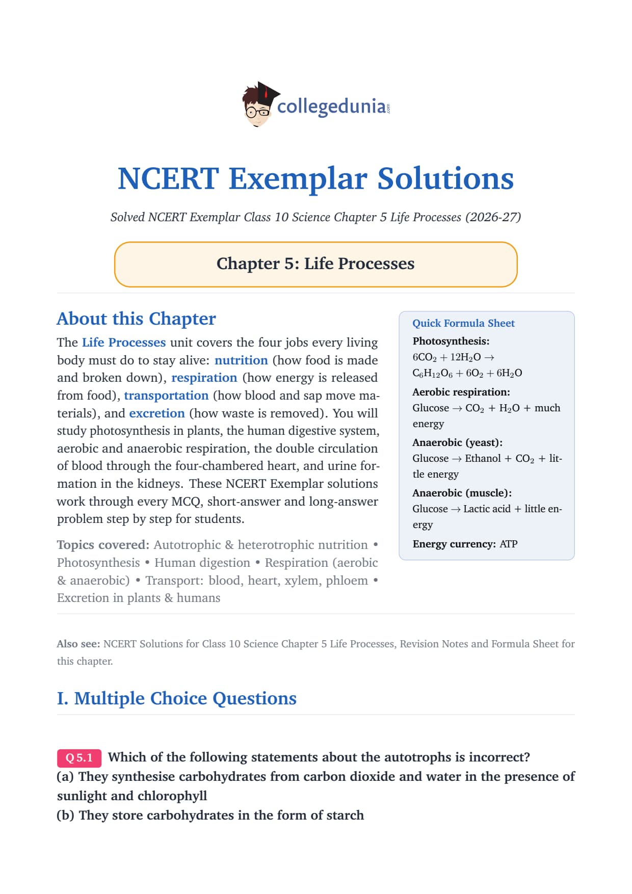
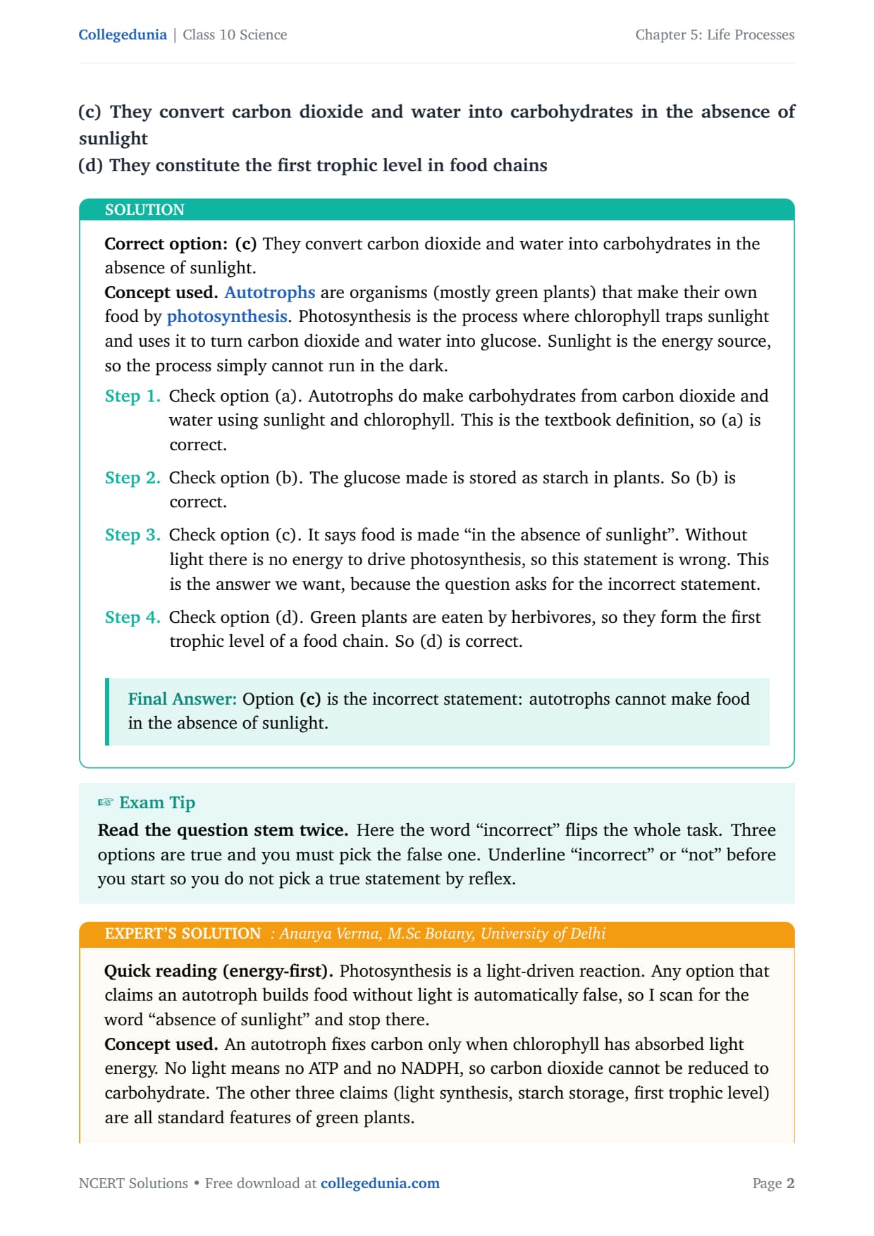
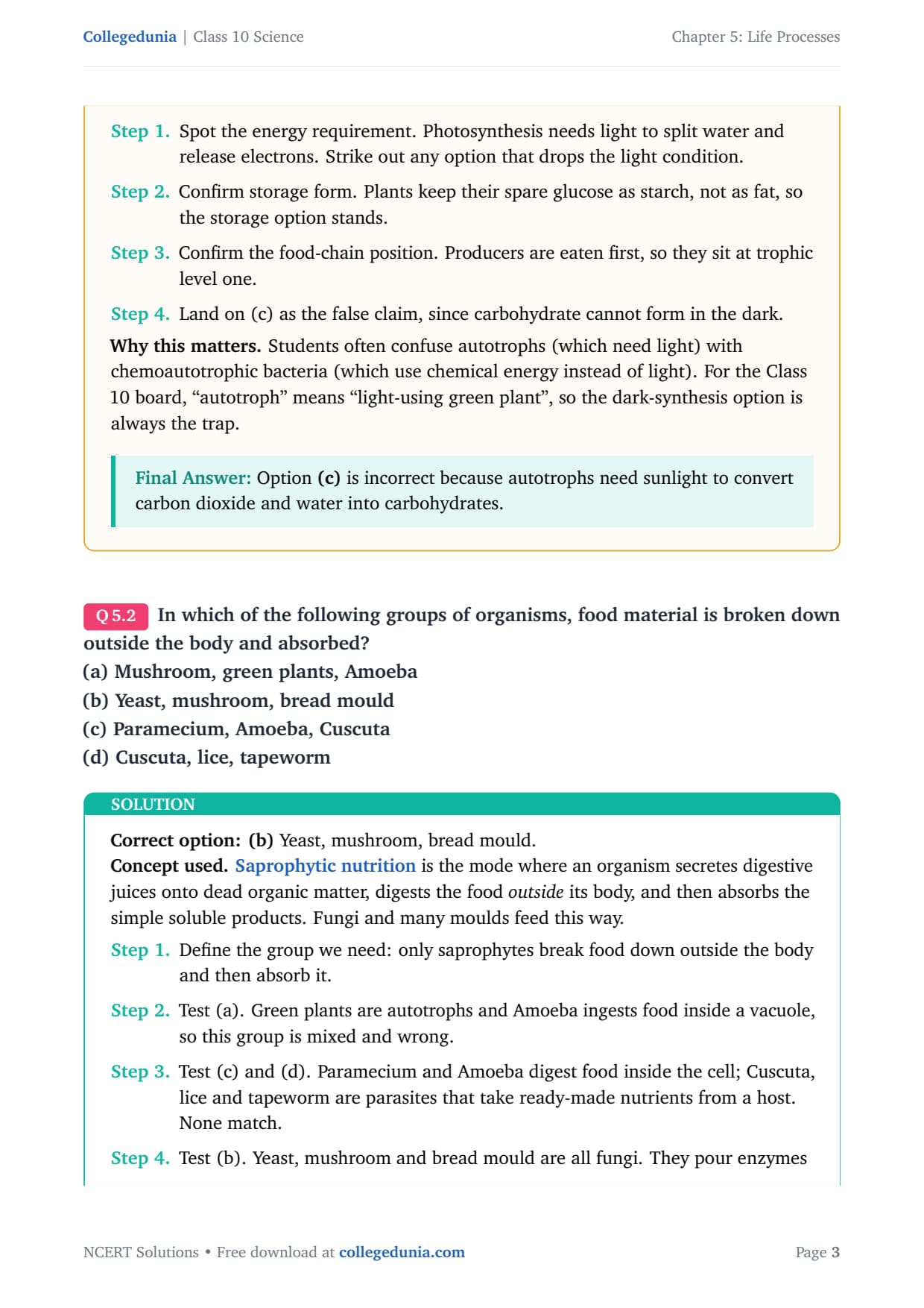
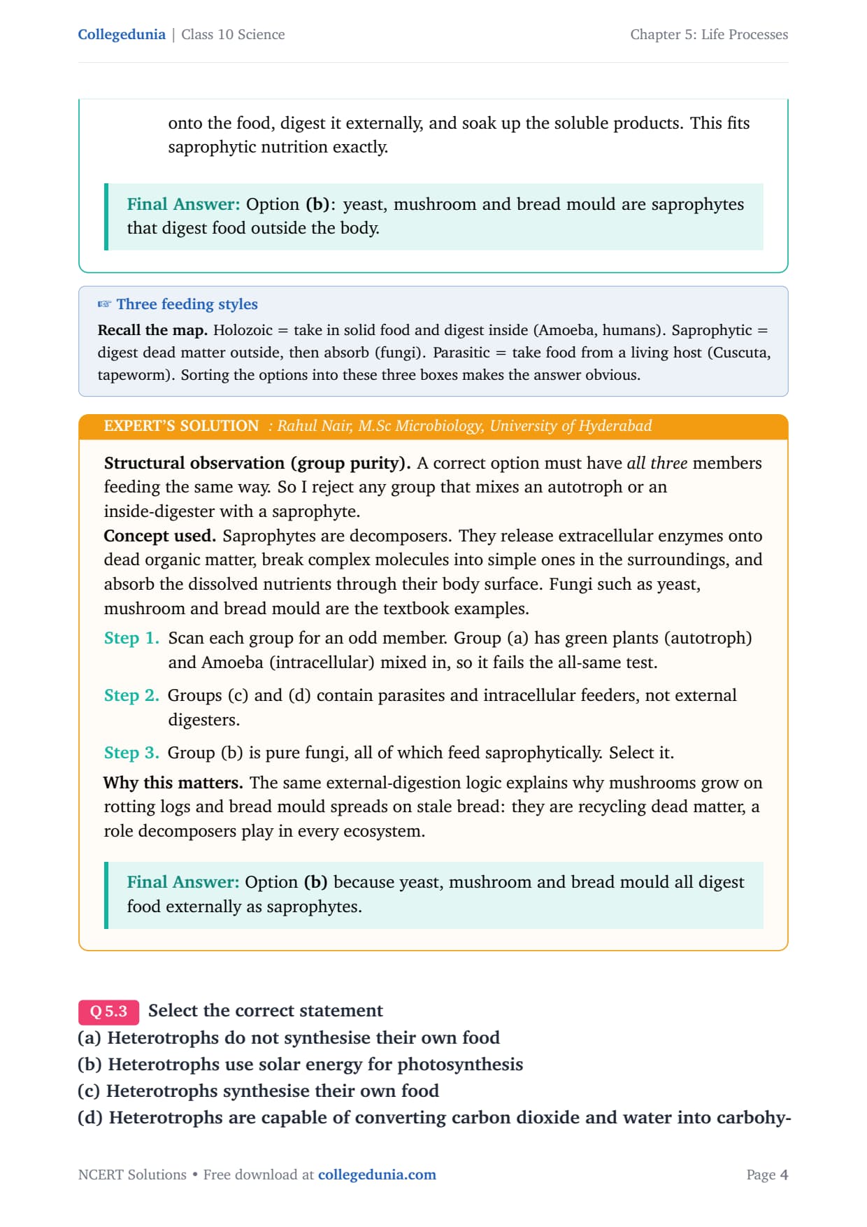
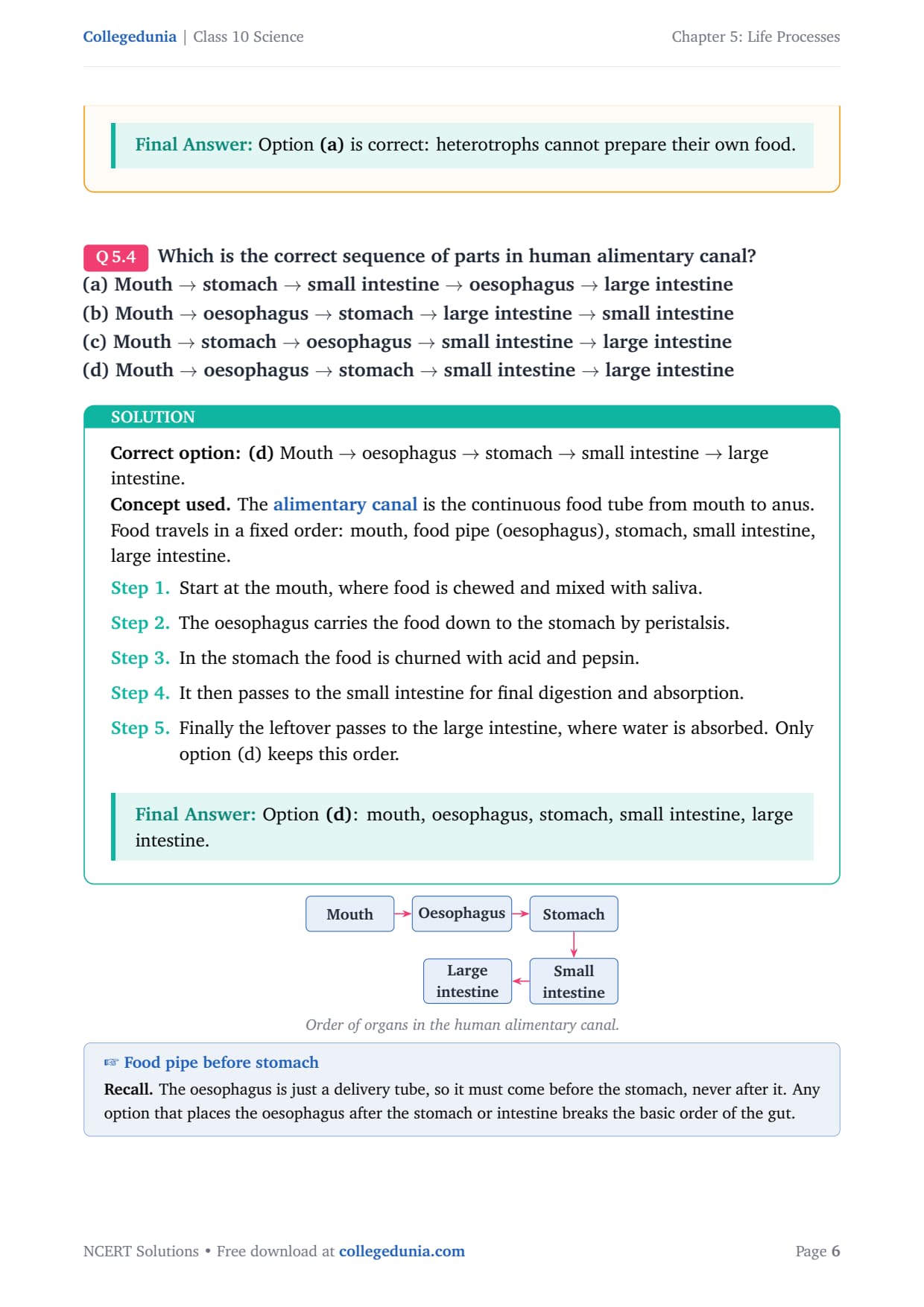
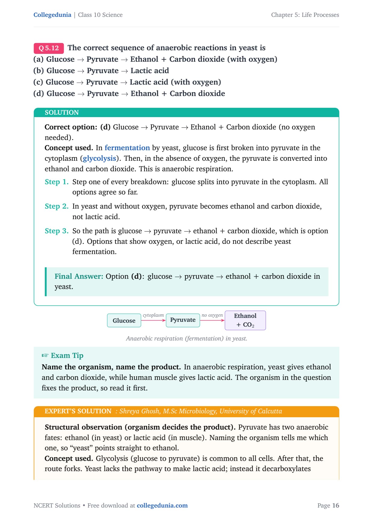
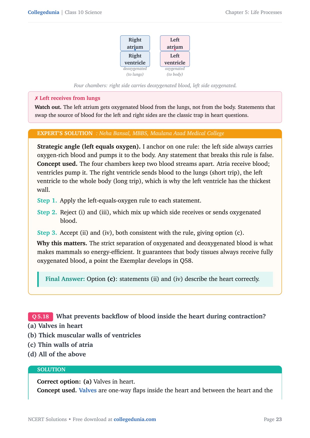
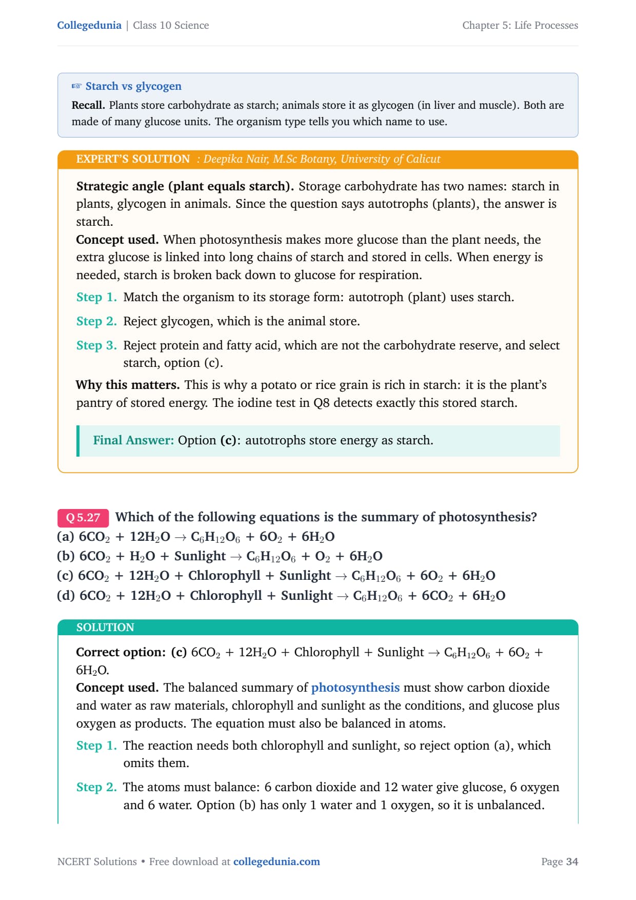
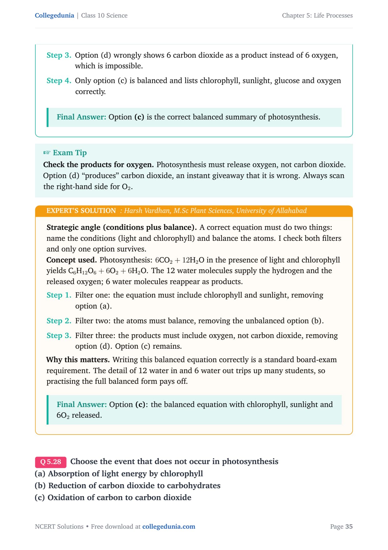
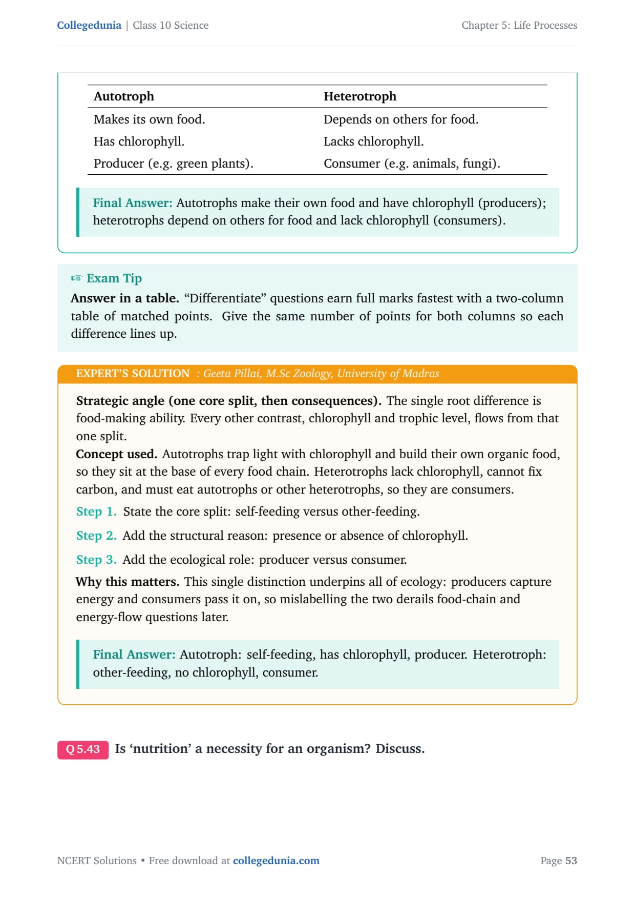
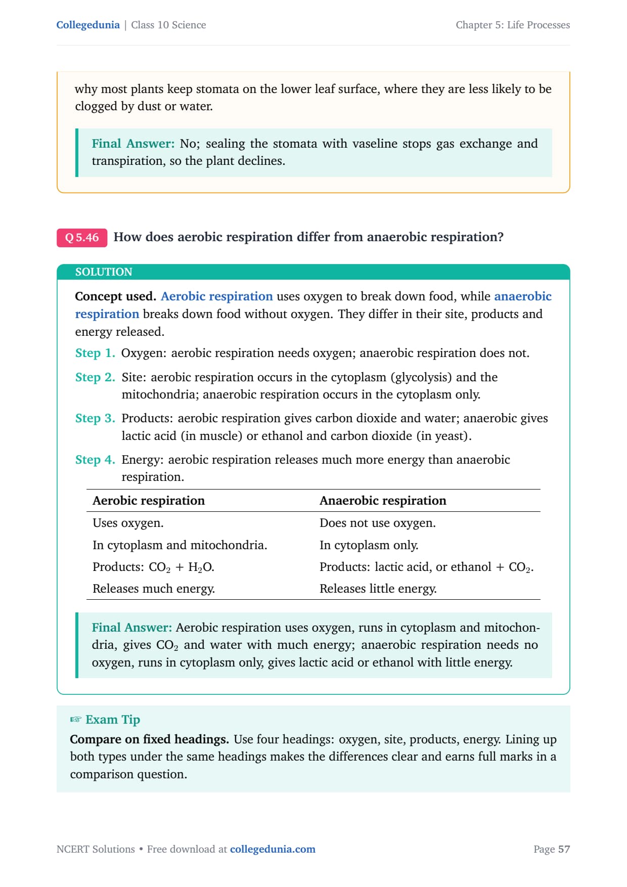
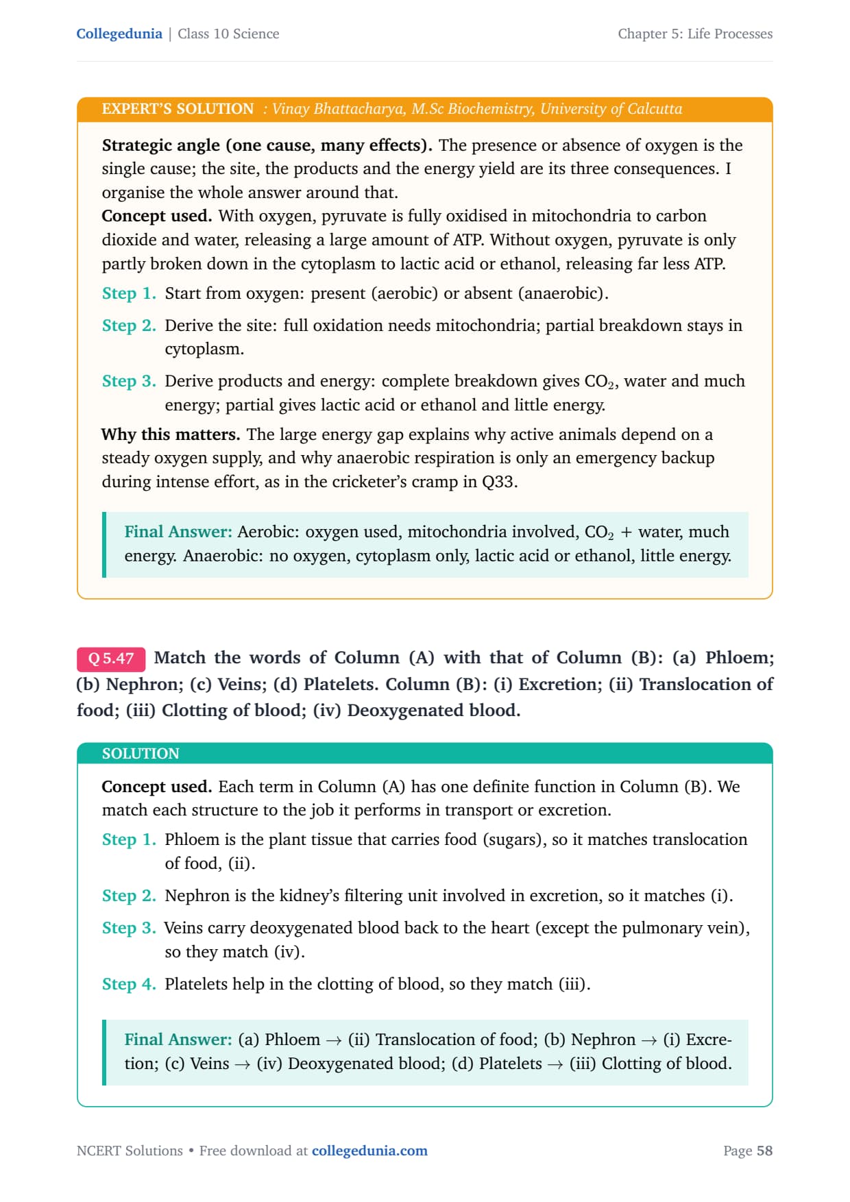
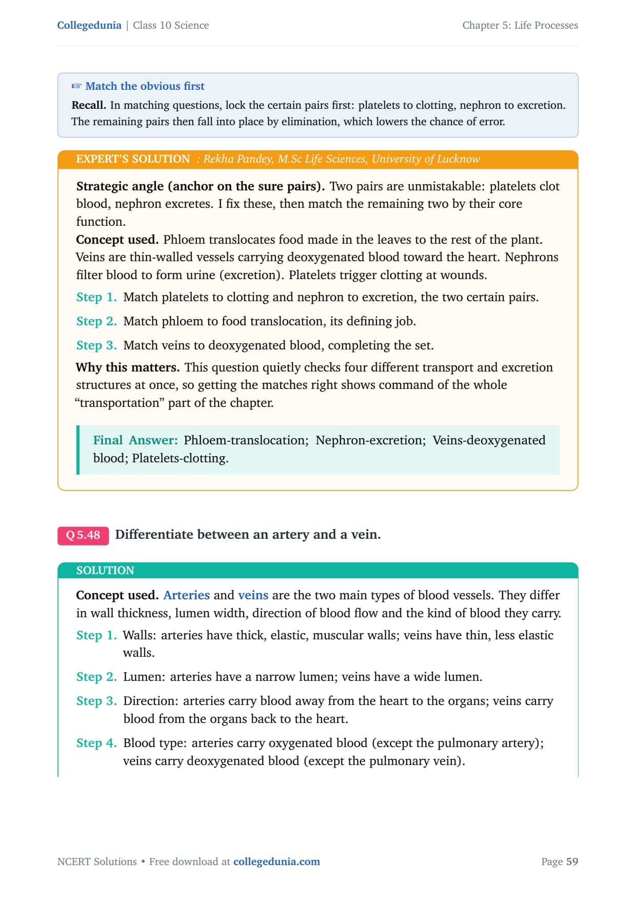
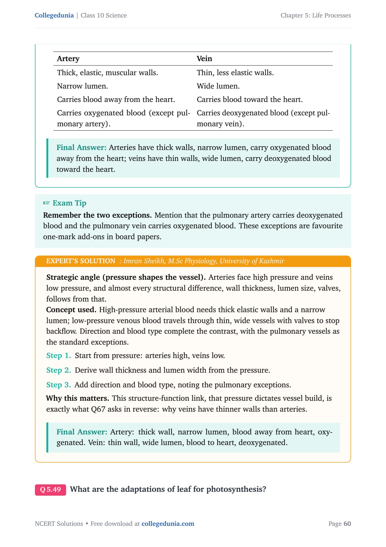
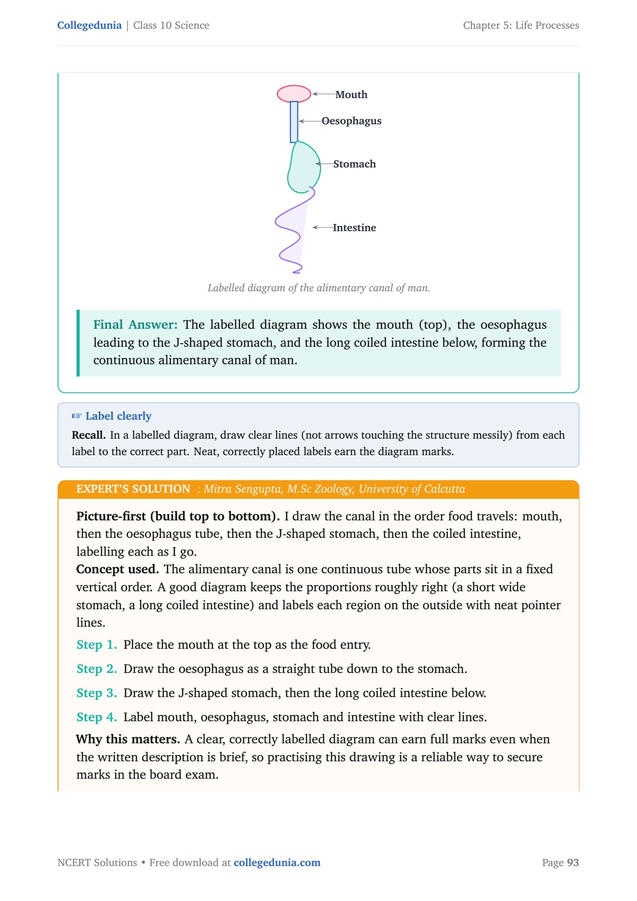
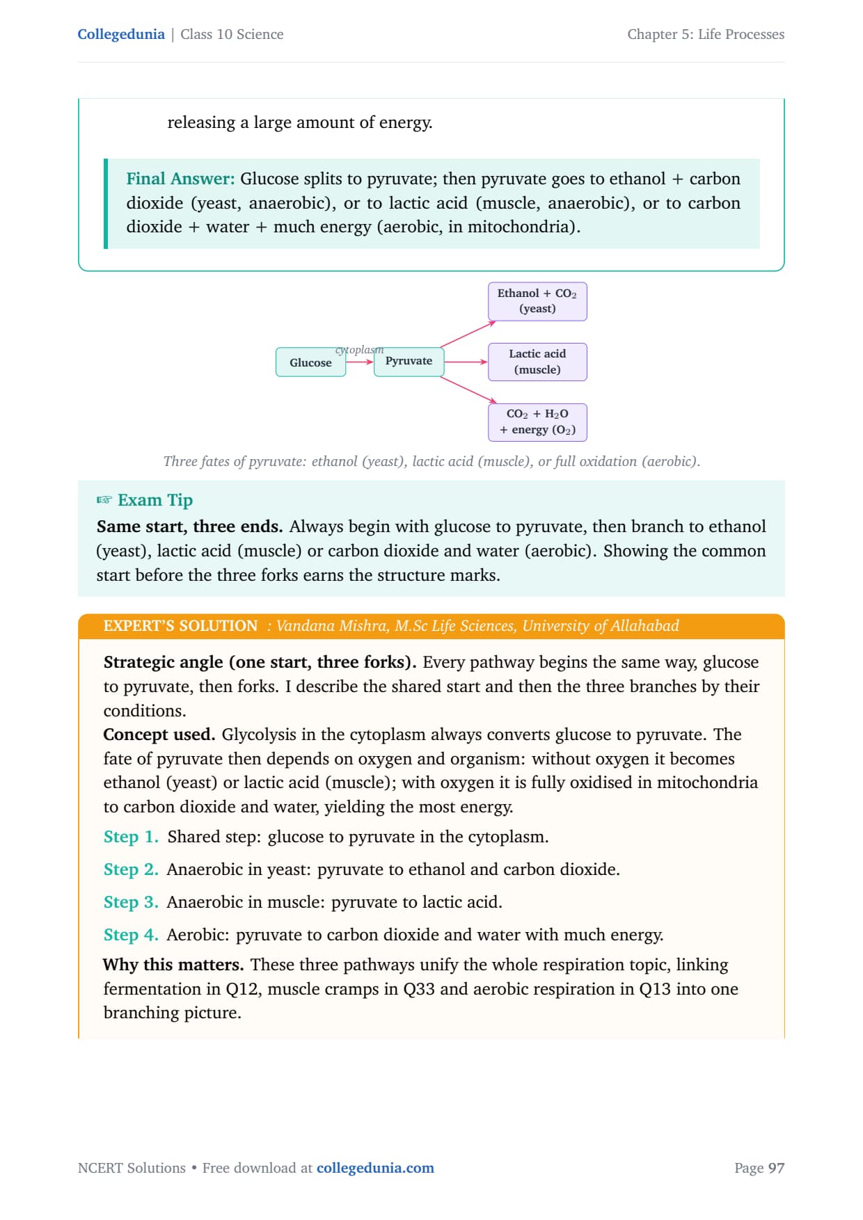
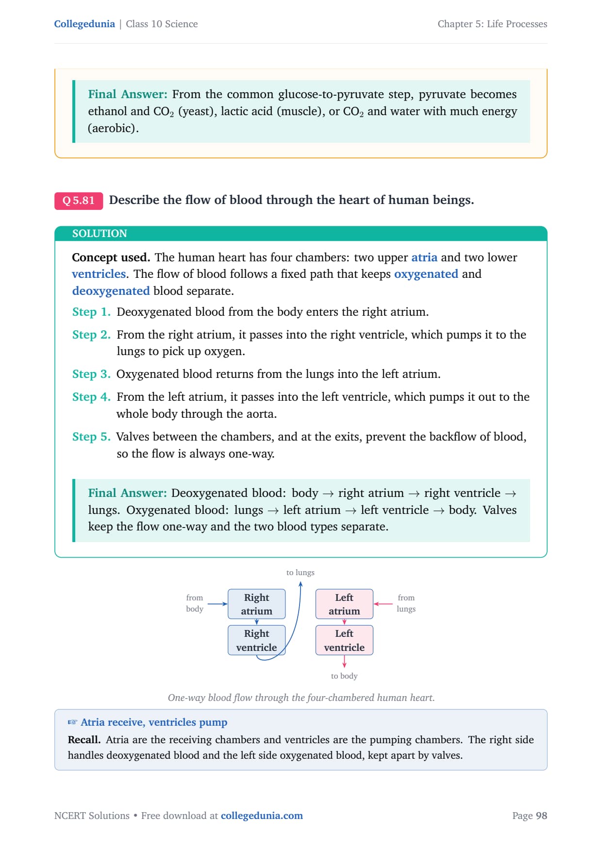
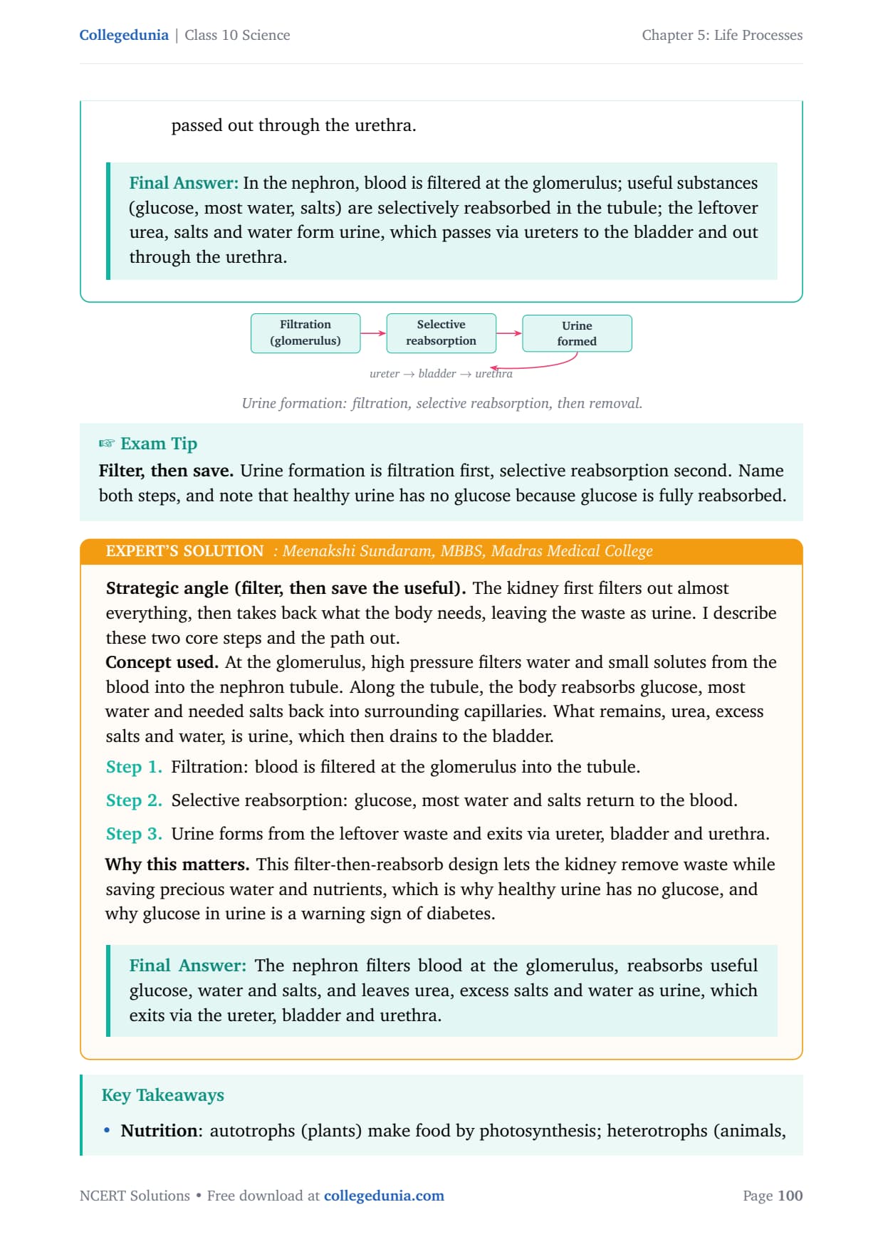



Comments