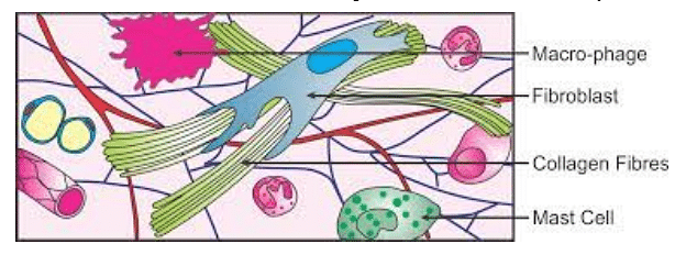
Areolar tissue is defined as simple loose connective tissue. Areolar tissue is a type of connective tissue that is present throughout the body. It provides support to the human body and protects human organs. Areolar tissue is made up of numerous different types of loosely organized fibers in a semi-fluid ground substance.
- It binds the skin to the underlying tissues.
- Areolar tissue encircles and supports the blood vessels.
- It fills the spaces between different organs.
- The areolar tissue is composed of 4 types of cells.
- It includes fibroblasts, mast cells, macrophages and flat cells.
Also read: NCERT Solutions for Class 11 Biology Structural Organisation in Animals
| Table of Content |
Key Terms: Areolar tissues, Areolar, Connective tissue, Matrix, Fibroblasts, Macrophages, Adipose tissue, Ligaments, Tendons, Elastin
Areolar Connective Tissues
[Click Here for Sample Questions]
Areolar tissue is the connective tissue that is found most widely throughout the body.
- The areolar tissue is made up of a translucent, jelly-like matrix.
- The matrix is packed with many fibers, different cell types, and mucin.
- This tissue can be found in numerous visceral organs.
- For example, in the stomach, trachea, skin, and artery walls.
- It occurs between the lobes of compound glands as well as the stroma of solid organs.
- This connective tissue can allow materials to diffuse through it.
- It forms the epithelium's basement membrane.
Types of Fibers Present in Areolar Tissues
[Click Here for Sample Questions]
Mainly two types of fibers are found in areolar tissues.
1. White fibers
The collagen-based fascia, which contains white fibers, are found in bundles. The toughness of the tissue is provided by collagen fibres.
- A protein called collagen resembles rope and gives connective tissue strength.
- These fibers are widely distributed in the tendons and ligaments
- Pepsin helps collagen break down.
2. Yellow fibers
In comparison to white fibers, yellow fibers are both rarer and thicker. They only exist as single, straight objects. They are elastic, branching, and malleable.
- An asymmetric network is created when the branches connect with one another.
- Elastin makes up their structure and gives them their shape.
- Trypsin can break down elastic fibers.
- Elasticity is added to the tissue by the yellow filaments.
Cells Present in Areolar Tissues
[Click Here for Sample Questions]
Cells that are present in the areolar tissues are fibroblasts, macrophages, mast cells and flat type of cells.
1. Fibroblasts
The primary cells of the areolar tissues are fibroblasts. They exude the substance used to create the fibers. These are in charge of producing extracellular matrix materials like tropo-collagen. This protein further externally polymerizes to form collagen and its secretion. The cells, known as fibrocytes, become less active as the tissue ages and stops growing.
2. Macrophages
The number of macrophages is almost equal to that of fibroblasts. They are flexible in size, robust, and ovoid-shaped amoeboid cells. Lysosomes and endocytic vesicles are abundant in macrophages. They aid in the fight against infection and are phagocytic cells. These are also known as "scavenger cells."
3. Mast cells
Mast cells are typically found along nerves, tiny blood veins, and lymph vessels. They are small, oval cells. Mast cells are primarily focused on secreting active substances such as-
1. Heparin
In addition to being an anticoagulant, it is a carbohydrate. It ensures that blood remains fluid and inhibits blood clotting inside the vessels.
2. Histamine
It is an amine that is created by decarboxylating the amino acid histidine. Its purpose is to start an inflammatory reaction and it takes part in things like swellings, burns, and aches. Blood vessels also enlarge (relax) as a result of it.
3. Serotonin
It is a protein and its main function is to contract the blood vessels (vasoconstrictor).
4. Lymphocytes
The cells in the matrix are tiny and move around freely. Because they are capable of producing antibodies, they typically appear where inflammation is present.
5. Adipose cells
They are substantial fat-storing cells that have one or more fat globules in their cytoplasm.
6. Plasma cells
Similar to lymphocytes, they are tiny cells involved in bodily defence.
Also Read: Muscular Tissue
Location of Areolar Tissues
[Click Here for Sample Questions]
All body systems with external openings have areolar tissue, which is located beneath the dermis layer and the epithelial tissue.
- It makes up the stroma of glands and is found in the hypodermis of the skin.
- Mucous membranes of the reproductive and urinary systems also contain areolar tissues.
- Areolar tissue is also found in the lamina propria of the digestive and respiratory tracts.
- The mesentery, which encircles the intestine, also contains areolar tissue.
- Loose connective tissue is a significant site of inflammatory and immunological reactions.

Areolar Tissue
Function of Areolar Tissues
[Click Here for Sample Questions]
Under mechanical stress, the tenacity of white fibers and the flexibility of yellow fibers prevent tissue and organ displacement and damage.
- It holds the muscles together.
- It secures the skin to the muscles.
- The tissue connects the blood vessels and nerves to the tissues around them.
- It fastens the peritoneum to the body wall and viscera.
- It creates the sub-mucosa in the wall of the alimentary canal as well as the dermis.
- Areolar tissues' cells aid in the defence against infection.
- Heparin helps to stop blood clotting in vessels.
Types of Areolar Tissues
[Click Here for Sample Questions]
Loose connective tissue and dense connective tissue are two different forms of areolar tissues.
- The tissue that surrounds and affixes to organs is called loose connective tissue.
- Organs, anatomical structures, and tissues are held in place by loose connective tissue.
- The extracellular matrix is the most important component In loose connective tissue.
- Dense connective tissue holds bones, muscles, and other tissues and organs in place.
- It provides support and protection for them.
- Dense connective tissue is found in the sclera (the white outer layer of the eye), tendons, inner skin, and ligaments.
- It is referred to as fibrous connective tissue.
Common Disorder of Areolar Tissue
[Click Here for Sample Questions]
Collagen and elastin become inflamed in a patient with a connective tissue disorder. Connective tissue disorders include-
1. Rheumatoid Arthritis (RA)
RA is one of the most prevalent connective tissue disorders. The autoimmune nature of RA causes the immune system to target the affected area. The membrane around joints is attacked and inflamed in this systemic illness by immune cells.
2. Scleroderma
It is an autoimmune disorder that results in the formation of scar tissue in the skin, internal organs (such as the GI tract), and small blood vessels. Women are affected by it three times more frequently than men are.
3. Ehlers-Danlos syndrome
A group of disorders known as Ehlers-Danlos syndrome impair the connective tissues that support the skin, bones, blood vessels, and numerous other organs and tissues.
4. Granulomatosis with polyangiitis (GPA)
Blood vessels swell up in Granulomatosis with Polyangiitis (GPA). It is an uncommon diseased condition. Major body organs suffer damage due to GPA.
5. Polymyositis/dermatomyositis
It is a condition that causes muscle deterioration and inflammation. The condition is known as dermatomyositis when the skin is also affected.
Also Read:
Things to Remember
- Areolar tissue is a loose connective tissue.
- It is found in the skin and in other organ systems of the human body.
- The main function of areolar tissue is to support and protect organs.
- White and yellow fibers are found in areolar connective tissue.
- Elastin and collagen are the main proteins of this tissue.
- Cells present in areolar tissue are fibroblast, macrophages, mast and flat cells.
- This tissue has a mesh structure made of collagen, reticular, and elastic fibers.
- It is located between skin and muscles, around blood arteries and nerves.
- It is also found in the bone marrow, and in organs having external openings.
- The dense connective tissue is composed of ligaments and tendons.
- Ligaments link bones to other bones and tendons attach muscles to bones.
Sample Questions
Ques. What is a Tissue? (1 mark)
Ans. It is a group of fells, which are similar in origin, structure and function. The study of tissues is called histology.
Ques. Write about the differences between dense regular and dense irregular tissues. (2 marks)
Ans. Collagen fibers are found in rows between several parallel bundles of fiber in the dense regular connective tissue. Ligaments that join one bone to another and tendons that attach skeletal muscles to bones are two examples of tissue. Contrarily, dense irregular connective tissue has several variably oriented fibers and fibroblasts, as well as an irregular arrangement of fibers. The skin contains the tissue mentioned.
Ques. What are tendons and ligaments? (2 marks)
Ans. Tendons are fibrous, strong, thick tissues made of collagen fibers. These serve to join bones and muscles together. The other hand's ligaments, which join two bones together, are made of yellow elastin fibers.
Ques. Where do you find fibers in connective tissues? What are their functions? (2 marks)
Ans. The fibers are found in all connective tissue, excluding blood. It secretes collagen or elastin fibers, which are structural proteins. The tissue's fibres provide strength, elasticity, and flexibility.
Ques. What are the functions of the areolar connective tissue? (4 marks)
Ans. Areolar tissue has a role in the following functions.
- It establishes a defence system.
- Contains mast cells that aid in the prevention of infection.
- The collagen fibers in the areolar tissue give strength and rigidity.
- The skin's flexibility and elasticity are maintained by the areolar connective tissue, which is located deep under the epidermis.
- It offers an anti-friction cushioning layer.
Ques. Describe the roles played by the cells that make up the areolar connective tissue. (5 marks)
Ans. 3 major types of cells along with adipose tissue in areolar tissue plays an important role in the body function.
- Fibroblasts: These aid in ground substance formation, extracellular matrix anchoring and strengthening, and tissue repair.
- Macrophages: They are phagocytic cells that eliminate invading infections.
- Mast cells: These cells release histamine and other chemicals, which cause allergic and immunological reactions. Additionally, they discharge chemoattractants into the environment.
- Lymphocytes: The matrix's microscopic, mobile cells are very small. Since they have the ability to produce antibodies, they frequently manifest themselves in areas of inflammation.
- Adipose tissue: They are large fat-storing cells with a globule or globules of fat in their cytoplasm.
- Plasma cell: They are tiny cells involved in bodily defence, much like lymphocytes.
Ques. What are white fibers? (3 marks)
Ans. Bundles of the collagen-based fascia, which include the white fibers, can be seen. Collagen fibers. It gives the tissue its tensile strength. Collagen is a rope-like protein that gives connective tissue strength and can bend but not stretch.
The tendons and ligaments that connect muscles to the bone and to one another, respectively, are widely dispersed with these fibers
Ques. What are connective tissues? (3 marks)
Ans. A type of tissue that supports, shields, and gives structure to the body's other tissues and organs. In addition to storing fat, helping to transport nutrients and other substances between tissues and organs, and aiding in tissue healing, connective tissue also helps move fat. Cells, fibers, and a gel-like substance make up connective tissue. Bone, cartilage, fat, blood, and lymphatic tissue are all examples of connective tissue types.
Ques. What are different disorders related to Connective Tissues? (3 marks)
Ans. There are many different types of connective tissue disorders, such as-
- Rheumatoid arthritis (RA)
- Scleroderma
- Granulomatosis with polyangiitis (GPA)
- Churg-Strauss syndrome
- Lupus
- Microscopic polyangiitis
- Polymyositis/dermatomyositis
- Marfan syndrome
Ques. What is Adipose tissue? (4 marks)
Ans. Adipose tissue is a connective tissue that covers the entire body. Mesenchymal stem cells during fetal development differentiate into adipocytes, a specific type of connective tissue known as adipose tissue. Mesenchymal stem cells are pluripotent cells that have the capacity to differentiate into a variety of cell types, including, but not limited to, muscle, fat, bone, and cartilage cells.
Adipose tissue can be divided into two functionally distinct tissues based on the type of adipocytes present: white adipose tissue, which is largely made up of beige and white adipocytes, and brown adipose tissue, which is made up of brown adipocytes.
For Latest Updates on Upcoming Board Exams, Click Here: https://t.me/class_10_12_board_updates
Do Check Out:


Comments