Biology Mentor, NEET Topper | Updated 3+ months ago
Human reproduction is a sexual mode of reproduction involving gamete formation, fertilisation, embryonic development, and parturition. Class 12 Biology Chapter 2 Human Reproduction is a scoring chapter for the CBSE Board exam and guarantees 4-5 marks for NEET.
This page contains the step-by-step human reproduction class 12 NCERT solutions PDF, the year-wise question pattern, and an answer-writing aligned to the 2026-27 NCERT.
CBSE Weightage: 4 to 6 marks, usually one short-answer plus one diagram-based question
NEET Weightage: 4 to 5 questions per year, among the highest in the Reproduction unit
Concept: Spermatogenesis and oogenesis repeat almost every year. Draw the gametogenesis flow, label every cell stage, and write the chromosome number beside each. Examiners give a separate mark for the labelled flow.
Human Reproduction Video Lecture for Class 12 Biology
Human Reproduction Class 12 Biology Previous Year Questions and Their Frequency
This table maps how Chapter 2 appeared across CBSE Board, NEET, and CUET in the last five sessions, newest first. Biology is not tested in JEE. Empty cells mean no standalone question that year.
Year
CBSE Board
NEET
CUET (UG)
2025
Menstrual cycle phases (5 marks)
Spermiogenesis vs spermiation (1 Q)
Placenta function (1 Q)
2024
Placenta as an endocrine gland (3 marks)
Graafian follicle structure (1 Q)
Fertilisation site (1 Q)
2023
Oogenesis labelled diagram (5 marks)
Capacitation and acrosomal reaction (1 Q)
-
2022
Hormonal control of male reproduction (3 marks)
Blastocyst and implantation (1 Q)
Seminiferous tubule cells (1 Q)
2021
-
Layers of the embryo sac (1 Q)
-
Spermatogenesis, oogenesis, and the menstrual cycle make up nearly two-thirds of every CBSE 5-marker on this chapter.
How Collegedunia's NCERT Solutions Help You with Human Reproduction
This Human Reproduction Class 12 NCERT Solutions PDF is built around the exact phrasing CBSE awards full marks on, with a NEET recall framing alongside.
2026-27 NCERT alignment: every answer matches the current Class 12 Biology syllabus.
Labelled diagrams and step marking: figures are drawn the way examiners expect, with each label graded.
Answer-writing discipline: each long answer is split into mark-bearing points.
Sample Fully-Solved Question: Menstrual Cycle Hormonal Control (Q10)
One repeated 5-marker is solved end to end here, so you can see the expected answer structure.
Question (Q10): Describe the role of hormones in the regulation of the menstrual cycle in a human female.
Step 1 (Follicular phase): FSH stimulates the primary follicle to grow into a Graafian follicle, which secretes oestrogen.
Step 2 (Ovulation): Rising oestrogen triggers an LH surge around day 14, rupturing the follicle to release the secondary oocyte.
Step 3 (Luteal phase): LH converts the ruptured follicle into the corpus luteum, which secretes progesterone to maintain the endometrium.
Step 4 (Menstruation): If fertilisation fails, the corpus luteum degenerates, progesterone falls, and the endometrium breaks down.
Step-by-Step Mark Annotation on a CBSE-Style Human Reproduction Answer
The 5-mark spermatogenesis question is among the most repeated in Class 12 Biology. The table shows how each mark is earned.
Mark
What earns it
Common gap that loses it
Mark 1
Defining spermatogenesis as sperm formation in seminiferous tubules at puberty
Skipping the site or the puberty trigger
Mark 2
Multiplication phase: spermatogonia (2n) divide by mitosis
Not stating the chromosome number 2n
Mark 3
Meiosis I and II producing four haploid spermatids
Writing two cells instead of four spermatids
Mark 4
Spermiogenesis: spermatids transform into spermatozoa
Confusing spermiogenesis with spermiation
Mark 5
Role of Sertoli cells and hormonal control by FSH and testosterone
Omitting Sertoli (nurse) cells
Quick Tip: Spermiogenesis is the transformation of spermatids into sperm; spermiation is the release of mature sperm into the tubule lumen. Mixing the two is the most common one-mark loss in this chapter.
Common Mistakes Students Make in Human Reproduction
Most marks here are lost to imprecise terms and unlabelled diagrams, not weak concepts.
Spermiogenesis vs spermiation: reversed by many students, costing a guaranteed recall mark.
Corpus luteum confusion: writing that it secretes oestrogen instead of progesterone breaks the luteal-phase answer.
Unlabelled gametogenesis flow: drawing the flow without the chromosome number (2n or n) forfeits the diagram mark.
Placenta called only nutritive: it is also an endocrine gland (hCG, hPL, oestrogen, progesterone); omitting this loses a mark.
Most Common Question Stems CBSE Uses in Human Reproduction
CBSE recycles a small set of phrasings. Spotting the stem tells you which answer structure to use.
Question Stem
Expected Answer Structure
"Describe the role of hormones in..."
Name hormone, source, target, effect, in four points
"Draw a labelled diagram of..."
Clean figure, every part labelled, one-line function note
"Differentiate between spermatogenesis and oogenesis"
Tabular, point-wise, minimum four bases of comparison
List the hormones it secretes with one function each
Top Concept Recall List for Human Reproduction Class 12 Biology
These five recall items are the ones NEET and CUET test on repeat.
Concept
One-Line Recall
Spermatogenesis site
Seminiferous tubules, begins at puberty under FSH and testosterone
Oogenesis timing
Begins in foetal life, arrested at prophase I until puberty
Fertilisation site
Ampullary-isthmic junction of the fallopian tube
Implantation stage
Blastocyst embeds in the endometrium around day 7
Pregnancy hormone marker
hCG, secreted by the trophoblast, basis of pregnancy tests
How to Study Human Reproduction for Class 12 Biology Boards
This chapter rewards a diagram-first approach. A focused three-session plan makes it a reliable scorer.
Session 1 (90 min): reproductive systems with fully labelled diagrams; practise the testis and ovary figures from memory.
Session 2 (90 min): gametogenesis and the menstrual cycle; build spermatogenesis and oogenesis flowcharts with chromosome numbers marked.
Session 3 (60 min): fertilisation, implantation, pregnancy, parturition, and lactation; solve five years of CBSE 5-markers against the annotation above.
That is about five hours total, realistic in two days before the Class 12 Biology exam.
All NCERT Solutions for Human Reproduction with Step-by-Step Working
Every NCERT textbook question for Class 12 Biology Chapter 2 Human Reproduction is listed below with its full Solution and Expert Solution inside collapsible tabs. Click Check Solution for the steps; click Expert Solution for the expanded explanation.
Questions
Q 2.1
Fill in the blanks:
(a) Humans reproduce 2.2cm0.4pt (asexually/sexually).
(b) Humans are 2.2cm0.4pt (oviparous, viviparous, ovoviviparous).
(c) Fertilisation is 2.2cm0.4pt in humans (external/internal).
(d) Male and female gametes are 2.2cm0.4pt (diploid/haploid).
(e) Zygote is 2.2cm0.4pt (diploid/haploid).
(f) The process of release of ovum from a mature follicle is called 2.2cm0.4pt.
(g) Ovulation is induced by a hormone called 2.2cm0.4pt.
(h) The fusion of male and female gametes is called 2.2cm0.4pt.
(i) Fertilisation takes place in 2.2cm0.4pt.
(j) Zygote divides to form 2.2cm0.4pt which is implanted in uterus.
(k) The structure which provides vascular connection between foetus and uterus is called 2.2cm0.4pt.
Concept used. Each blank tests one core fact about human sexual reproduction. Sexual reproduction means two parents make haploid gametes that fuse. A viviparous animal gives birth to live young that developed inside the mother's body. Internal fertilisation means the sperm meets the egg inside the female body. Haploid (n) means one set of chromosomes; diploid (2n) means two sets. We answer each blank from these definitions.
(a) sexually. Humans have two sexes. A male and a female each contribute a gamete, so reproduction is sexual, not asexual.
(b) viviparous. The young one develops inside the mother's uterus and is born alive, so humans are viviparous (not egg-laying like birds).
(c) internal. Sperm are deposited inside the female tract and the egg is fertilised inside the body, so fertilisation is internal.
(d) haploid. Gametes are formed by meiosis. A human sperm and a human ovum each carry 23 chromosomes, so both gametes are haploid (n).
(e) diploid. A haploid sperm (n = 23) fuses with a haploid ovum (n = 23). The zygote therefore has 23 + 23 = 46 chromosomes, so it is diploid (2n).
(f) ovulation. The release of the secondary oocyte (ovum) from a mature Graafian follicle is called ovulation.
(g) Luteinising Hormone (LH). A sharp rise in LH (the LH surge) from the anterior pituitary triggers ovulation.
(h) fertilisation (syngamy). The fusion of the male gamete (sperm) and the female gamete (ovum) is fertilisation, also called syngamy.
(i) the ampulla of the fallopian tube. Fertilisation occurs at the ampullary–isthmic junction of the oviduct, that is, in the ampulla region of the fallopian tube.
(j) blastocyst. The zygote divides (cleavage) into a morula and then forms a hollow blastocyst, which implants in the uterine wall.
(k) placenta. The placenta is the structure that links the foetal blood supply with the maternal blood supply (a vascular connection between foetus and uterus).
Quick reading. Read each blank as a yes/no fact, then anchor it to one definition so you never guess. I group the eleven blanks into three families: type of reproduction (a, b, c), ploidy (d, e), and events and structures (f to k).
Type family: humans need two parents (sexual), keep the young inside and deliver live offspring (viviparous), and join gametes inside the body (internal).
Ploidy family: meiosis halves the chromosome number, so gametes are haploid; fusion restores it, so the zygote is diploid.
Events family: ovum release is ovulation; the trigger is the LH surge; gamete fusion is fertilisation; it happens in the ampulla; the zygote becomes a blastocyst; the foeto-maternal bridge is the placenta.
Fix the ploidy logic once. Human body cells are 2n = 46. Meiosis in testis and ovary gives gametes with n = 46/2 = 23. So (d) is haploid.
Fertilisation reverses this. Sperm (23) + ovum (23)⇒ zygote (46) = 2n. So (e) is diploid. Check: 23 + 23 = 46 (correct), the normal human number.
Walk the timeline for (f) to (k): mature follicle bursts (ovulation, f) under the LH surge (g); sperm fuses with ovum (fertilisation, h) in the ampulla (i); the zygote cleaves to a blastocyst (j) that implants; the placenta (k) then forms.
Why this matters. The whole chapter is one storyline: make haploid gametes, fuse them inside the body, grow a diploid embryo, support it through the placenta. Every blank here is a checkpoint on that single path.
Draw a labelled diagram of male reproductive system.
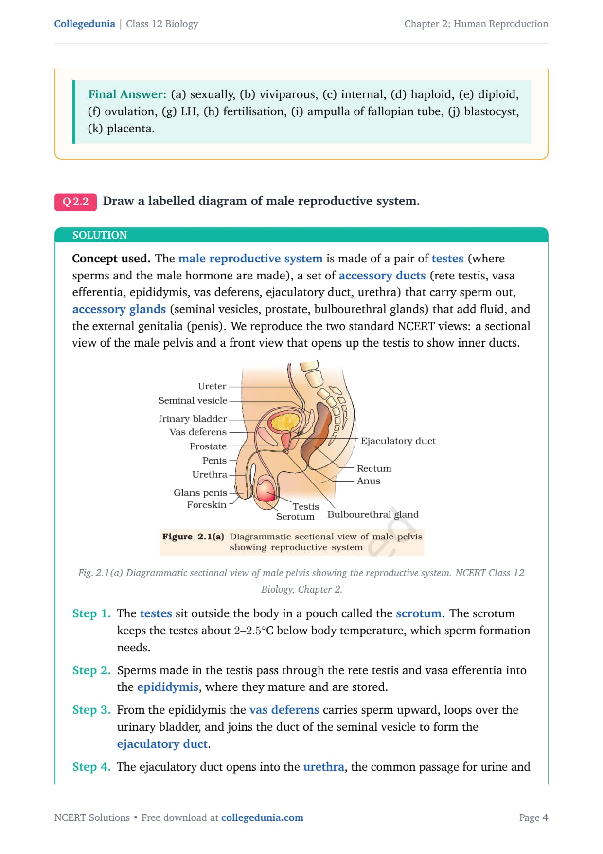
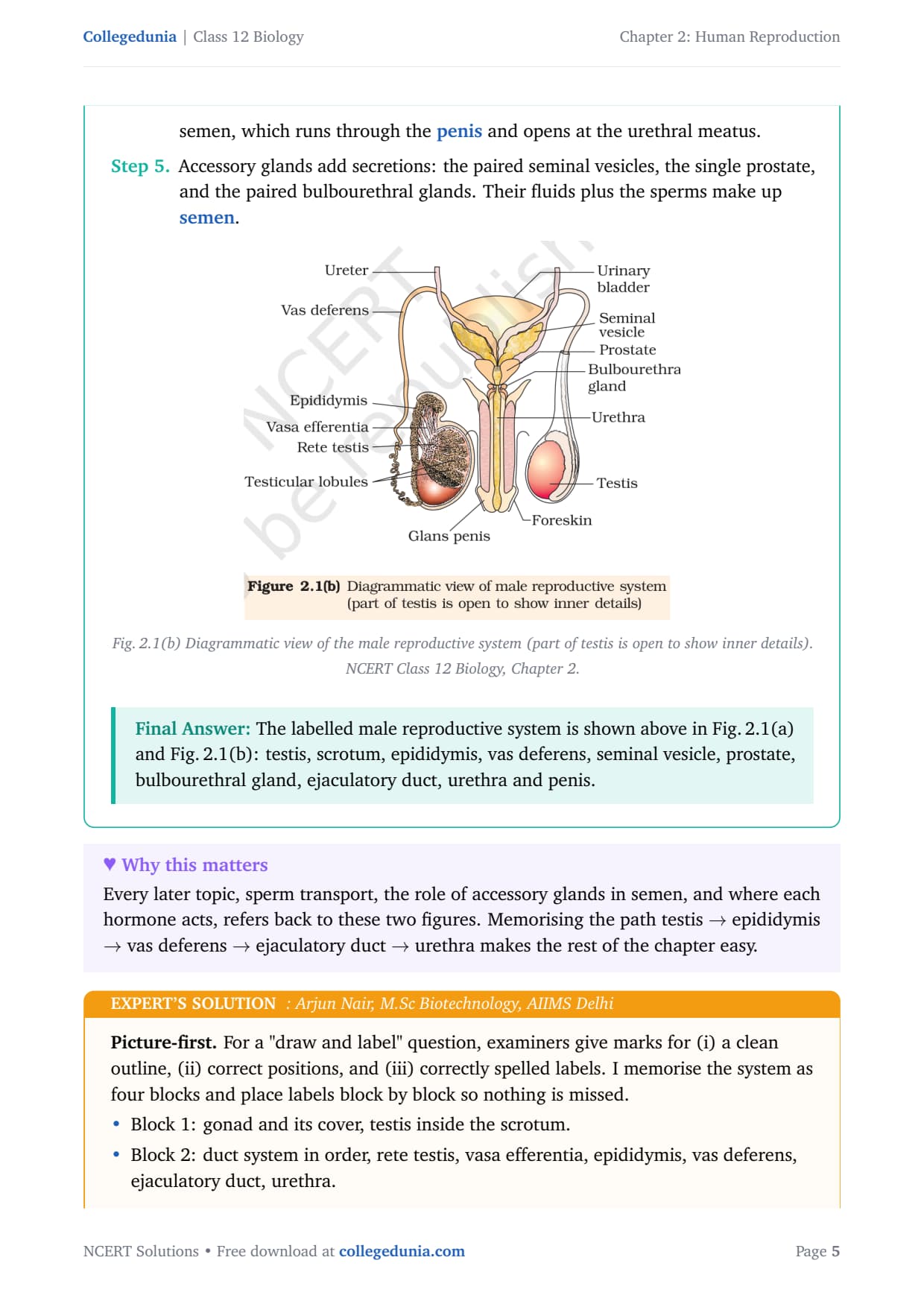
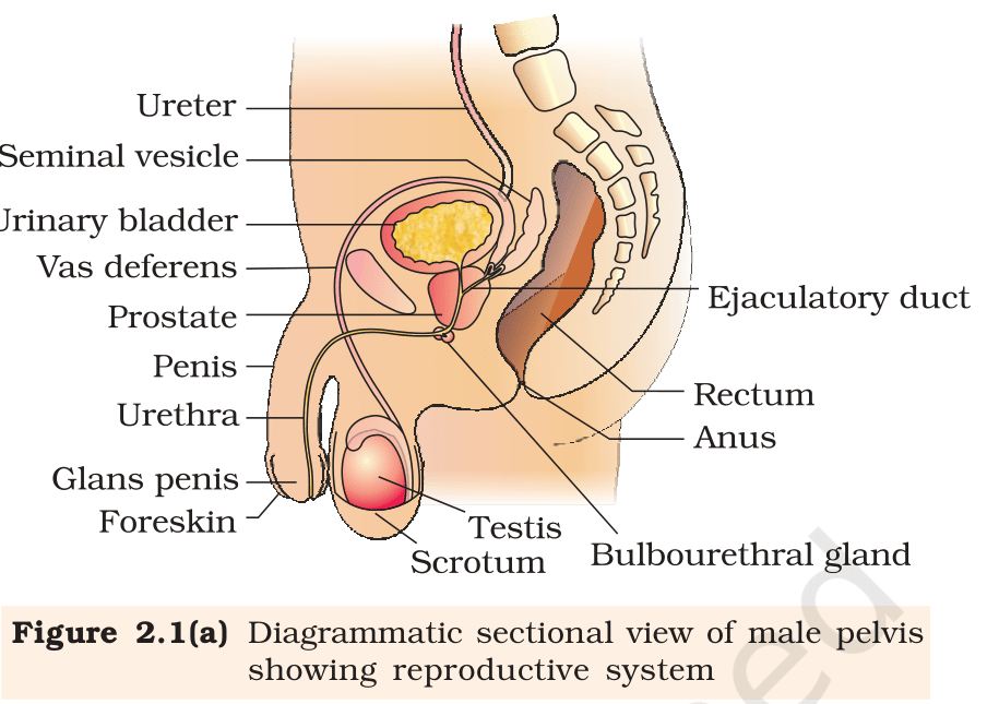
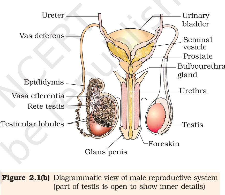
Concept used. The male reproductive system is made of a pair of testes (where sperms and the male hormone are made), a set of accessory ducts (rete testis, vasa efferentia, epididymis, vas deferens, ejaculatory duct, urethra) that carry sperm out, accessory glands (seminal vesicles, prostate, bulbourethral glands) that add fluid, and the external genitalia (penis). We reproduce the two standard NCERT views: a sectional view of the male pelvis and a front view that opens up the testis to show inner ducts.
Fig. 2.1(a) Diagrammatic sectional view of male pelvis showing the reproductive system. NCERT Class 12 Biology, Chapter 2.
The testes sit outside the body in a pouch called the scrotum. The scrotum keeps the testes about 2--2.5∘C below body temperature, which sperm formation needs.
Sperms made in the testis pass through the rete testis and vasa efferentia into the epididymis, where they mature and are stored.
From the epididymis the vas deferens carries sperm upward, loops over the urinary bladder, and joins the duct of the seminal vesicle to form the ejaculatory duct.
The ejaculatory duct opens into the urethra, the common passage for urine and semen, which runs through the penis and opens at the urethral meatus.
Accessory glands add secretions: the paired seminal vesicles, the single prostate, and the paired bulbourethral glands. Their fluids plus the sperms make up semen.
Fig. 2.1(b) Diagrammatic view of the male reproductive system (part of testis is open to show inner details). NCERT Class 12 Biology, Chapter 2.
The labelled male reproductive system is shown above in Fig. 2.1(a) and Fig. 2.1(b): testis, scrotum, epididymis, vas deferens, seminal vesicle, prostate, bulbourethral gland, ejaculatory duct, urethra and penis.
AN
Arjun Nair
M.Sc Biotechnology, AIIMS Delhi
Verified Expert
Picture-first. For a "draw and label" question, examiners give marks for (i) a clean outline, (ii) correct positions, and (iii) correctly spelled labels. I memorise the system as four blocks and place labels block by block so nothing is missed.
Block 1: gonad and its cover, testis inside the scrotum.
Block 2: duct system in order, rete testis, vasa efferentia, epididymis, vas deferens, ejaculatory duct, urethra.
Block 3: three accessory glands, seminal vesicle, prostate, bulbourethral gland.
Block 4: external genitalia, the penis with glans and foreskin.
Start with the body outline and mark the scrotum low and outside, then draw the oval testis inside it.
Trace the duct from the testis upward: epididymis hugging the testis, vas deferens looping over the bladder, joining the seminal vesicle duct to form the ejaculatory duct, then the urethra down the penis.
Add the three glands at their points of entry and label the penis parts. Cross-check every label against Fig. 2.1(a) and 2.1(b) so spelling and position both score.
Why this matters. A correctly ordered duct system is the backbone answer that examiners look for first; getting the sequence right earns most of the marks even before fine detail.
A correctly labelled diagram (as in Fig. 2.1a, b) with testis, scrotum, epididymis, vas deferens, seminal vesicle, prostate, bulbourethral gland, ejaculatory duct, urethra and penis is the complete answer.
Q 2.3
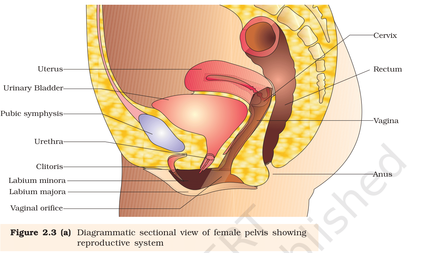
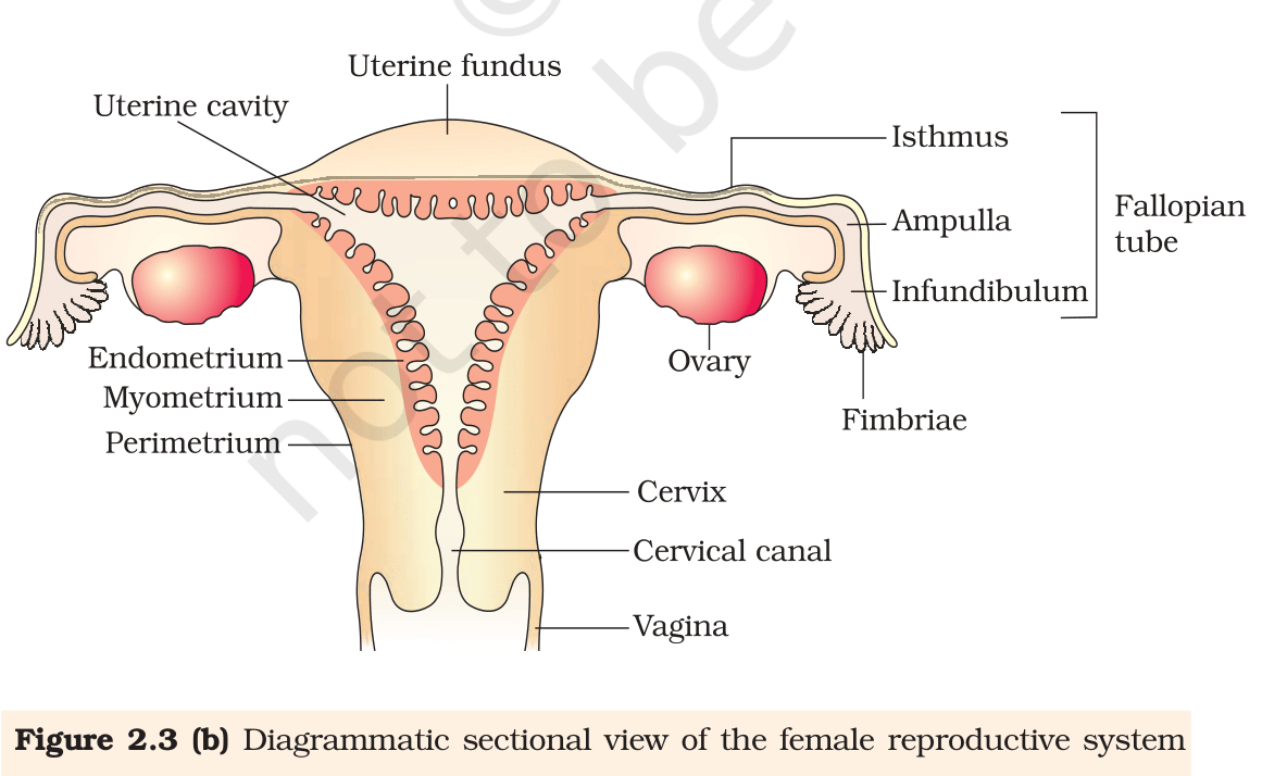
Draw a labelled diagram of female reproductive system.
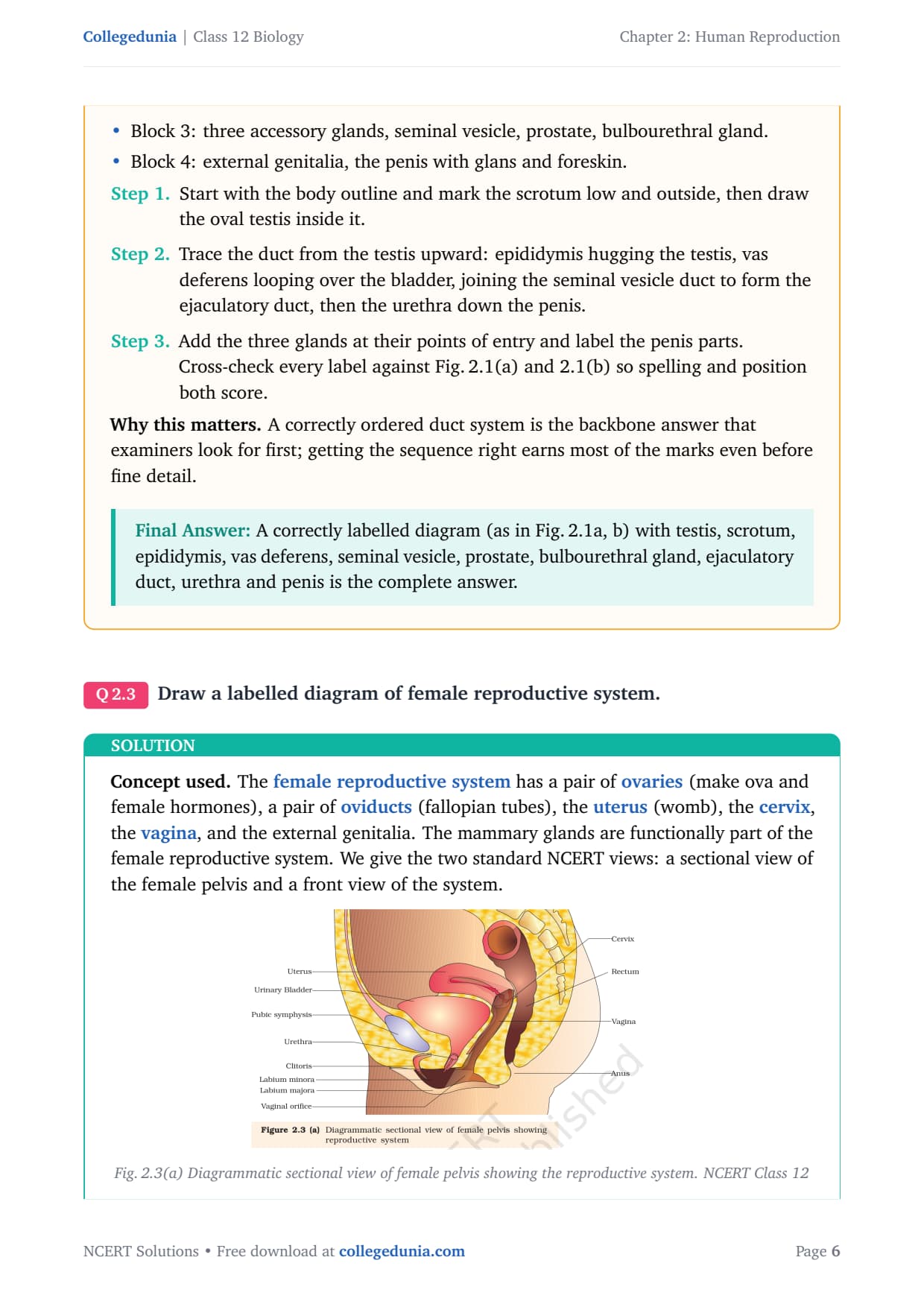
Concept used. The female reproductive system has a pair of ovaries (make ova and female hormones), a pair of oviducts (fallopian tubes), the uterus (womb), the cervix, the vagina, and the external genitalia. The mammary glands are functionally part of the female reproductive system. We give the two standard NCERT views: a sectional view of the female pelvis and a front view of the system.
Fig. 2.3(a) Diagrammatic sectional view of female pelvis showing the reproductive system. NCERT Class 12 Biology, Chapter 2.
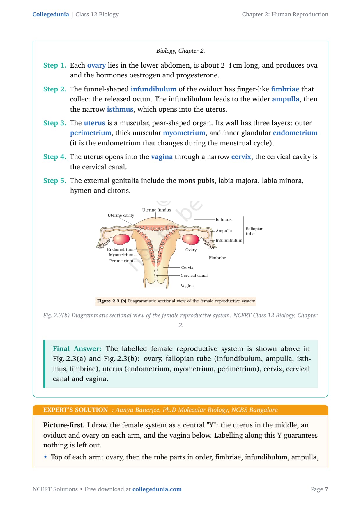
Each ovary lies in the lower abdomen, is about 2--4 cm long, and produces ova and the hormones oestrogen and progesterone.
The funnel-shaped infundibulum of the oviduct has finger-like fimbriae that collect the released ovum. The infundibulum leads to the wider ampulla, then the narrow isthmus, which opens into the uterus.
The uterus is a muscular, pear-shaped organ. Its wall has three layers: outer perimetrium, thick muscular myometrium, and inner glandular endometrium (it is the endometrium that changes during the menstrual cycle).
The uterus opens into the vagina through a narrow cervix; the cervical cavity is the cervical canal.
The external genitalia include the mons pubis, labia majora, labia minora, hymen and clitoris.
Fig. 2.3(b) Diagrammatic sectional view of the female reproductive system. NCERT Class 12 Biology, Chapter 2.
The labelled female reproductive system is shown above in Fig. 2.3(a) and Fig. 2.3(b): ovary, fallopian tube (infundibulum, ampulla, isthmus, fimbriae), uterus (endometrium, myometrium, perimetrium), cervix, cervical canal and vagina.
AB
Aanya Banerjee
Ph.D Molecular Biology, NCBS Bangalore
Verified Expert
Picture-first. I draw the female system as a central "Y": the uterus in the middle, an oviduct and ovary on each arm, and the vagina below. Labelling along this Y guarantees nothing is left out.
Top of each arm: ovary, then the tube parts in order, fimbriae, infundibulum, ampulla, isthmus.
Centre: uterus with its three wall layers named from inside out, endometrium, myometrium, perimetrium.
Base: cervix, then cervical canal, then vagina.
Sketch the uterus first as a hollow pear, then add the two oviducts curving up to the two ovaries.
Label the tube parts from the ovary inward so the order fimbriae → infundibulum → ampulla → isthmus is clearly shown (this order matters for the fertilisation question later).
Name the three uterine layers and finish with cervix, cervical canal and vagina. Verify each label against Fig. 2.3(a) and 2.3(b).
Why this matters. Knowing the tube order tells you exactly where fertilisation happens (ampulla) and where the embryo travels to implant (uterus), which links straight to Questions 17 and the fertilisation pathway.
A correctly labelled diagram (as in Fig. 2.3a, b) with ovary, oviduct (fimbriae, infundibulum, ampulla, isthmus), uterus (endometrium, myometrium, perimetrium), cervix and vagina is the complete answer.
Q 2.4
Write two major functions each of testis and ovary.
Concept used. A gonad has two jobs: a gametogenic job (making gametes) and an endocrine job (making sex hormones). The testis is the male gonad and the ovary is the female gonad. We state the two jobs for each.
Testis, function 1 (gametogenic). The seminiferous tubules of the testis produce male gametes, spermatozoa, by the process of spermatogenesis.
Testis, function 2 (endocrine). The Leydig (interstitial) cells of the testis secrete male sex hormones called androgens, mainly testosterone.
Ovary, function 1 (gametogenic). The ovary produces the female gamete, the ovum, by the process of oogenesis.
Ovary, function 2 (endocrine). The ovary secretes the female sex hormones oestrogen and progesterone, which control the menstrual cycle and support pregnancy.
Testis: (1) produces sperms (spermatogenesis); (2) secretes androgens (testosterone). Ovary: (1) produces ova (oogenesis); (2) secretes oestrogen and progesterone.
KD
Krishna Desai
M.Sc Microbiology, JNU
Verified Expert
Structural observation. Every gonad is a "factory plus a gland". Spotting this pattern lets you answer the same way for testis and ovary without rote learning.
Identify the factory part. In the testis these are the seminiferous tubules (make sperm); in the ovary these are the follicles (make ova).
Identify the gland part. In the testis these are the Leydig cells (make androgens); in the ovary the follicle and corpus luteum (make oestrogen and progesterone).
State one point from each part for each organ, giving exactly the two major functions asked for.
Why this matters. The same "gamete + hormone" template answers many short questions in this chapter and in Reproductive Health, so it is worth fixing firmly.
Testis: makes sperms and secretes androgens. Ovary: makes ova and secretes oestrogen and progesterone.
Q 2.5
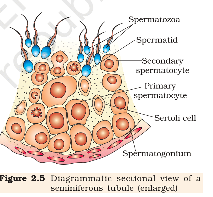
Describe the structure of a seminiferous tubule.
Concept used. A seminiferous tubule is the coiled tube inside each testicular lobule where sperms are made. To describe it we name its lining cells (germ cells and Sertoli cells) and the tissue around it (interstitial Leydig cells), and say what each does.
Fig. 2.5 Diagrammatic sectional view of a seminiferous tubule (enlarged). NCERT Class 12 Biology, Chapter 2.
Each testicular lobule has 1--3 highly coiled seminiferous tubules. The inner lining of each tubule is made of two kinds of cells: male germ cells (spermatogonia) and Sertoli cells.
The spermatogonia lie near the wall of the tubule. They divide and mature into primary spermatocytes, secondary spermatocytes, spermatids and finally spermatozoa, moving toward the centre (lumen) of the tubule.
The Sertoli cells are tall cells that extend from the wall to the lumen. They provide nourishment and support to the developing germ cells (so they are also called nurse cells).
The regions outside the seminiferous tubules, between them, contain small blood vessels and Leydig cells (interstitial cells). The Leydig cells synthesise and secrete the testicular androgens.
A few other immunologically competent cells are also present in the interstitial spaces.
A seminiferous tubule is lined by spermatogonia (which form sperms) and supporting Sertoli (nurse) cells; the spaces around the tubules contain Leydig cells that secrete androgens.
AS
Aditi Sharma
M.Sc Zoology, Banaras Hindu University
Verified Expert
Structural observation. Describe the tubule from the wall inward, then step just outside it. This "inside-out" order keeps the answer complete and ordered.
At the wall: spermatogonia (germ cells) and the base of Sertoli cells.
Toward the lumen: maturing stages, primary and secondary spermatocytes, spermatids, then spermatozoa.
Just outside the tubule: Leydig (interstitial) cells and blood vessels.
Name the two cell types lining the tubule: germ cells and Sertoli cells, and state where each sits.
Trace the germ-cell maturation gradient from wall to lumen so the description shows movement, not just a list.
Step outside the tubule and add the Leydig cells, noting their hormone role, which completes the picture for full marks.
Why this matters. The wall-to-lumen gradient you describe here is exactly the spermatogenesis sequence asked in Question 6, so one clear picture answers two questions.
Tubule lined by spermatogonia and Sertoli (nurse) cells, with germ cells maturing from wall to lumen; Leydig cells lie outside and make androgens.
Q 2.6
What is spermatogenesis? Briefly describe the process of spermatogenesis.
Concept used.Spermatogenesis is the process by which immature male germ cells (spermatogonia) become mature spermatozoa in the testis. It involves mitosis (to multiply cells), meiosis (to halve the chromosome number from 2n to n) and a final change of shape called spermiogenesis.
Multiplication. At puberty, spermatogonia on the inside wall of the seminiferous tubules increase in number by mitosis. Each spermatogonium is diploid and has 46 chromosomes.
Growth into primary spermatocyte. Some spermatogonia, called primary spermatocytes, periodically undergo growth. A primary spermatocyte is still diploid (2n = 46).
First meiotic division. A primary spermatocyte completes the first meiotic (reduction) division to form two equal haploid secondary spermatocytes, each with 23 chromosomes (n).
Second meiotic division. Each secondary spermatocyte undergoes the second meiotic division to give four equal haploid spermatids (each n = 23).
Spermiogenesis. The spermatids are transformed into spermatozoa (sperms) by spermiogenesis. After this, sperm heads become embedded in Sertoli cells and are finally released into the lumen by spermiation.
Spermatogenesis is the formation of haploid spermatozoa from diploid spermatogonia: spermatogonium → primary spermatocyte → (meiosis I) → secondary spermatocytes → (meiosis II) → spermatids → (spermiogenesis) → spermatozoa.
RV
Rohit Verma
M.Sc Biotechnology, AIIMS Delhi
Verified Expert
Quick reading. Track one number, the chromosome count, through the pathway. When the number halves you know meiosis happened; this makes the whole process easy to recall.
Spermatogonium: 2n = 46. Mitosis only multiplies cells, the number stays 46.
Primary spermatocyte: still 2n = 46. It then enters meiosis I.
After meiosis I: two secondary spermatocytes, each n = 46/2 = 23. The halving marks the reduction division.
After meiosis II: four spermatids, each still n = 23 (meiosis II does not change the number). Spermiogenesis then reshapes each spermatid into a motile sperm; spermiation releases it.
Why this matters. The same chromosome-tracking trick works for oogenesis in Question 12; mastering it here makes that question almost free.
46 → 46 → (meiosis I) → 23,23 → (meiosis II) → 23×4 spermatids → sperms; this chain is spermatogenesis.
Q 2.7
Name the hormones involved in regulation of spermatogenesis.
Concept used. Spermatogenesis starts at puberty and is under the control of the hypothalamus–pituitary–testis axis. We name the hormones in the order they act along this axis.
GnRH. At puberty the hypothalamus releases increased amounts of Gonadotropin Releasing Hormone (GnRH).
LH and FSH. GnRH acts on the anterior pituitary and stimulates secretion of two gonadotropins: Luteinising Hormone (LH) and Follicle Stimulating Hormone (FSH).
Action of LH. LH acts on the Leydig cells and stimulates them to secrete androgens (mainly testosterone). Androgens in turn stimulate the process of spermatogenesis.
Action of FSH. FSH acts on the Sertoli cells and stimulates secretion of factors that help the process of spermiogenesis.
The hormones are GnRH (from the hypothalamus), LH and FSH (from the anterior pituitary), and androgens / testosterone (from the Leydig cells).
VR
Vivaan Rao
Ph.D Molecular Biology, NCBS Bangalore
Verified Expert
Strategic angle. Answer along the control axis, not as a loose list. Each hormone has a clear "from where" and "acts on what", and naming both makes the answer precise.
Hypothalamus: GnRH.
Anterior pituitary (released because of GnRH): LH and FSH.
Testis (responding to LH): androgens / testosterone.
State the source hormone GnRH and its origin (hypothalamus).
Name the two gonadotropins LH and FSH and their origin (anterior pituitary).
Add the target effects: LH → Leydig cells → androgens; FSH → Sertoli cells → spermiogenesis support. This gives every hormone the question wants.
Why this matters. The same hypothalamus-pituitary-gonad axis controls the menstrual cycle in Question 17, so this layout transfers directly.
GnRH, LH, FSH and androgens (testosterone) regulate spermatogenesis.
Q 2.8
Define spermiogenesis and spermiation.
Concept used. These are two successive late events of spermatogenesis. Spermiogenesis is a change of shape; spermiation is a release. We define each separately.
Spermiogenesis. It is the process by which spermatids (round, non-motile cells) are transformed into spermatozoa (sperms) that have a head, neck, middle piece and tail. No cell division occurs here; only the shape and parts change.
Spermiation. After spermiogenesis the sperm heads remain embedded in the Sertoli cells for a while. Spermiation is the process by which these mature sperms are finally released from the Sertoli cells into the lumen of the seminiferous tubule.
Spermiogenesis: transformation of spermatids into spermatozoa (shape change). Spermiation: release of the mature spermatozoa from the Sertoli cells into the seminiferous tubule lumen.
TP
Tara Pillai
M.Sc Zoology, Banaras Hindu University
Verified Expert
Quick reading. Two words that sound alike but mean different things. I anchor each to one key verb: spermiogenesis = make the shape; spermiation = set it free.
Spermiogenesis: input is a spermatid, output is a fully formed sperm. The change is structural (grows a tail, condenses the head), not numerical.
Spermiation: input is a sperm stuck to a Sertoli cell, output is a free sperm in the tubule lumen. The change is positional (release), not structural.
Why this matters. Exam questions often pair these terms to test whether you know the order, shape change first, then release.
Spermiogenesis = spermatid → sperm (shape). Spermiation = sperm released from Sertoli cell into tubule lumen.
Q 2.9
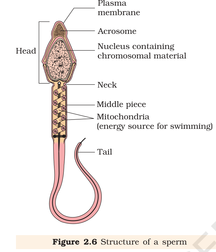
Draw a labelled diagram of sperm.
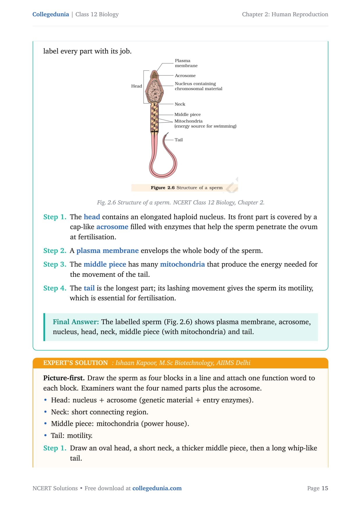
Concept used. A sperm is a microscopic single cell made of a head, a neck, a middle piece and a tail, all wrapped by a plasma membrane. We draw the NCERT figure and label every part with its job.
Fig. 2.6 Structure of a sperm. NCERT Class 12 Biology, Chapter 2.
The head contains an elongated haploid nucleus. Its front part is covered by a cap-like acrosome filled with enzymes that help the sperm penetrate the ovum at fertilisation.
A plasma membrane envelops the whole body of the sperm.
The middle piece has many mitochondria that produce the energy needed for the movement of the tail.
The tail is the longest part; its lashing movement gives the sperm its motility, which is essential for fertilisation.
The labelled sperm (Fig. 2.6) shows plasma membrane, acrosome, nucleus, head, neck, middle piece (with mitochondria) and tail.
IK
Ishaan Kapoor
M.Sc Biotechnology, AIIMS Delhi
Verified Expert
Picture-first. Draw the sperm as four blocks in a line and attach one function word to each block. Examiners want the four named parts plus the acrosome.
Head: nucleus + acrosome (genetic material + entry enzymes).
Neck: short connecting region.
Middle piece: mitochondria (power house).
Tail: motility.
Draw an oval head, a short neck, a thicker middle piece, then a long whip-like tail.
Mark the acrosome as a cap on the front of the head and label the nucleus inside it.
Show mitochondria coiled around the middle piece and label the plasma membrane around the whole cell, then check every label against Fig. 2.6.
Why this matters. The acrosome and the mitochondria-rich middle piece directly explain how the sperm reaches and enters the ovum, which connects to the fertilisation pathway.
A four-part labelled sperm (head with acrosome and nucleus, neck, middle piece with mitochondria, tail) is the complete answer.
Q 2.10
What are the major components of seminal plasma?
Concept used.Semen = sperms + seminal plasma. The seminal plasma is the fluid part of semen, contributed by the male accessory glands (seminal vesicles, prostate, bulbourethral glands). We list what it is rich in and why.
Seminal plasma is rich in fructose, a sugar that provides energy to the sperms for their motility.
It contains calcium and certain enzymes (secreted mainly by the prostate and seminal vesicles).
It also contains other ions and secretions that nourish the sperms and keep the medium suitable for their movement and survival.
Seminal plasma is rich in fructose, calcium and certain enzymes (it is the secretion of the seminal vesicles, prostate and bulbourethral glands that nourishes and activates the sperms).
MJ
Meera Joshi
M.Sc Microbiology, JNU
Verified Expert
Quick reading. Remember the seminal plasma by its job: feed and activate sperms. The components follow from that job.
Energy supply: fructose (sugar) gives sperms fuel for swimming.
Activation and medium: calcium ions and enzymes help activate the sperms and maintain a favourable medium.
Source check: these come from the accessory glands, not the testis, which is why a vasectomy still allows fluid but no sperms.
Why this matters. Knowing what seminal plasma does explains the role of the accessory glands asked in Question 11.
Fructose, calcium and certain enzymes are the major components of seminal plasma.
Q 2.11
What are the major functions of male accessory ducts and glands?
Concept used. The male system has accessory ducts (rete testis, vasa efferentia, epididymis, vas deferens) and accessory glands (paired seminal vesicles, a prostate, paired bulbourethral glands). We state the function of each group.
Function of accessory ducts. The accessory ducts store the sperms and transport them from the testis to the outside through the urethra. In the epididymis the sperms also mature and gain motility.
Function of accessory glands (energy). The seminal vesicles and prostate secrete a fluid rich in fructose, which nourishes the sperms.
Function of accessory glands (medium and motility). Their secretions also provide calcium and enzymes, activate the sperms and make a fluid medium that is essential for the easy transport and motility of sperms.
Accessory ducts store, mature and transport sperms; accessory glands secrete a fructose-, calcium- and enzyme-rich fluid that nourishes, activates and provides a transport medium for the sperms.
DC
Dev Chatterjee
M.Sc Biotechnology, AIIMS Delhi
Verified Expert
Structural observation. Split the answer into "ducts = plumbing" and "glands = support fluid". Each half then has a clean, markable point.
Ducts: a one-way path, store → mature (epididymis) → transport.
Glands: make the carrier fluid, energy (fructose), activation (calcium, enzymes), lubrication.
State the duct functions together: storage, maturation in the epididymis, and transport to the urethra.
State the gland functions together: nourishment (fructose), activation (calcium, enzymes), and a fluid medium for motility.
Conclude that ducts move the sperm while glands keep it alive and motile, the complete two-part answer.
Why this matters. This division (move vs. support) explains why semen has both cells and a rich fluid, linking back to Question 10.
Ducts: store, mature and transport sperms. Glands: provide a fructose-, calcium- and enzyme-rich nourishing, activating fluid medium.
Q 2.12
What is oogenesis? Give a brief account of oogenesis.
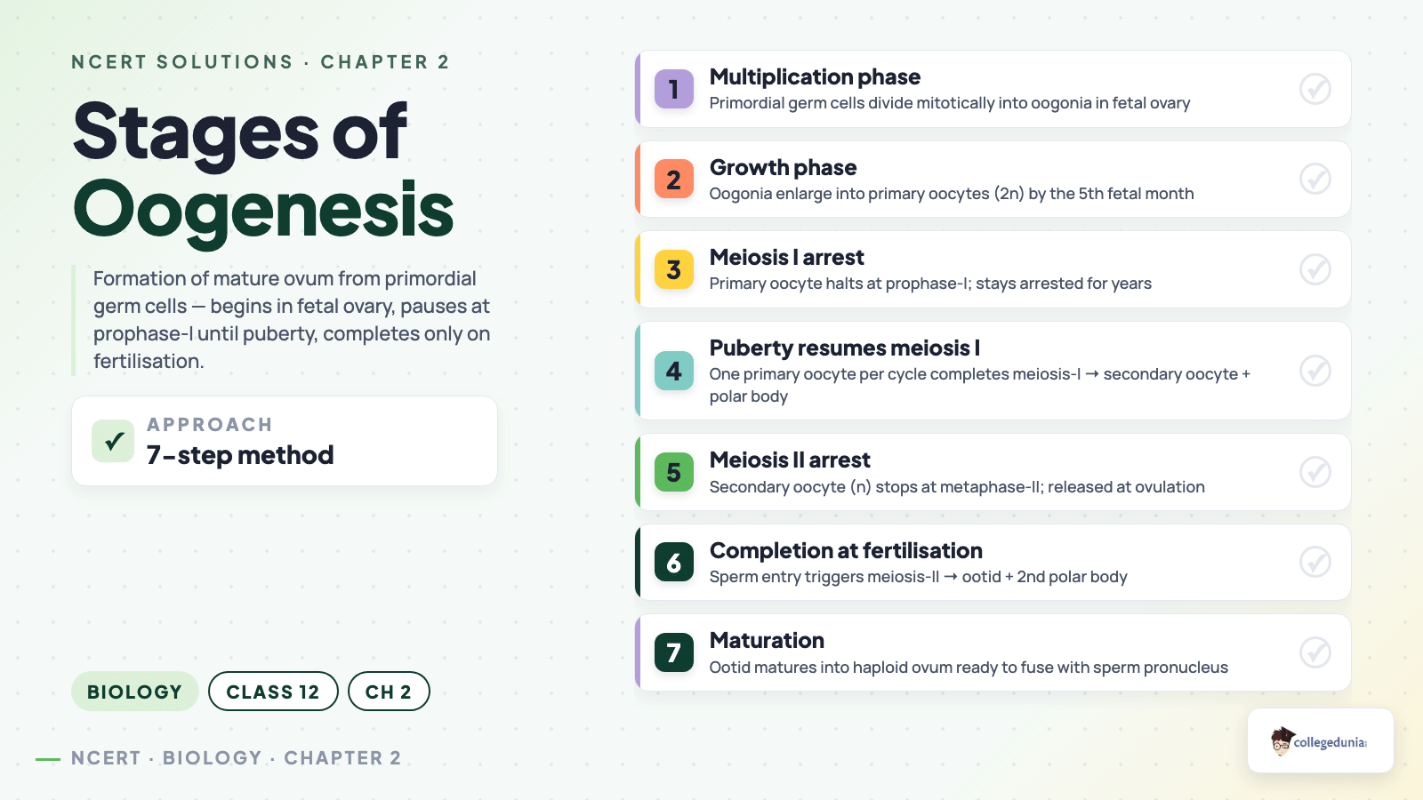
Concept used.Oogenesis is the process of formation of a mature female gamete (ovum) from the female germ cells in the ovary. Like spermatogenesis it uses mitosis and meiosis, but it begins in the embryo, pauses for years, and produces only one ovum per cycle.
Starts in foetal life. Oogenesis is initiated during the embryonic (foetal) stage. Gamete mother cells, the oogonia, are formed in the foetal ovary; no more oogonia are formed after birth.
Primary oocyte, arrested. Oogonia enter prophase I of the first meiotic division and get arrested at that stage as primary oocytes. Each primary oocyte gets surrounded by a layer of granulosa cells, forming the primary follicle.
Follicle development. The primary follicle becomes a secondary follicle and then a tertiary follicle, which has a fluid-filled cavity called the antrum. The primary oocyte in the tertiary follicle completes meiosis I.
Unequal division. Meiosis I gives a large haploid secondary oocyte and a tiny first polar body; the secondary oocyte keeps the bulk of the nutrient-rich cytoplasm.
Graafian follicle and ovulation. The tertiary follicle becomes the mature Graafian follicle, which ruptures to release the secondary oocyte (ovulation). Meiosis II of the secondary oocyte completes only if a sperm enters.
Oogenesis is the formation of a haploid ovum from oogonia in the ovary: oogonia → primary oocyte (arrested in meiosis I) → secondary oocyte + first polar body → ovum (meiosis II completed at fertilisation).
AR
Ananya Reddy
Ph.D Molecular Biology, NCBS Bangalore
Verified Expert
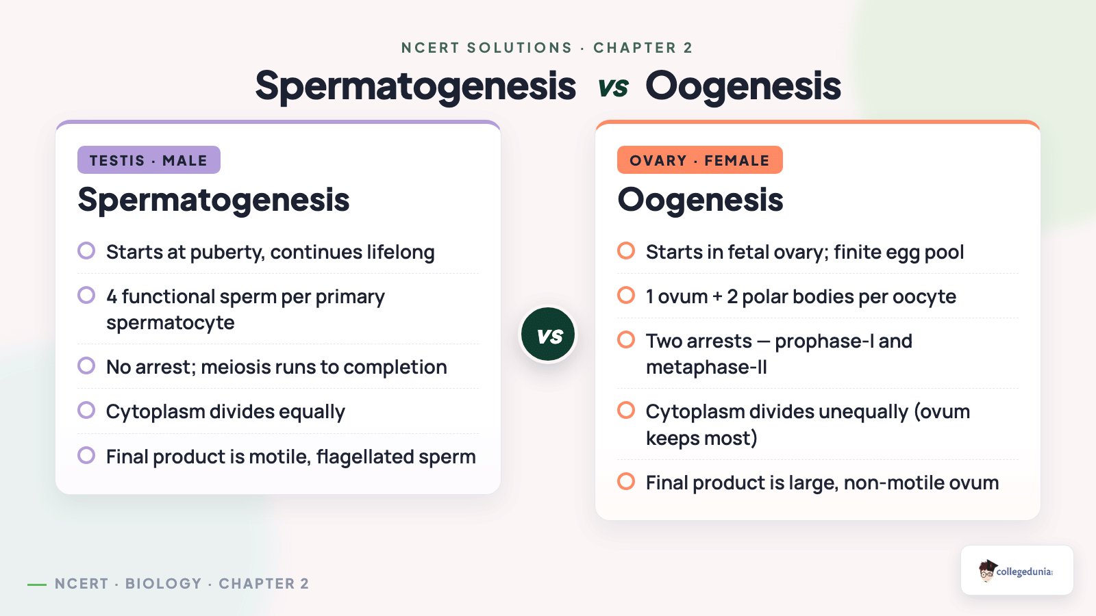
Quick reading. Compare against spermatogenesis to remember the three differences: starts before birth, pauses, and gives one ovum (not four).
Timing: oogonia form in foetal life only; this is unlike spermatogonia, which keep dividing after puberty.
Arrest: primary oocytes stop in prophase I for years and resume one at a time each cycle.
Unequal split: meiosis I gives one big secondary oocyte (n) and a small polar body; meiosis II (at fertilisation) gives the ovum and another polar body, so only one ovum results.
Why this matters. The "one ovum per cycle" point is exactly what Question 20 about identical and fraternal twins tests.
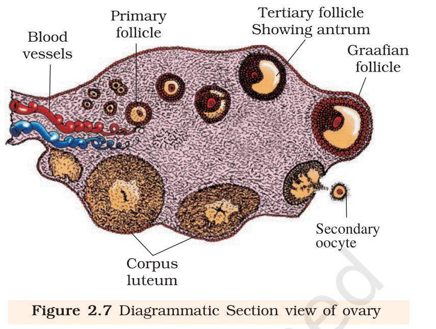
Draw a labelled diagram of a section through ovary.
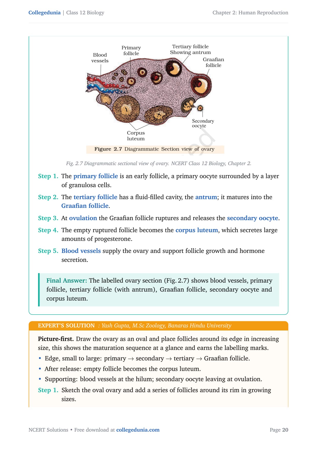
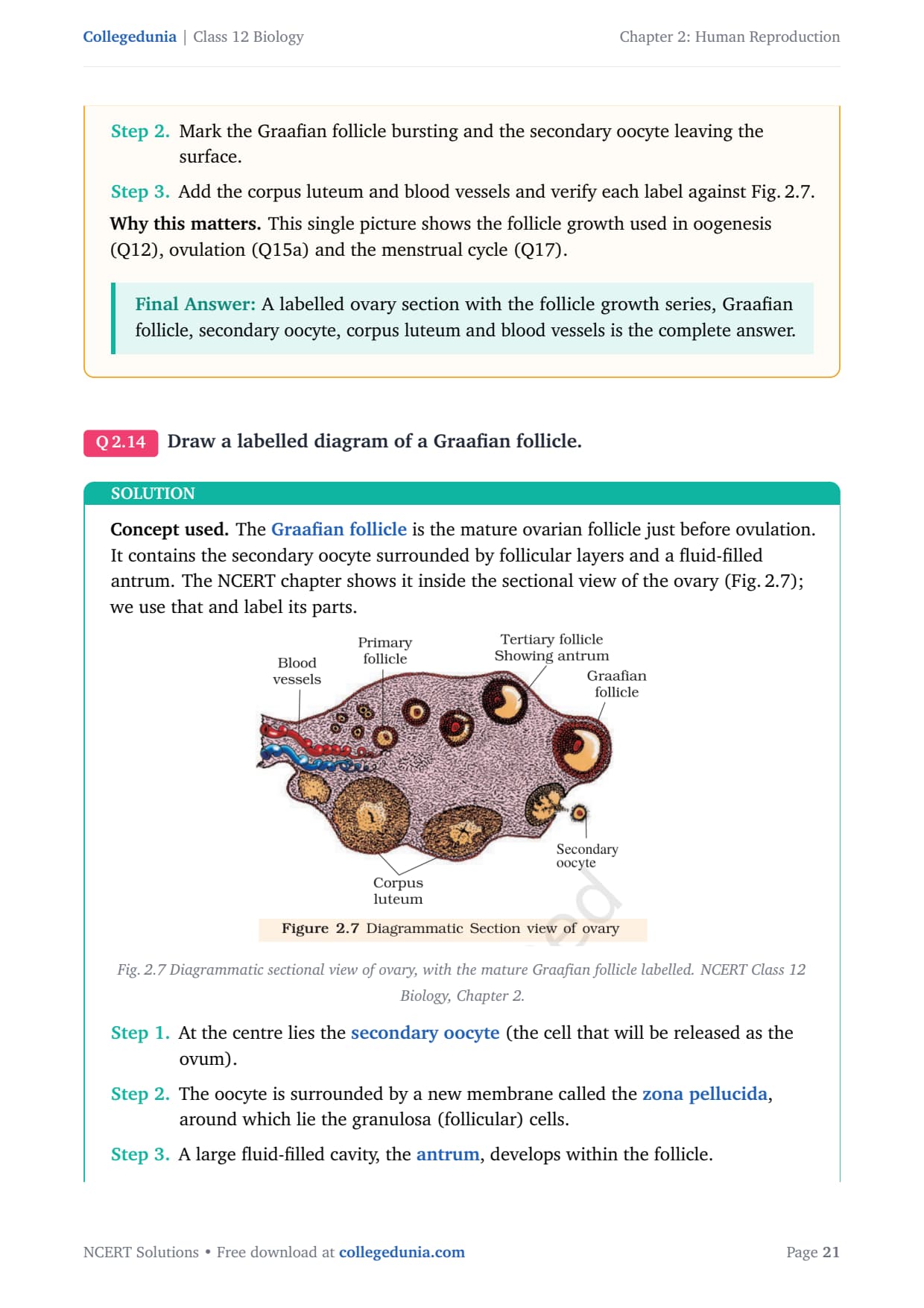
Concept used. A section through the ovary shows follicles at different stages of growth, a corpus luteum, blood vessels and the secondary oocyte being released. We embed and label the NCERT sectional view.
Fig. 2.7 Diagrammatic sectional view of ovary. NCERT Class 12 Biology, Chapter 2.
The primary follicle is an early follicle, a primary oocyte surrounded by a layer of granulosa cells.
The tertiary follicle has a fluid-filled cavity, the antrum; it matures into the Graafian follicle.
At ovulation the Graafian follicle ruptures and releases the secondary oocyte.
The empty ruptured follicle becomes the corpus luteum, which secretes large amounts of progesterone.
Blood vessels supply the ovary and support follicle growth and hormone secretion.
The labelled ovary section (Fig. 2.7) shows blood vessels, primary follicle, tertiary follicle (with antrum), Graafian follicle, secondary oocyte and corpus luteum.
YG
Yash Gupta
M.Sc Zoology, Banaras Hindu University
Verified Expert
Picture-first. Draw the ovary as an oval and place follicles around its edge in increasing size, this shows the maturation sequence at a glance and earns the labelling marks.
Edge, small to large: primary → secondary → tertiary → Graafian follicle.
After release: empty follicle becomes the corpus luteum.
Supporting: blood vessels at the hilum; secondary oocyte leaving at ovulation.
Sketch the oval ovary and add a series of follicles around its rim in growing sizes.
Mark the Graafian follicle bursting and the secondary oocyte leaving the surface.
Add the corpus luteum and blood vessels and verify each label against Fig. 2.7.
Why this matters. This single picture shows the follicle growth used in oogenesis (Q12), ovulation (Q15a) and the menstrual cycle (Q17).
A labelled ovary section with the follicle growth series, Graafian follicle, secondary oocyte, corpus luteum and blood vessels is the complete answer.
Q 2.14
Draw a labelled diagram of a Graafian follicle.
Concept used. The Graafian follicle is the mature ovarian follicle just before ovulation. It contains the secondary oocyte surrounded by follicular layers and a fluid-filled antrum. The NCERT chapter shows it inside the sectional view of the ovary (Fig. 2.7); we use that and label its parts.
Fig. 2.7 Diagrammatic sectional view of ovary, with the mature Graafian follicle labelled. NCERT Class 12 Biology, Chapter 2.
At the centre lies the secondary oocyte (the cell that will be released as the ovum).
The oocyte is surrounded by a new membrane called the zona pellucida, around which lie the granulosa (follicular) cells.
A large fluid-filled cavity, the antrum, develops within the follicle.
The whole follicle is enclosed by layers of theca cells. At maturity this Graafian follicle bursts to release the secondary oocyte, the event called ovulation.
The Graafian follicle (mature follicle in Fig. 2.7) shows the secondary oocyte, zona pellucida, granulosa cells, the fluid-filled antrum and the theca layers; it ruptures at ovulation.
PM
Priya Mehta
M.Sc Biotechnology, AIIMS Delhi
Verified Expert
Picture-first. Draw the Graafian follicle as a ring of cells around a big fluid space, with the oocyte sitting to one side. Label from the centre outward.
Place the secondary oocyte off-centre and draw the zona pellucida as a clear ring around it.
Draw the granulosa cells surrounding the oocyte and the large antrum (fluid cavity) filling most of the follicle.
Enclose everything in the theca layers and note that rupture of this follicle is ovulation; cross-check labels with Fig. 2.7.
Why this matters. Knowing the Graafian follicle's parts explains both ovulation (Q15a) and how the corpus luteum forms.
A labelled Graafian follicle (secondary oocyte, zona pellucida, granulosa cells, antrum, theca layers) is the complete answer.
Q 2.15
Name the functions of the following:
(a) Corpus luteum (b) Endometrium (c) Acrosome
(d) Sperm tail (e) Fimbriae
Concept used. Each of these is a named reproductive structure; the question asks only its function. We give one clear function per structure, naming the hormone or process involved.
(a) Corpus luteum. The corpus luteum secretes large amounts of progesterone, which is essential for maintaining the endometrium and supporting pregnancy.
(b) Endometrium. The endometrium is the inner lining of the uterus; it undergoes cyclic changes and is the site of implantation of the blastocyst and is essential for pregnancy.
(c) Acrosome. The acrosome is the cap over the sperm head; it is filled with enzymes that help the sperm penetrate and fertilise the ovum.
(d) Sperm tail. The tail provides motility: its lashing movement propels the sperm so it can reach the ovum (this motility is essential for fertilisation).
(e) Fimbriae. The fimbriae are finger-like projections of the infundibulum; they help in collection of the ovum after ovulation.
(a) secretes progesterone (maintains endometrium/pregnancy); (b) site of implantation and supports pregnancy; (c) enzymes for sperm to penetrate the ovum; (d) motility to reach the ovum; (e) collect the released ovum.
AS
Aditya Singh
M.Sc Zoology, Banaras Hindu University
Verified Expert
Quick reading. Each part needs just one verb. I pick the single most important action for each structure so the answer is sharp and complete.
Corpus luteum → secretes progesterone.
Endometrium → supports implantation.
Acrosome → enzymes to enter the ovum.
Sperm tail → swimming (motility).
Fimbriae → catch the ovum.
Read each structure and recall whether it is hormonal (corpus luteum), structural-supportive (endometrium, fimbriae) or sperm-related (acrosome, tail).
Assign the one defining function to each, using the hormone or process name where there is one.
State the five functions as a labelled list so each scores independently.
Why this matters. These five functions reappear across fertilisation, the menstrual cycle and pregnancy, so a one-line function for each is high-value revision.
Identify True/False statements. Correct each false statement to make it true.
(a) Androgens are produced by Sertoli cells. (True/False)
(b) Spermatozoa get nutrition from Sertoli cells. (True/False)
(c) Leydig cells are found in ovary. (True/False)
(d) Leydig cells synthesise androgens. (True/False)
(e) Oogenesis takes place in corpus luteum. (True/False)
(f) Menstrual cycle ceases during pregnancy. (True/False)
(g) Presence or absence of hymen is not a reliable indicator of virginity or sexual experience. (True/False)
Concept used. We test each statement against a single known fact and, if it is false, replace only the wrong word so the statement becomes true. Key facts: Leydig cells (not Sertoli cells) make androgens; Sertoli cells nourish germ cells; Leydig cells are in the testis; oogenesis occurs in the ovary; ovarian cycling stops during pregnancy; the hymen is not a virginity indicator.
(a) False. Androgens are produced by the Leydig cells, not Sertoli cells. Corrected: ``Androgens are produced by Leydig cells.''
(b) True. Sertoli cells (nurse cells) provide nutrition to the developing spermatozoa.
(c) False. Leydig cells are found in the testis, not the ovary. Corrected: ``Leydig cells are found in the testis.''
(d) True. Leydig cells synthesise and secrete the testicular androgens.
(e) False. Oogenesis takes place in the ovary (in the germ cells/follicles), not in the corpus luteum. Corrected: ``Oogenesis takes place in the ovary.''
(f) True. During pregnancy the menstrual cycle ceases, because high progesterone prevents new cycles.
(g) True. The presence or absence of the hymen is not a reliable indicator of virginity or sexual experience.
Quick reading. For true/false-and-correct, do not rewrite the whole sentence, swap only the single wrong term. This is faster and scores the correction mark cleanly.
Cell-role pairs: Sertoli = nurse; Leydig = hormone. Use this to settle (a), (b), (d).
Location: Leydig in testis, oogenesis in ovary. Settles (c), (e).
Whole-body facts: pregnancy halts cycling (f); hymen is not a virginity test (g), both true as stated.
What is menstrual cycle? Which hormones regulate menstrual cycle?
Concept used. The menstrual cycle is the cyclic change in the reproductive tract of primate females, repeating about every 28 days. It is controlled by the same hypothalamus–pituitary–ovary axis: pituitary hormones (FSH, LH) and ovarian hormones (oestrogen, progesterone).
Definition. The menstrual cycle is the monthly cycle of changes (in the ovary and uterine endometrium) in human females from puberty (menarche) up to menopause; one cycle is the period from one menstruation to the next, about 28 days.
Phases. It has the menstrual phase (breakdown of the endometrium, bleeding for 3--5 days), the follicular (proliferative) phase, ovulation (around day 14), and the luteal (secretory) phase.
Pituitary hormones.FSH stimulates follicle growth; a mid-cycle surge of LH (the LH surge) induces ovulation.
Ovarian hormones. The growing follicle secretes oestrogen; after ovulation the corpus luteum secretes progesterone, which maintains the endometrium.
Falling oestrogen and progesterone (if no pregnancy) cause the endometrium to break down, starting the next cycle.
The menstrual cycle is the ∼ 28-day cyclic change in the female reproductive tract; it is regulated by the pituitary hormones FSH and LH and the ovarian hormones oestrogen and progesterone (under hypothalamic GnRH).
SK
Sanya Kumar
Ph.D Molecular Biology, NCBS Bangalore
Verified Expert
Strategic angle. Tie each hormone to the phase it drives. Hormones are not a loose list, they form a timeline across the 28 days.
Days 1–5 (menstrual): low oestrogen and progesterone cause endometrial shedding.
Days 6–13 (follicular): FSH grows the follicle, which secretes oestrogen and rebuilds the endometrium.
Day 14 (ovulation): the LH surge bursts the Graafian follicle.
Days 15–28 (luteal): the corpus luteum secretes progesterone to maintain the endometrium; if no pregnancy it regresses and the cycle restarts.
Why this matters. This timeline links directly to ovulation (Q15) and to why pregnancy stops the cycle (Q16f).
Menstrual cycle = ∼ 28-day reproductive cycle; regulated by FSH, LH (pituitary) and oestrogen, progesterone (ovary).
Q 2.18
What is parturition? Which hormones are involved in induction of parturition?
Concept used.Parturition is the process of delivery of the foetus, that is, childbirth, at the end of pregnancy. It is induced by a complex neuroendocrine mechanism involving signals from the fully developed foetus and the placenta.
Definition. Parturition is the expulsion of the fully developed foetus from the mother's uterus at the end of gestation, brought about by vigorous contractions of the uterus.
Trigger. Signals from the fully developed foetus and the placenta produce mild uterine contractions called the foetal ejection reflex.
Oxytocin. This reflex triggers release of oxytocin from the maternal pituitary. Oxytocin acts on the uterine muscle and causes stronger uterine contractions.
Positive feedback. Stronger contractions stimulate more oxytocin secretion, and the cycle of contraction and oxytocin release continues (positive feedback) until the baby is delivered, followed by expulsion of the placenta. Relaxin also assists by relaxing the pelvic ligaments.
Parturition is childbirth, the expulsion of the foetus from the uterus at the end of pregnancy; it is mainly induced by oxytocin (with relaxin assisting), driven by a foetus-and-placenta signal in a positive-feedback loop.
PI
Pranav Iyer
M.Sc Biotechnology, AIIMS Delhi
Verified Expert
Strategic angle. Explain parturition as a positive-feedback loop. Naming oxytocin and the loop is the high-mark content.
State what parturition is: delivery of the fully developed foetus by uterine contractions.
Name the start signal: the foetal ejection reflex from the mature foetus and placenta.
Name the key hormone and loop: oxytocin causes contractions; contractions cause more oxytocin (positive feedback) until delivery; relaxin softens the pelvis to help.
Why this matters. Parturition is the textbook example of a positive-feedback hormonal loop, a concept reused across physiology chapters.
Parturition = childbirth; induced mainly by oxytocin (plus relaxin) through a foetal-signal positive-feedback contraction loop.
Q 2.19
In our society the women are often blamed for giving birth to daughters. Can you explain why this is not correct?
Concept used. The sex of a child is decided by the sex chromosomes. Human females are XX and males are XY. The mother can give only an X; the father gives either X or Y. So it is the father's sperm, not the mother, that determines the child's sex.
All eggs (ova) made by the mother carry one X chromosome, because the mother is XX. The mother can never pass a Y.
The father is XY, so he makes two kinds of sperm: half carry an X chromosome and half carry a Y chromosome.
If an X-bearing sperm fertilises the egg, the zygote is XX, a girl. If a Y-bearing sperm fertilises the egg, the zygote is XY, a boy. Xovum + Xsperm → XX (girl); Xovum + Ysperm → XY (boy).
Therefore the sex of the baby depends entirely on which sperm (X or Y) of the father fertilises the egg. The mother has no role in deciding it, so blaming the woman for the child's sex is scientifically wrong and unjust.
The mother contributes only an X chromosome; the father's sperm (X or Y) decides the child's sex. So the sex of the child is determined by the father, not the mother, and blaming women for the birth of daughters is biologically incorrect.
RJ
Riya Joshi
M.Sc Zoology, Banaras Hindu University
Verified Expert
Strategic angle. Settle it with a one-line genetic cross. The mother's contribution is fixed; only the father's varies, so the "blame" is misplaced.
Write the parents: mother = XX, father = XY.
Write the gametes: every ovum = X; sperm = X or Y in equal numbers.
Combine: X= XX (girl), X= XY (boy). Since the variable gamete comes only from the father, the father determines the sex; the mother cannot influence it.
Why this matters. A simple Punnett-style argument turns a social misconception into a clear scientific fact, useful for value-based exam questions too.
Sex is set by the father's X- or Y-sperm; the mother always gives X. Blaming the woman is biologically incorrect.
Q 2.20
How many eggs are released by a human ovary in a month? How many eggs do you think would have been released if the mother gave birth to identical twins? Would your answer change if the twins born were fraternal?
Concept used. A human female is normally uniparous: one secondary oocyte (egg) is released per menstrual cycle (per month). Identical (monozygotic) twins come from one fertilised egg that splits; fraternal (dizygotic) twins come from two eggs fertilised by two sperms.
Eggs per month. In humans, generally only one ovum is released by one of the two ovaries in one menstrual cycle, so one egg is released per month.
Identical twins. Identical twins develop from a single fertilised egg (zygote) that divides into two embryos. So even for identical twins, only one egg was released.
Fraternal twins. Fraternal twins develop from two different eggs, each fertilised by a different sperm. So if the twins were fraternal, two eggs must have been released. Hence the answer does change: one egg for identical, two eggs for fraternal.
One egg is released per month. For identical twins, still only one egg (it splits after fertilisation). For fraternal twins the answer changes to two eggs (two eggs fertilised by two sperms).
NK
Neha Kapoor
M.Sc Zoology, Banaras Hindu University
Verified Expert
Quick reading. Separate "eggs released" from "embryos formed". One egg can still give two identical babies if it splits.
Baseline: humans release one ovum per cycle, so one egg per month.
Identical twins: one egg, fertilised once, then the zygote splits into two, egg count stays one.
Fraternal twins: two eggs released, each fertilised by its own sperm, so the count becomes two; therefore the answer does change with twin type.
Why this matters. This is the practical test of the "one ovum per cycle" fact from oogenesis (Q12) and explains the next question about a dog.
How many eggs do you think were released by the ovary of a female dog which gave birth to 6 puppies?
Concept used. A dog is a polyovulatory (multiparous) animal: unlike humans, its ovaries release many eggs in one cycle, so it can produce a litter. Each puppy normally develops from its own fertilised egg.
In multiparous animals such as the dog, more than one ovum is released from the ovary at the time of ovulation.
Each puppy in the litter develops from a separate egg fertilised by a separate sperm.
Since the female dog gave birth to 6 puppies, at least 6 eggs must have been released by its ovary (assuming one puppy per fertilised egg).
At least 6 eggs were released, because the dog is a polyovulatory animal and each of the 6 puppies develops from a separate fertilised egg.
AM
Ankit Mehta
M.Sc Zoology, Banaras Hindu University
Verified Expert
Quick reading. Match egg count to offspring count for polyovulatory animals: one egg per offspring, so six puppies need six eggs.
Recognise the species type: dogs are multiparous (polyovulatory), releasing several eggs per cycle.
Conclude at least 6 eggs were ovulated (more may be released but 6 is the minimum needed for 6 puppies).
Why this matters. Contrasting the dog (polyovulatory) with the human (uniparous, Q20) sharply fixes the idea that egg number depends on the species, not on having a litter.
Six eggs (one per puppy) were released by the dog's ovary.
What a Class 12 Biology Survey Says About Chapter 2 Human Reproduction
In a recent survey of 14,500 Class 12 Biology students, the menstrual cycle hormone-graph was rated the hardest sub-topic, even though it carries the highest single-question marks in CBSE and NEET.
Student Feedback
75% of students marked the menstrual cycle hormone-graph as the hardest sub-topic.
68% lost 1-2 marks on differentiating spermatogenesis from oogenesis.
The average student took 6.2 hours for the first read and 2.7 hours for a revision pass.
Source: 2025-26 Class 12 Biology survey of 14,500 students across 18 states.
Other Resources for Human Reproduction Class 12 Biology
Human Reproduction Class 12 Biology NCERT Solutions FAQs
Ques. Where can I download Human Reproduction Class 12 NCERT Solutions PDF?
Ans. You can download the Human Reproduction Class 12 Biology NCERT Solutions PDF directly from this page. Both the Normal and HD versions are free and cover every one of the 17 textbook exercise questions.
Ques. Are these Human Reproduction NCERT Solutions aligned with the 2026-27 NCERT?
Ans. Yes. The solutions follow the current 2026-27 syllabus for Class 12 Biology. Chapter 2 Human Reproduction is retained in full in the new NCERT edition, so every exercise question and diagram still applies.
Ques. How many questions are there in Class 12 Biology Chapter 2 Human Reproduction?
Ans. The NCERT textbook has a single exercise of 17 numbered questions, including a fill-in-the-blank set, diagram-based questions, and long answers on the menstrual cycle and pregnancy.
Ques. How many pages is the Class 12th Biology Human Reproduction NCERT Solutions PDF?
Ans. The Solutions PDF runs approximately 30 to 38 pages and covers all 17 exercise answers with fully labelled diagrams of the reproductive systems, gametogenesis, and the menstrual cycle.
Ques. Which topics in Human Reproduction are most important for NEET and CBSE?
Ans. Spermatogenesis, oogenesis, and the hormonal control of the menstrual cycle are the highest-yield topics. Fertilisation, implantation, and the placenta as an endocrine tissue are the next most frequently asked.
Ques. What is the difference between spermiogenesis and spermiation in this chapter?
Ans. Spermiogenesis is the transformation of spermatids into mature spermatozoa. Spermiation is the release of those mature sperm from the Sertoli cells into the lumen of the seminiferous tubule. The solutions explain both with a labelled flow.
Ques. Is Chapter 2 Human Reproduction good for scoring in Class 12 Biology?
Ans. Yes. It carries 4 to 6 marks in the CBSE Board exam and 4 to 5 questions in NEET. Because the questions are diagram and recall based, it is one of the most reliable scoring chapters when revised with the marking pattern shown on this page.








Comments