




The class 11 biology NCERT solutions chapter 17 Locomotion and Movement cover every back-exercise question, according to the latest 2026-27 CBSE syllabus, and help students prepare for the CBSE Boards, NEET and CUET. Each answer is worked step by step, from the three types of cell movement and the build of a sarcomere, through the sliding filament theory of muscle contraction, to the 206 bones of the human skeleton and the joints that let them move.
This chapter sits in the coordination half of Class 11 Biology, and the muscle and bone vocabulary students learn here is tested again in the neural and chemical control chapters that follow.
- CBSE Weightage: 3 to 4 marks, part of the Human Physiology unit that carries the largest share of the Class 11 paper.
- Topics covered: types of movement, the three muscle types, the sarcomere and its bands, contractile proteins, the sliding filament theory, red and white fibres, the skeletal system, and the classification of joints.
- Exercise count: 10 back-exercise questions, a mix of diagram-drawing, describe, distinguish, match, true-or-false and fill-in-the-blank tasks.
These class 11 biology NCERT solutions chapter 17 Locomotion and Movement are curated by subject experts, based on the 2026-27 NCERT textbook, and checked against the last five years of CBSE Board and NEET papers.
Why Locomotion and Movement Matters and What the Chapter Covers
Every deliberate action, from lifting a pen to running for a bus, is movement, and movement that carries the whole body from one place to another is locomotion. NCERT makes the point that all locomotion is movement but not all movement is locomotion, and that locomotion needs the coordinated activity of the muscular, skeletal and neural systems working together. This chapter builds the language of that machinery, from a single contracting filament to the entire framework of bones.
- Everyday role: muscles moving against bones at joints are what let students walk, write, chew and even keep an upright posture.
- Core skill: tracing a contraction from a nerve signal, through calcium release, to the sliding of filaments that shortens a muscle.
- Why it matters for NEET: the sarcomere, the sliding filament theory, the bone count and the joint types are among the most frequently tested facts in the Human Physiology unit.
Almost every question in this chapter comes back to one idea: filaments that slide past each other to shorten a muscle, and a bony frame that turns that pull into motion. Students who understand that single mechanism can answer the sarcomere, contraction and muscle-type questions from the same base. The class 11 biology NCERT solutions chapter 17 Locomotion and Movement below follow the same order as the NCERT textbook so students can check their working line by line.
Types of Movement and the Three Muscle Types
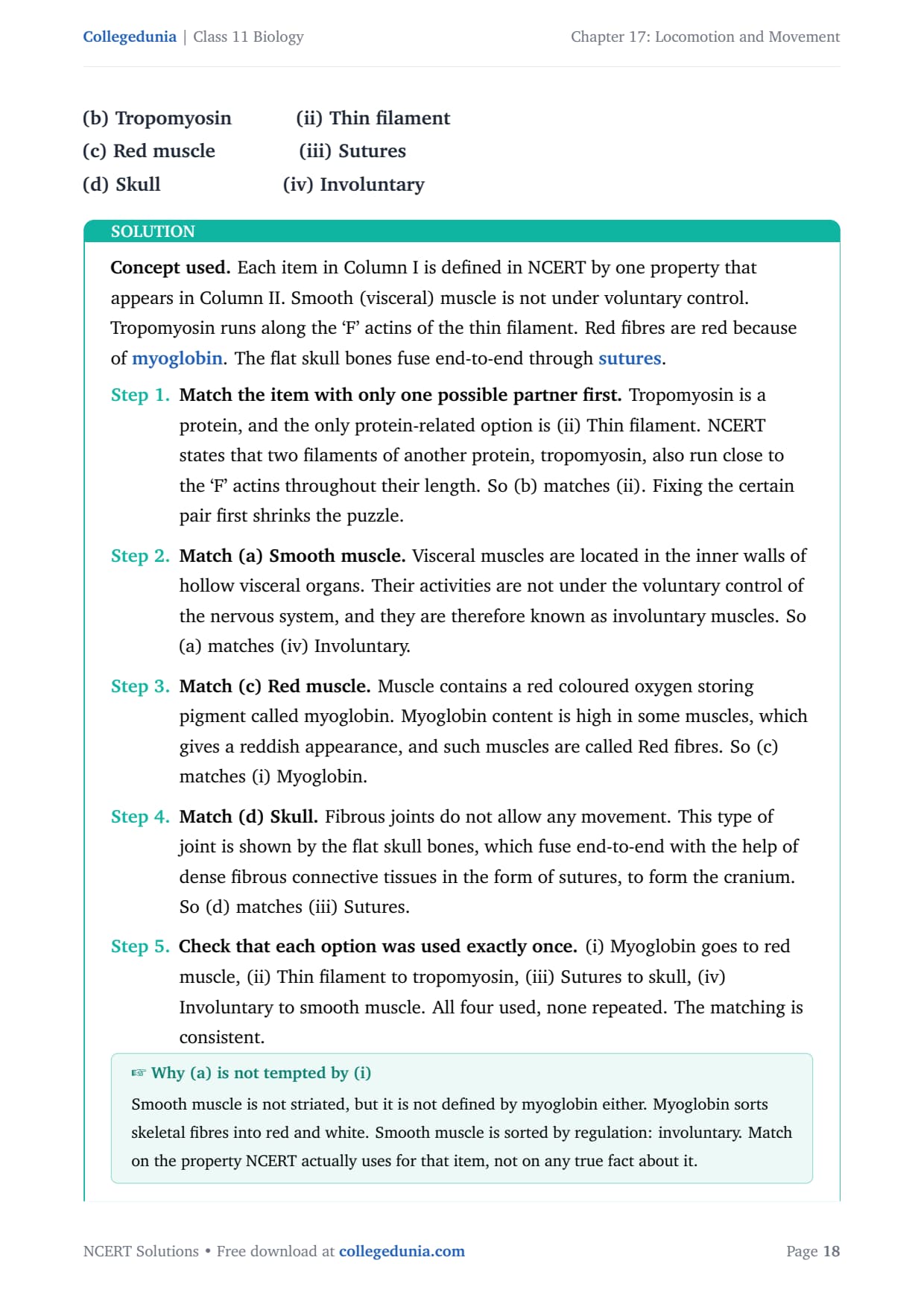
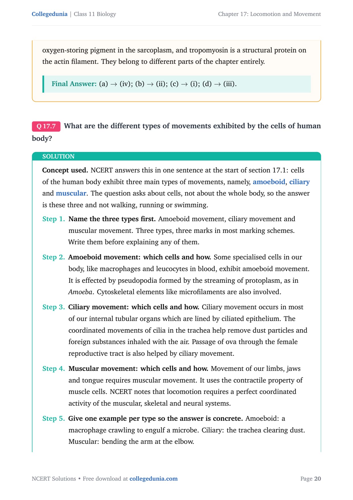
NCERT opens the chapter by naming three types of movement shown by the cells of the human body: amoeboid, ciliary and muscular. Amoeboid movement uses pseudopodia formed by the streaming of protoplasm, as in the macrophages and leucocytes of blood. Ciliary movement occurs in the internal tubular organs lined by ciliated epithelium, so the trachea clears dust and the oviduct moves the ovum. Muscular movement uses the contractile property of muscle cells and moves the limbs, jaws and tongue.
| Muscle type | Location and features | Regulation |
|---|---|---|
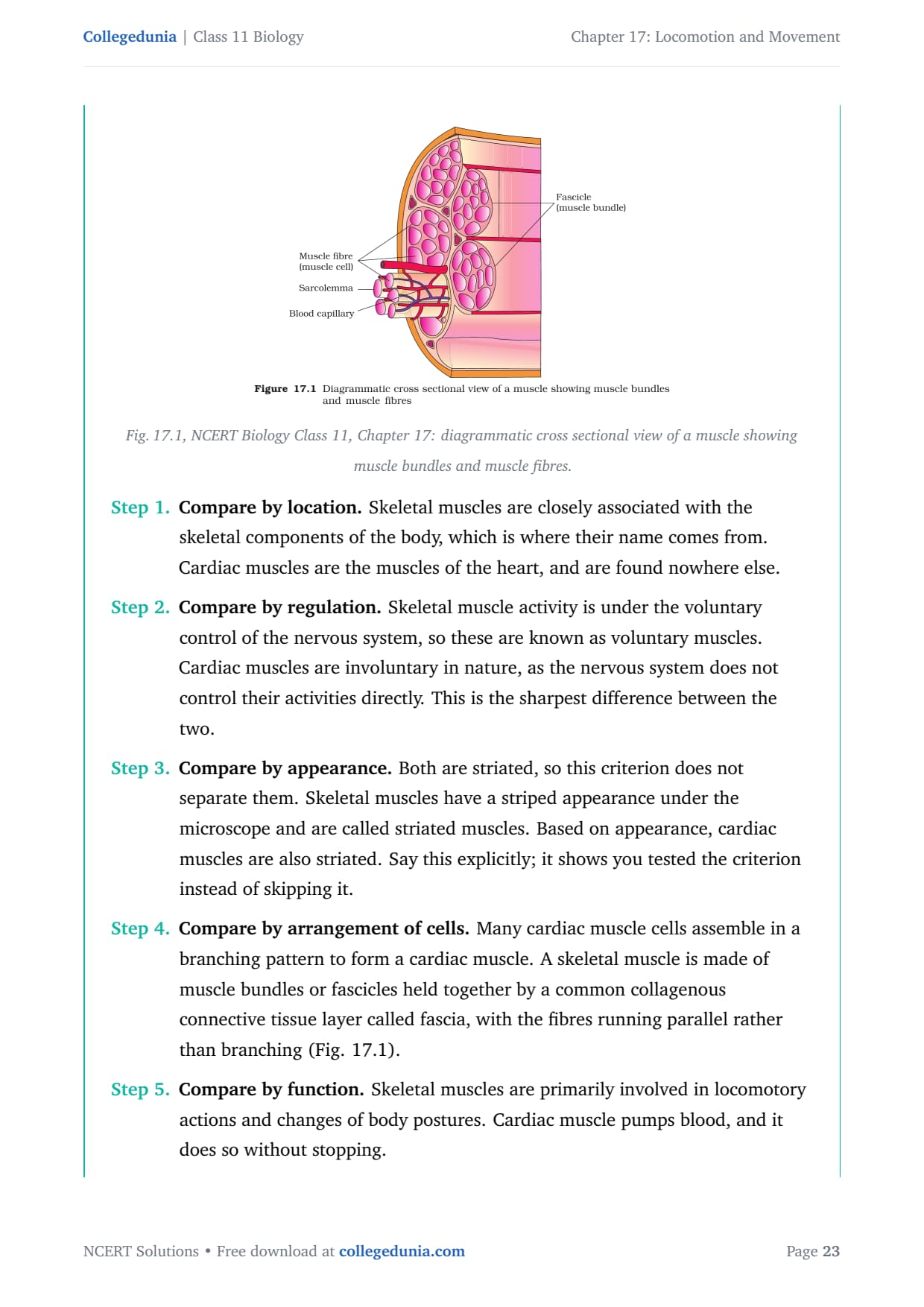
| Skeletal (striated) | Attached to skeletal components; striped fibres held in bundles by fascia; drives locomotion and posture | Voluntary |
| Visceral (smooth) | Walls of hollow visceral organs; spindle-shaped, non-striated cells | Involuntary |
| Cardiac | Found only in the heart; striated, branching interconnected cells; works without fatigue | Involuntary |
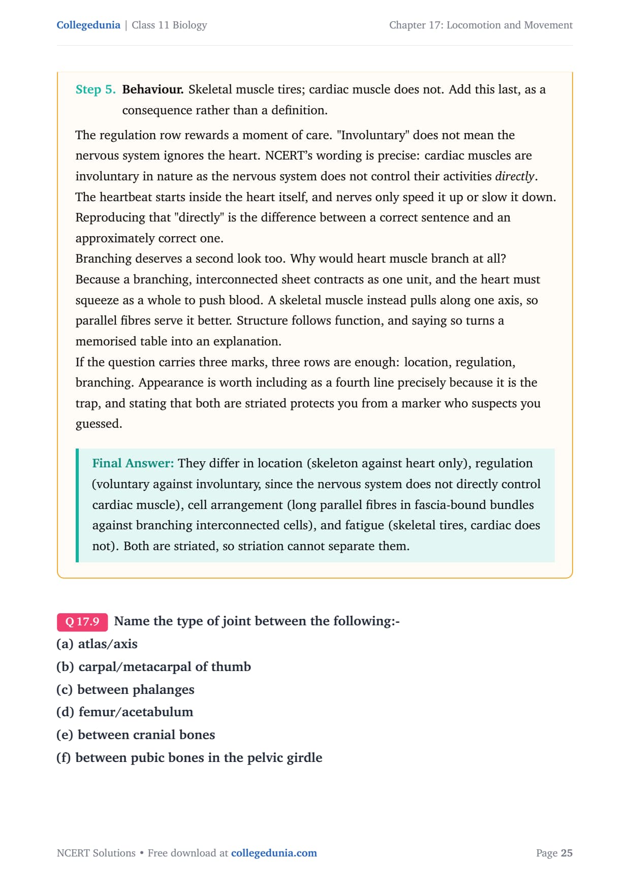
Cardiac muscle is striated, so striation alone never separates it from skeletal muscle: the true separators are location, regulation and the branching pattern of cardiac cells. Muscle makes up 40 to 50 per cent of adult body weight and is mesodermal in origin. See Exercise Q7 and Q8 for the full answers on cell movements and on distinguishing skeletal from cardiac muscle.
The Sarcomere and the Structure of a Muscle Fibre
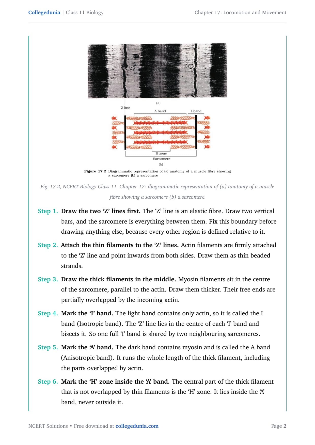
A muscle fibre is packed with parallel myofibrils, and the functional unit of contraction inside each is the sarcomere, the portion of the myofibril between two successive 'Z' lines. It is built from two proteins arranged as parallel rods: thin actin filaments attached to the 'Z' lines and thick myosin filaments lying in the centre. Every band that students must label in Exercise Q1 is simply a statement about which filaments are present there.
| Region | What it contains |
|---|---|
| 'Z' line | Elastic fibre in the centre of the 'I' band; the thin filaments are anchored to it. |
| 'I' band (isotropic) | Light band with actin only; bisected by the 'Z' line, so one 'I' band is shared by two neighbouring sarcomeres. |
| 'A' band (anisotropic) | Dark band running the full length of the myosin, including where actin overlaps it. |
| 'H' zone | Central part of the thick filament not overlapped by actin; lies strictly inside the 'A' band. |
| 'M' line | Fibrous membrane holding the thick filaments together at the centre of the 'A' band. |
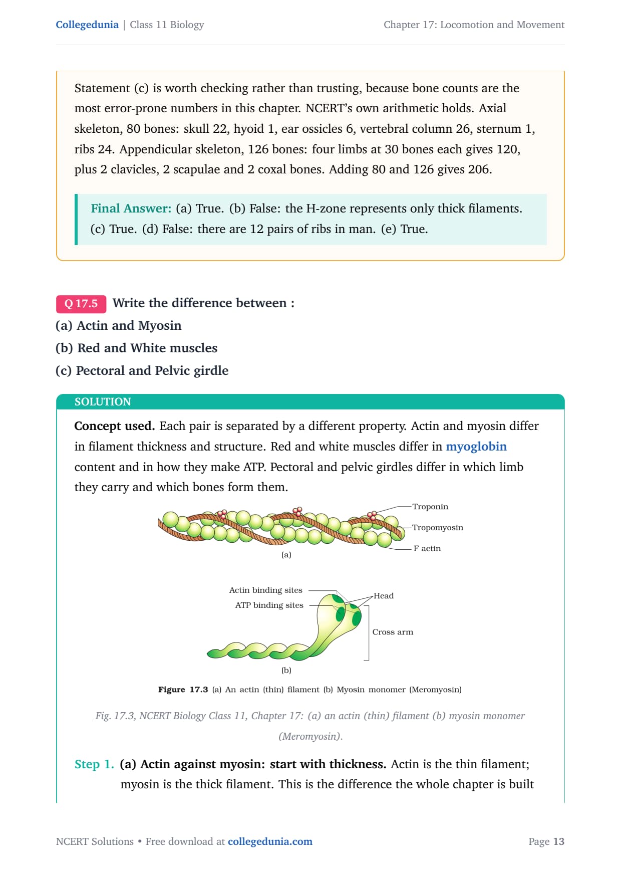
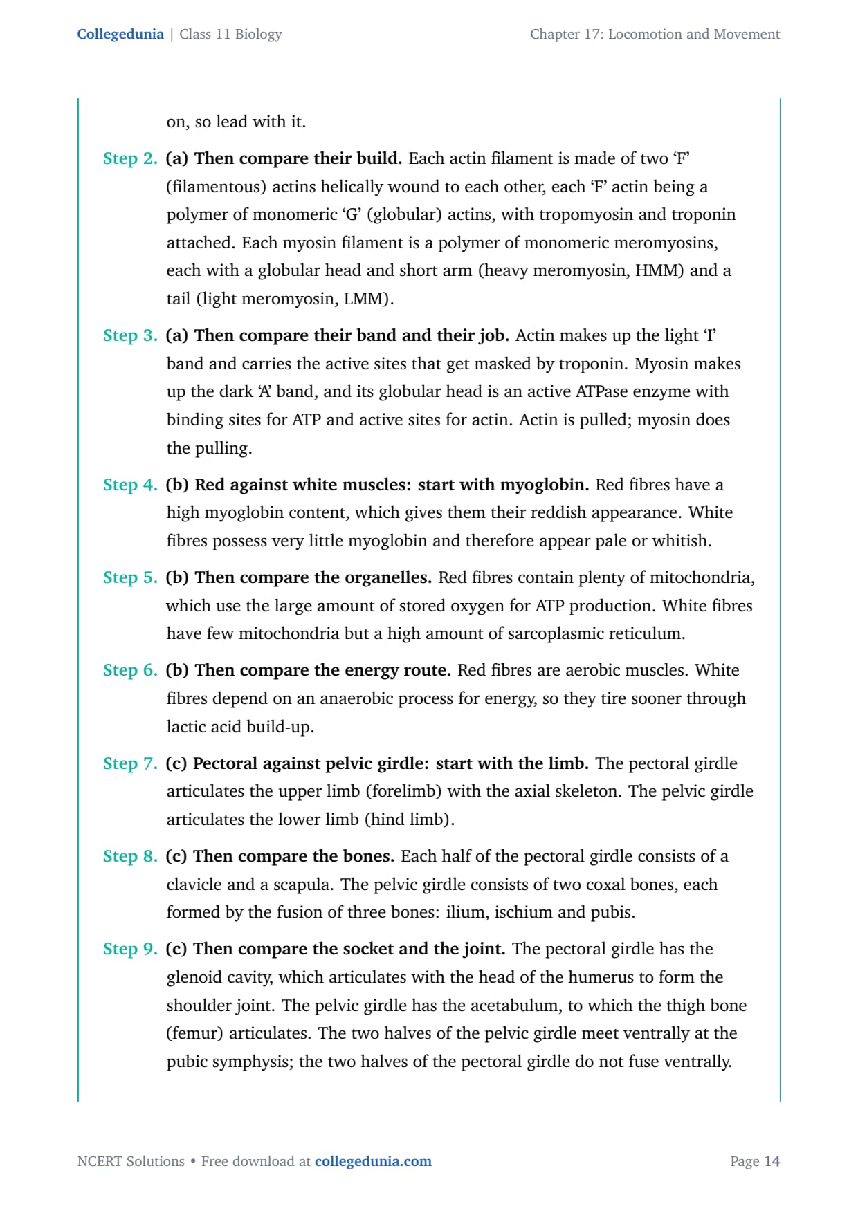
The thin filament is made of two 'F' (filamentous) actins helically wound together, each a polymer of globular 'G' actins, with tropomyosin running along them and troponin distributed at regular intervals on the tropomyosin. The thick filament is a polymer of meromyosins, each with a globular head and short arm (heavy meromyosin) and a tail (light meromyosin), and the head carries an ATPase and the binding site for actin. The sarcomere runs from one 'Z' line to the next, so it contains one full 'A' band and two half 'I' bands. Drawing a whole 'I' band inside the 'Z' lines is the commonest error in the diagram question.
Sliding Filament Theory and the Steps of Muscle Contraction
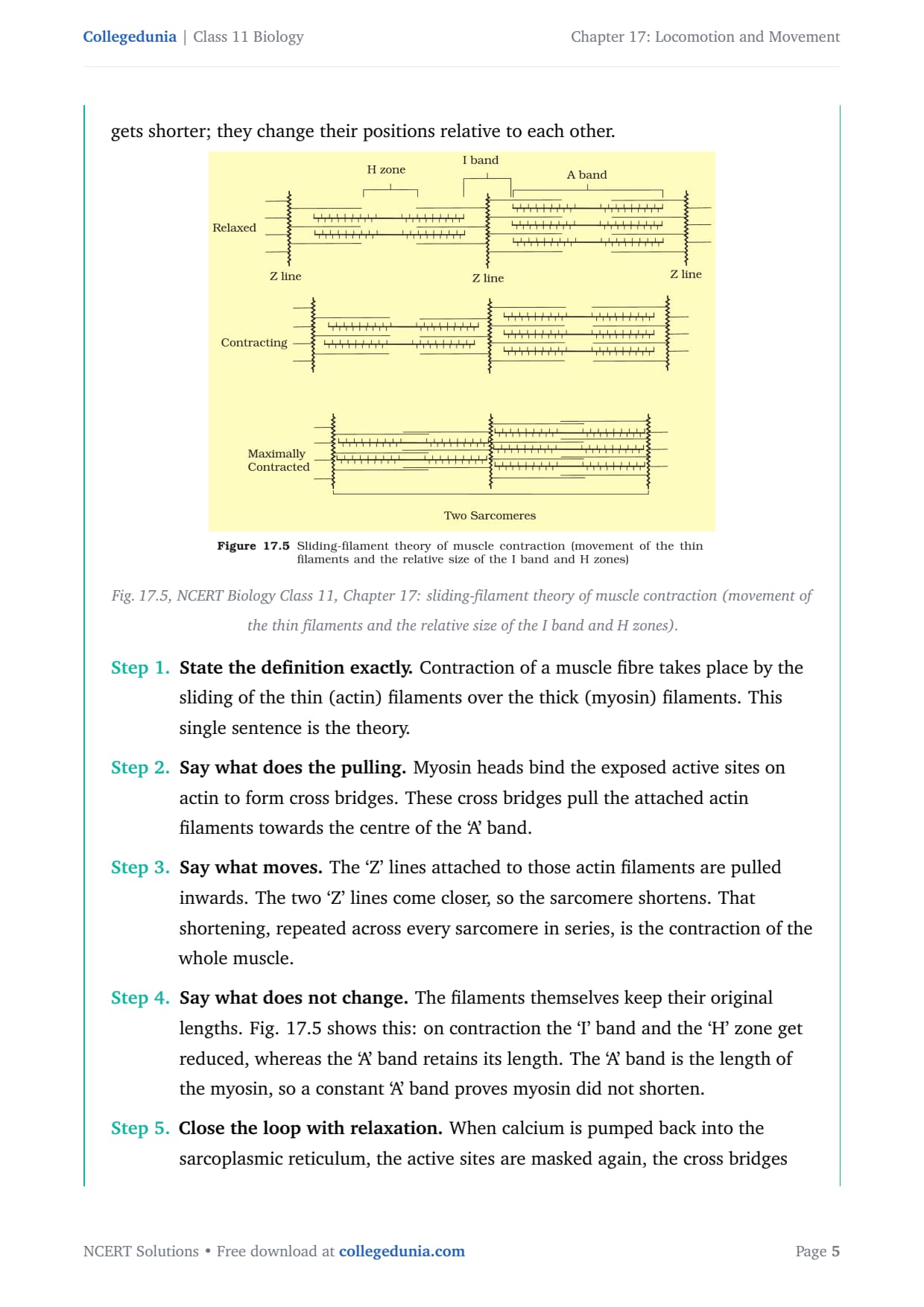
The sliding filament theory states that contraction of a muscle fibre takes place by the sliding of the thin filaments over the thick filaments. The word sliding does all the work: neither filament shortens, they change their positions relative to each other. Myosin cross bridges pull the actin towards the centre of the 'A' band, dragging the 'Z' lines inwards so the sarcomere shortens. Because the filaments keep their lengths, the 'I' band and 'H' zone shorten while the 'A' band stays constant, and that constant 'A' band is the proof that myosin did not shrink.
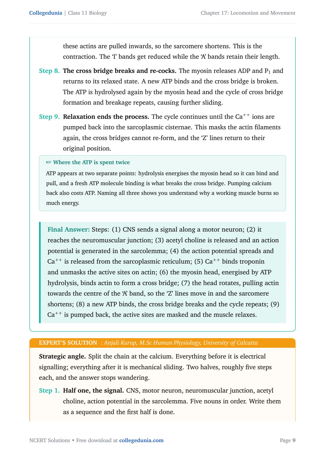
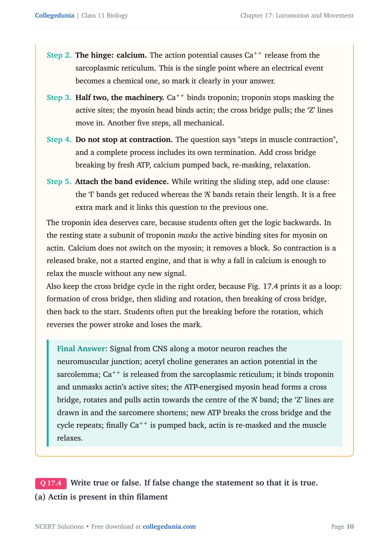
- The signal: the central nervous system sends an impulse along a motor neuron to the neuromuscular junction, where acetylcholine generates an action potential in the sarcolemma.
- The trigger: the action potential releases Ca++ from the sarcoplasmic reticulum; the calcium binds troponin and unmasks the active sites on actin.
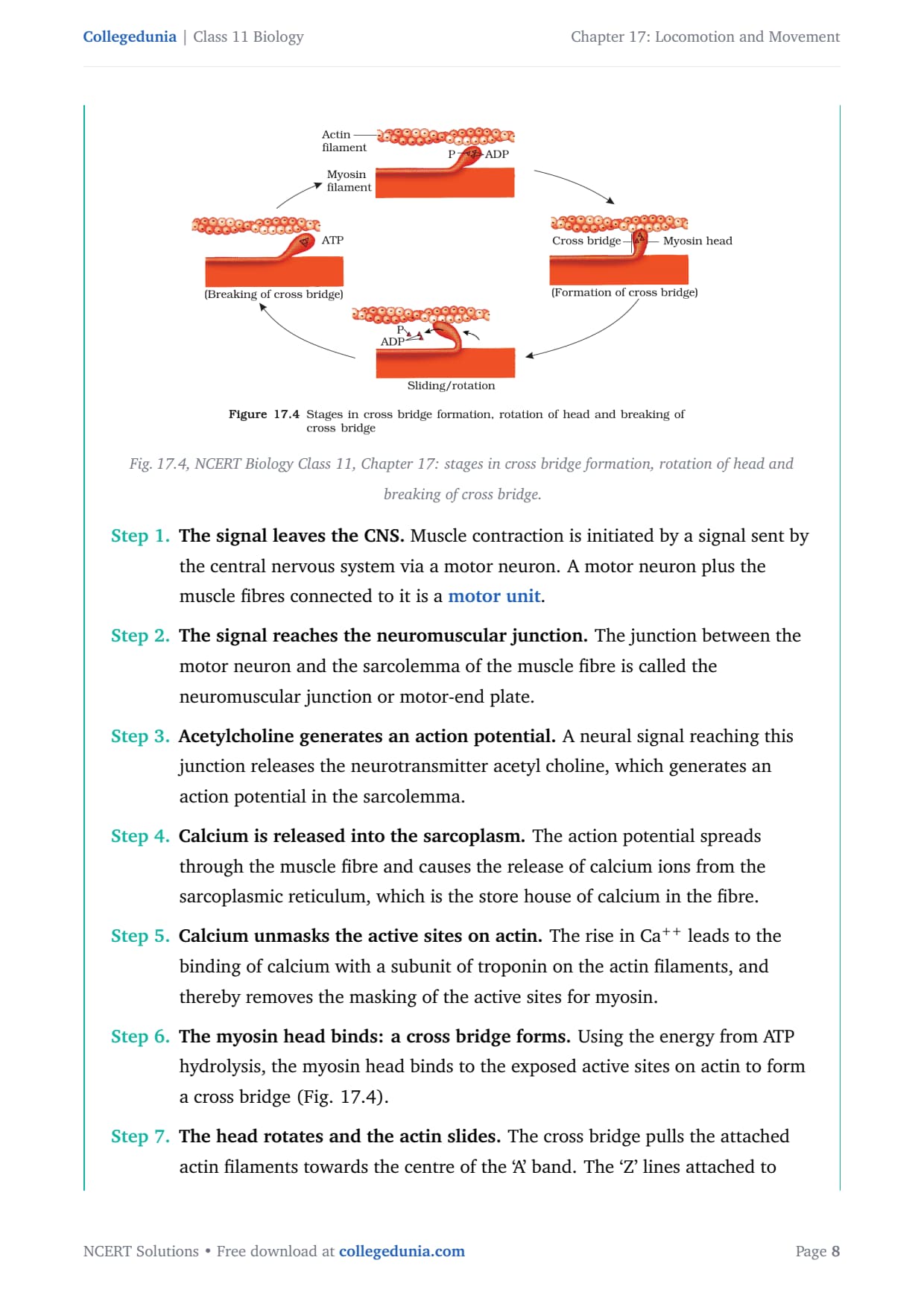
- The pull: the ATP-energised myosin head binds actin to form a cross bridge, rotates to slide the actin inward, then a fresh ATP breaks the cross bridge so the cycle repeats.
- Relaxation: Ca++ is pumped back into the sarcoplasmic reticulum, the active sites are masked again, and the 'Z' lines return to their original position.
ATP is spent at two separate points: its hydrolysis energises the myosin head to bind and pull, and a fresh ATP binding is what breaks the cross bridge, while pumping calcium back costs still more. That is why a working muscle burns so much energy. Repeated activation builds up lactic acid from the anaerobic breakdown of glycogen, which is what causes fatigue. The full step-by-step chain is worked out in Exercise Q2 and Q3.
Red and White Muscle Fibres, Actin and Myosin
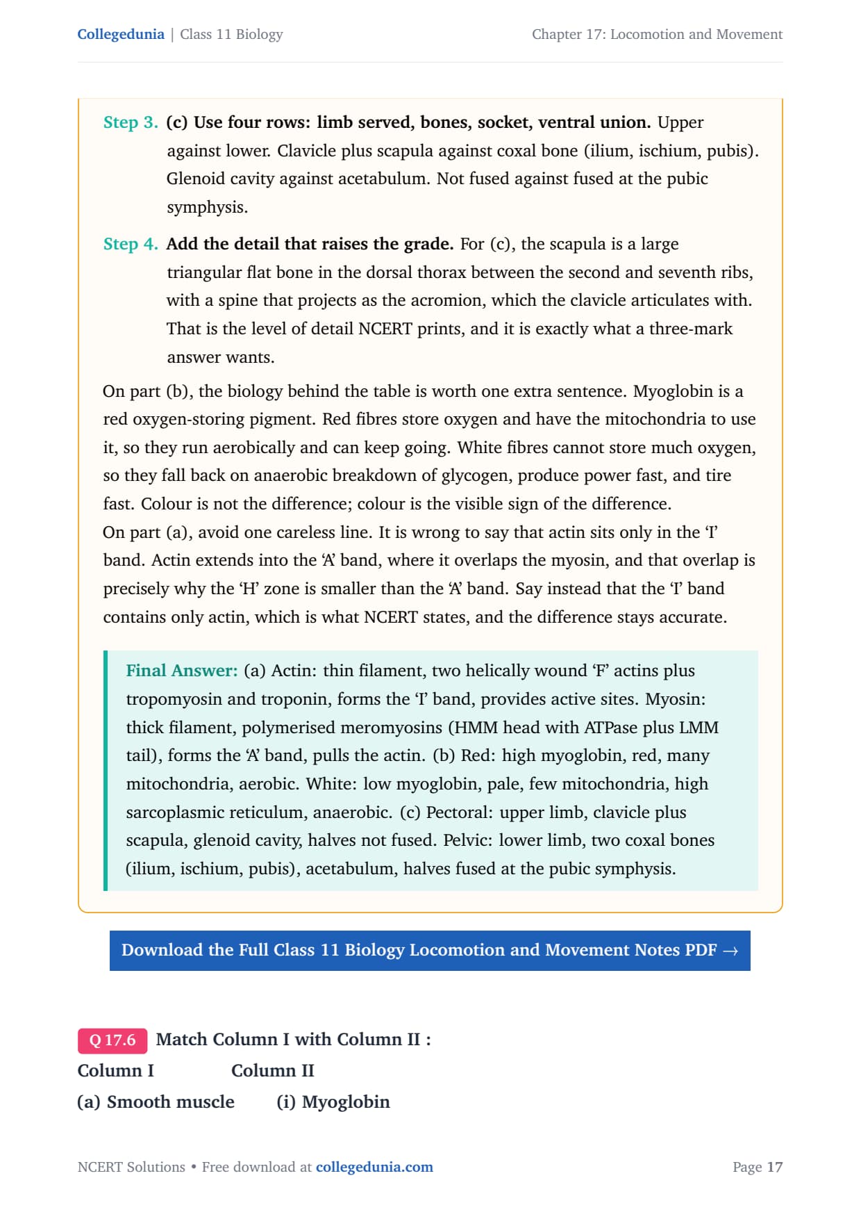
Skeletal muscle fibres are sorted into two kinds by their content of myoglobin, a red oxygen-storing pigment. Red fibres have a high myoglobin content and look reddish, while white fibres have very little and look pale. This single difference in a pigment sets up a whole chain of differences in how the two fibres make and spend energy, which is exactly what Exercise Q5 tests alongside the actin and myosin contrast.
| Feature | Red fibres | White fibres |
|---|---|---|
| Myoglobin | High, giving a reddish colour | Low, giving a pale colour |
| Mitochondria | Many | Few |
| Sarcoplasmic reticulum | Less | More |
| Respiration | Aerobic; resist fatigue | Anaerobic; tire quickly |
The actin and myosin contrast runs the other way, as a comparison of two filaments. Actin is the thin filament, forms the light 'I' band, and carries the active sites that troponin masks. Myosin is the thick filament, forms the dark 'A' band, and its globular head is an active ATPase that pulls the actin. Colour is not the difference between red and white fibres; colour is the visible sign of the difference, which is really about oxygen storage and how ATP is made. Red fibres store oxygen in myoglobin and have the mitochondria to use it aerobically, so they keep going, while white fibres fall back on the anaerobic breakdown of glycogen and tire fast.
The Skeletal System: Bones, Ribs and the Skull
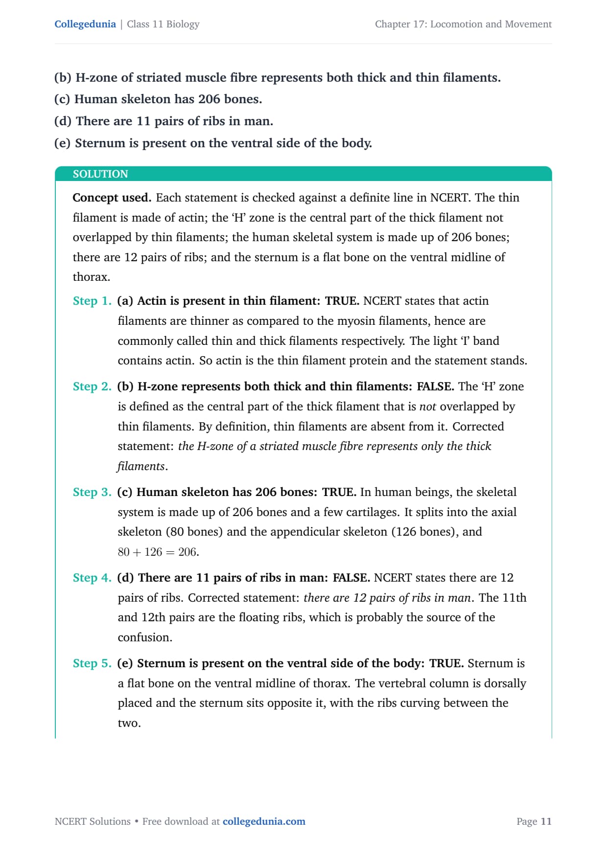
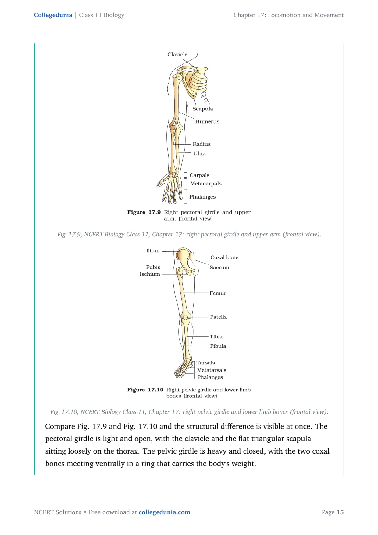
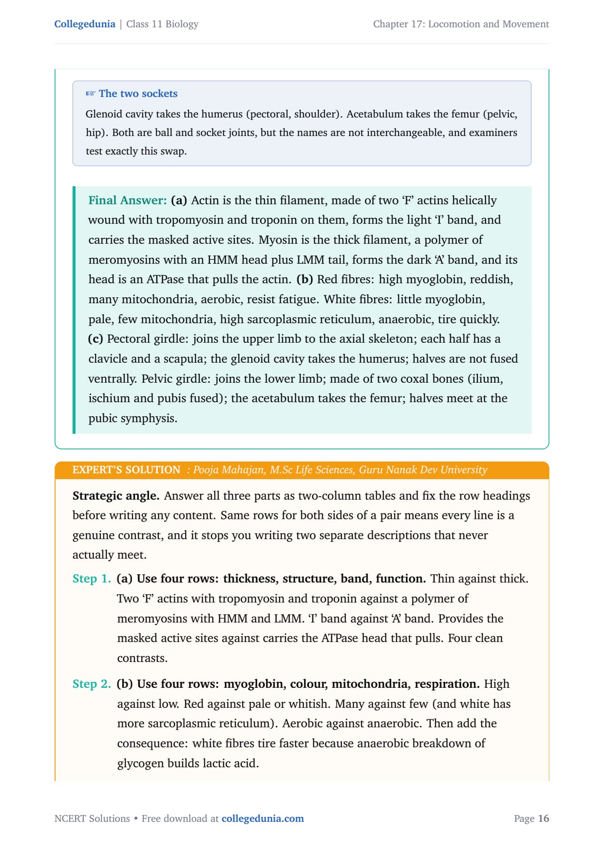
The human skeletal system is made up of 206 bones and a few cartilages, and it splits into the axial skeleton with 80 bones and the appendicular skeleton with 126 bones, so 80 plus 126 gives 206. The axial skeleton is the skull, vertebral column, sternum and ribs, while the appendicular skeleton is the limb bones and the two girdles. Several exact counts from this section are tested directly in Exercise Q4 and Q10.
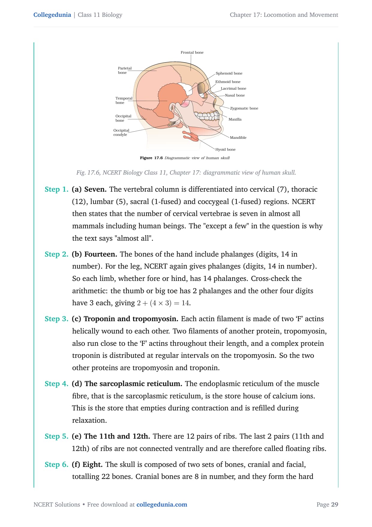
- The skull: 22 bones in total, of which 8 form the cranium (the braincase) and 14 form the facial region; the cranium is not the same as the whole skull.
- The vertebral column: 26 vertebrae, of which 7 are cervical in almost all mammals, including human beings and the giraffe.
- The ribs: 12 pairs, with the first 7 pairs true ribs, the 8th to 10th false (vertebrochondral) ribs, and the 11th and 12th pairs floating ribs that are not connected ventrally.
- The limbs: each limb has 14 phalanges, and the sternum is a flat bone on the ventral midline of the thorax.
The chapter deliberately places similar numbers close together, so read the noun each figure is attached to: skull is 22 but cranium is 8, ribs are 12 pairs but only the last 2 pairs float. Reading which word the question uses before writing a number is the whole test in the fill-in-the-blank and true-or-false questions. The 11 versus 12 slip on ribs, and the cranium versus skull slip on bone counts, are the two errors that cost the most marks in this section.
Joints and Their Types
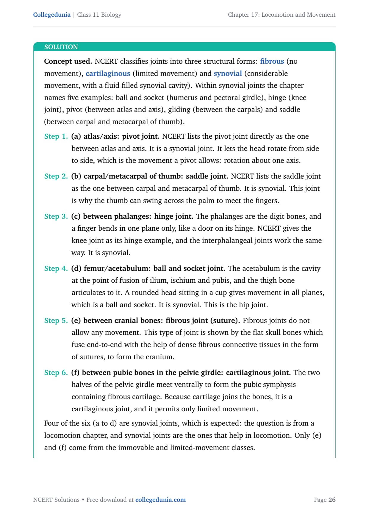
A joint is the point of contact between bones, and NCERT classifies joints into three structural forms by how much movement they allow. Fibrous joints allow no movement, cartilaginous joints allow limited movement, and synovial joints allow considerable movement because they have a fluid-filled synovial cavity. The naming question in Exercise Q9 works entirely from this three-way split, and most of its answers are synovial because synovial joints are the ones that help in locomotion.
| Joint class | Movement | Example from the chapter |
|---|---|---|
| Fibrous (suture) | None | Between the cranial bones of the skull |
| Cartilaginous | Limited | Pubic symphysis between the pubic bones |
| Synovial: ball and socket | All planes | Femur in the acetabulum (hip); humerus in the pectoral girdle |
| Synovial: hinge | One plane | Knee, and between the phalanges |
| Synovial: pivot | Rotation | Between the atlas and the axis |
| Synovial: saddle | Two planes | Between the carpal and metacarpal of the thumb |
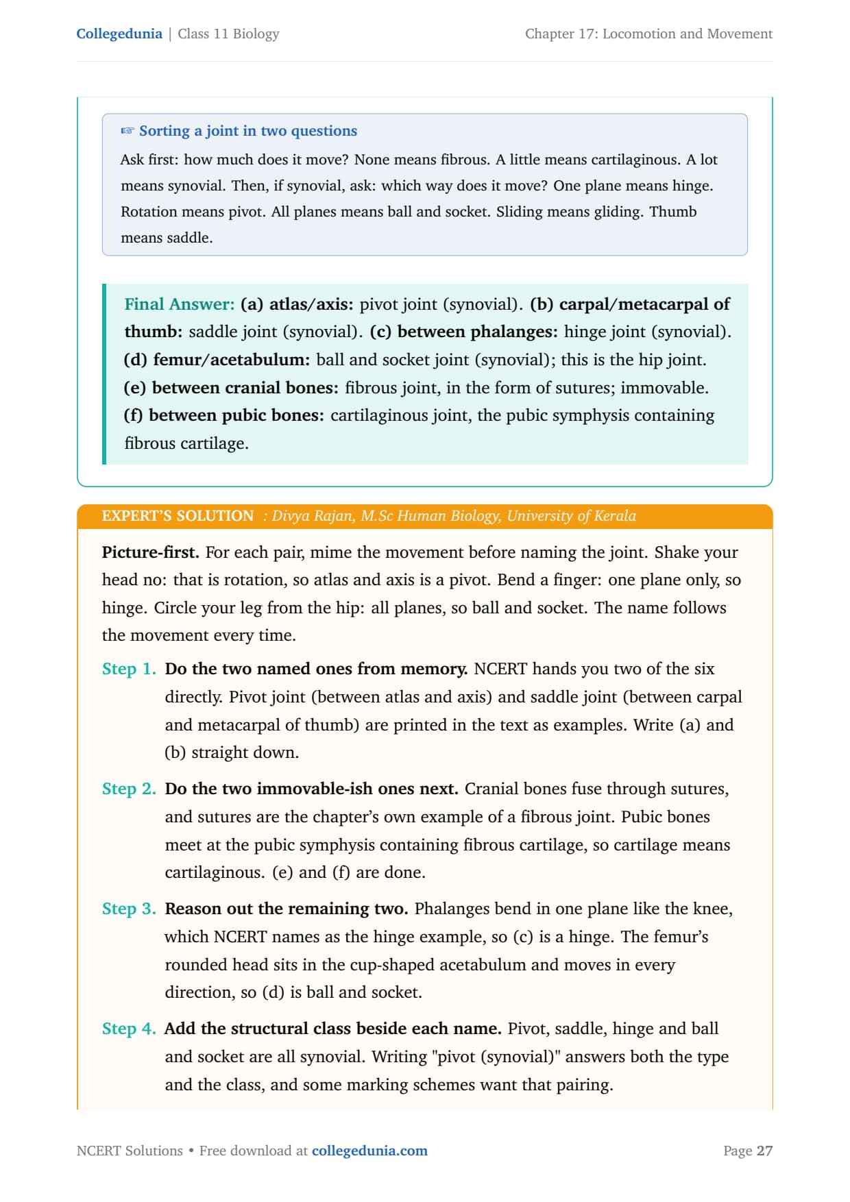
To sort any joint, ask first how much it moves: none means fibrous, a little means cartilaginous, a lot means synovial. Then, if synovial, ask which way it moves: one plane is a hinge, rotation is a pivot, all planes is ball and socket. The pubic symphysis is the trap, because it contains fibrous cartilage, so students see the word fibrous and answer fibrous joint. Read the noun, not the adjective: the tissue joining the bones is cartilage, so the joint is cartilaginous. The skull sutures are the reverse case, joined by dense fibrous connective tissue and allowing no movement at all.
Locomotion and Movement Exercise-wise Breakdown
The NCERT back exercise has 10 questions, and they mix diagram drawing, describe-and-distinguish answers, matching, true-or-false and fill-in-the-blank tasks. The table below maps the question blocks to their topics so students can plan their practice by theme.
| Question block | What it tests |
|---|---|
| Q 1 to Q 3 | Drawing the sarcomere, defining the sliding filament theory, and the steps of muscle contraction. |
| Q 4 and Q 10 | True-or-false and fill-in-the-blank on the bands, bone counts, ribs, cranium and the calcium store. |
| Q 5 and Q 8 | Differences between actin and myosin, red and white muscles, the two girdles, and skeletal against cardiac muscle. |
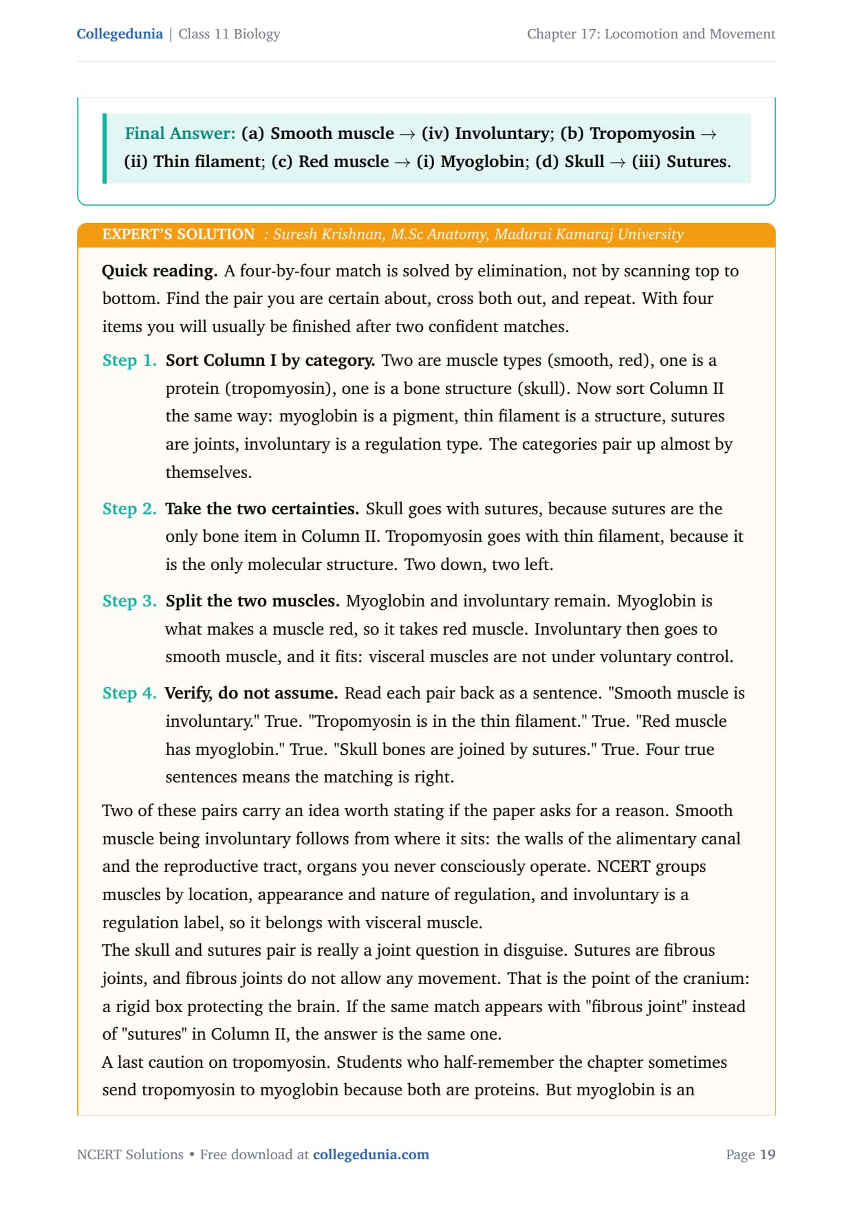
| Q 6 | Matching muscle types and structures with their defining property. |
| Q 7 and Q 9 | The three types of cell movement, and naming the joint between named bones. |
The chapter rewards precise wording, clean labelled diagrams and exact numbers rather than calculation. Learn the sarcomere bands as statements about which filaments are present, and fix the bone counts in blocks so the similar numbers stop being confusing. Every question in the class 11 biology NCERT solutions chapter 17 Locomotion and Movement PDF is solved with each step of reasoning shown, so students can compare their answers against the model working.
Practice the solved questions: Work through the full question bank with step-by-step answers and expert tips.
Common Mistakes Students Make in Locomotion and Movement
Most marks in this chapter are lost on small slips of wording and number, not on hard ideas. Each mistake below costs 1 to 2 marks, so watch for it at the exact step.
Mistake 1: Writing that the filaments contract or shorten during contraction. They slide past each other. If a filament shortened, the 'A' band would shrink, but NCERT states it retains its length.
Mistake 2: Saying cardiac muscle is not striated. Cardiac muscle is striated; only smooth (visceral) muscle is non-striated. Separate skeletal from cardiac on location, regulation and branching.
Mistake 3: Confusing the cranium with the skull. The cranium is 8 bones, the skull is 22 bones (8 cranial plus 14 facial). Read which word the question uses.
Mistake 4: Answering 11 pairs of ribs. There are 12 pairs; the 11th and 12th pairs are the floating ribs, which is where the number 11 belongs.
Student Feedback on Locomotion and Movement Solutions
What 12,240 students told us about their Locomotion and Movement preparation:
- 63% of students said keeping the bone counts straight, especially cranium against skull and the rib pairs, was the trickiest part of the chapter.
- Most-skipped detail: correcting a false statement in writing rather than only marking it false, missed by about 4 in 10 students.
- Students who drew the sarcomere from the filaments outward, rather than from the labels inward, said the band questions became automatic.
Source: 2026-27 Class 11 Biology student poll. Sample of 12,240 students from CBSE schools across 16 states, conducted before the 2026 boards.
Other Locomotion and Movement Class 11 Biology Resources
Pair these solutions with the revision notes, formula sheet and NCERT textbook PDF for the same chapter.
| Resource | Link |
|---|---|
| NCERT Notes | Locomotion and Movement Class 11 Notes |
| Formula Sheet | Locomotion and Movement Class 11 Formula Sheet |
| NCERT Book PDF | Locomotion and Movement Class 11 Book PDF |
NCERT Solutions for Class 11 Biology: All Chapters
Jump to the step-by-step solutions for any other Class 11 Biology chapter below.
| Chapter | NCERT Solutions |
|---|---|
| Chapter 1 | The Living World |
| Chapter 2 | Biological Classification |
| Chapter 3 | Plant Kingdom |
| Chapter 4 | Animal Kingdom |
| Chapter 5 | Morphology of Flowering Plants |
| Chapter 6 | Anatomy of Flowering Plants |
| Chapter 7 | Structural Organisation in Animals |
| Chapter 8 | Cell: The Unit of Life |
| Chapter 9 | Biomolecules |
| Chapter 10 | Cell Cycle and Cell Division |
| Chapter 11 | Photosynthesis in Higher Plants |
| Chapter 12 | Respiration in Plants |
| Chapter 13 | Plant Growth and Development |
| Chapter 14 | Breathing and Exchange of Gases |
| Chapter 15 | Body Fluids and Circulation |
| Chapter 16 | Excretory Products and their Elimination |
| Chapter 17 | Locomotion and Movement |
| Chapter 18 | Neural Control and Coordination |
| Chapter 19 | Chemical Coordination and Integration |
FAQs on Locomotion and Movement Class 11 NCERT Solutions
Locomotion and Movement NCERT Solutions - Frequently Asked Questions
Ques. What do the class 11 biology NCERT solutions chapter 17 Locomotion and Movement cover?
Ans. These solutions cover all 10 back-exercise questions, including the sarcomere diagram, the sliding filament theory, the steps of muscle contraction, differences between actin and myosin and between red and white muscles, skeletal against cardiac muscle, the three types of cell movement, the bone and rib counts, and naming the joints between named bones. Every question is solved step by step.
Ques. What is the sliding filament theory of muscle contraction?
Ans. The sliding filament theory states that a muscle fibre contracts by the sliding of the thin filaments over the thick filaments. Myosin cross bridges pull the actin towards the centre of the 'A' band, drawing the 'Z' lines closer so the sarcomere shortens. The filaments do not shorten, so the 'I' band and 'H' zone are reduced while the 'A' band retains its length.
Ques. How many bones are in the human skeleton and how are they divided?
Ans. The human skeletal system has 206 bones and a few cartilages. The axial skeleton has 80 bones (skull, vertebral column, sternum and ribs) and the appendicular skeleton has 126 bones (the limb bones and the two girdles), and 80 plus 126 equals 206. The skull has 22 bones, of which 8 form the cranium, and there are 12 pairs of ribs.
Ques. How is skeletal muscle different from cardiac muscle?
Ans. Skeletal muscle is attached to the skeleton, is voluntary and has long parallel fibres in fascia-bound bundles. Cardiac muscle is found only in the heart, is involuntary because the nervous system does not control it directly, and has branching interconnected cells that let it work without fatigue. Both are striated, so striation alone cannot separate them.
Ques. What are the three types of movement shown by human cells?
Ans. The cells of the human body show three types of movement. Amoeboid movement uses pseudopodia formed by streaming protoplasm, as in macrophages and leucocytes. Ciliary movement occurs in the ciliated epithelium of internal tubular organs, such as the trachea clearing dust. Muscular movement uses the contractile property of muscle cells to move the limbs, jaws and tongue.
Ques. What is the weightage of Locomotion and Movement in CBSE Class 11 Biology?
Ans. Locomotion and Movement carries about 3 to 4 marks in the CBSE Class 11 Biology paper, through diagram, distinguish, match and fill-in-the-blank questions from the Human Physiology unit. It is also heavily tested in NEET, where the sarcomere, the sliding filament theory, the bone count and the joint types appear regularly.



Comments