




NEET SS 2024 exam for Medical Group was conducted by NBEMS on 30th March 2025. NEET SS 2024 was conducted as part of the national-level super speciality exam for admissions into DM and DrNB programmes. Candidates are required to attempt 150 questions in 150 minutes, with a total of 600 marks. Each correct response carries 4 marks, while 1 mark is deducted for every incorrect answer. The NEET SS 2024 exam for Medical Group covers 40% of the questions from General Medicine and 60% from connected super-specialties like Cardiology, Neurology, Nephrology, Endocrinology, and Medical Gastroenterology.
NEET SS 2024 Medical Group Question Paper with Solutions
| NEET SS 2024 Medical Group Question Paper | Download PDF | Check Solutions |

A liver transplant patient presents with diarrhoea, colitis, a total leukocyte count of 1800, and a positive blood CMV PCR. What is the next step in management?
View Solution
Concept:
The clinical scenario describes a liver transplant recipient who has developed gastrointestinal symptoms, including diarrhea and colitis, along with profound leukopenia and a positive blood Cytomegalovirus (CMV) PCR test.
The primary clinical objective here is to determine the most definitive diagnostic step required to confirm tissue-invasive CMV disease as opposed to asymptomatic viremia, which is essential before initiating potentially toxic aggressive antiviral therapy.
Step-by-step Explanation:
- Cytomegalovirus (CMV) is recognized as one of the most significant opportunistic viral pathogens affecting solid organ transplant recipients, typically reactivating within the first one to four months post-transplant due to heavy immunosuppression.
- Clinically, CMV infection is broadly classified into two distinct categories: CMV viremia, where the virus is merely present and replicating in the bloodstream without causing specific organ damage, and tissue-invasive CMV disease, where the virus actively invades and damages target organs, such as causing colitis, pneumonitis, or hepatitis.
- A positive blood CMV PCR test is highly sensitive and effectively confirms the presence of CMV viremia, but it does not definitively prove that the patient's current colitis and gastrointestinal symptoms are directly caused by CMV end-organ invasion, as other post-transplant complications like acute cellular rejection, inflammatory bowel disease flares, or Clostridioides difficile infection can present with identical gastrointestinal symptoms.
- Therefore, to establish a definitive and irrefutable diagnosis of CMV gastrointestinal disease, direct visual examination of the affected gastrointestinal tissue and subsequent histological confirmation are absolutely required.
- Consequently, performing an endoscopy accompanied by a mucosal biopsy is considered the gold standard investigation in this clinical context.
- The obtained biopsy specimen will be meticulously evaluated by pathology for the hallmark viral cytopathic effects, such as the classic enlarged cells containing prominent intranuclear "owl's eye" inclusion bodies, and the diagnosis can be further corroborated with specific immunohistochemical staining for CMV antigens.
- Initiating empiric antifungal therapy (Option B) is entirely inappropriate and clinically unjustified as the patient's symptoms and the positive PCR findings strongly and specifically point towards a viral etiology rather than a fungal infection.
- Increasing the patient's baseline immunosuppression (Option C) is strongly contraindicated in this scenario because it would drastically exacerbate the underlying opportunistic viral infection, potentially leading to overwhelming systemic disease and death.
- Relying solely on observation and supportive care (Option D) is completely inadequate and dangerous, as untreated tissue-invasive CMV disease in a heavily immunocompromised transplant recipient can rapidly progress, leading to severe gastrointestinal bleeding, perforation, and ultimately a fatal outcome.
Final Answer:
The most appropriate and definitive next step in the management of this transplant patient is to perform an endoscopy and mucosal biopsy to histologically confirm CMV colitis.
Gastrointestinal involvement, particularly colitis, is the most common presentation of tissue-invasive CMV disease in solid organ transplant patients.
A 32-year-old male recovering from dengue fever initially showed clinical improvement but now presents with recurrence of high-grade fever, fatigue, and pancytopenia on day 10 of illness. He is hemodynamically stable with no bleeding manifestations. Which of the following investigations should be performed first to evaluate the cause of relapse and cytopenia?
View Solution
Concept:
A young adult patient exhibits a highly concerning biphasic clinical course, developing a sudden recurrence of high-grade fever and newly onset severe pancytopenia after an initial period of expected clinical recovery from Dengue fever.
This specific clinical trajectory and constellation of symptoms is highly suspicious for a post-infectious hyperinflammatory state, specifically secondary Hemophagocytic Lymphohistiocytosis (HLH), which is also frequently referred to as Macrophage Activation Syndrome (MAS) in certain rheumatological contexts.
Step-by-step Explanation:
- Dengue fever is an acute, mosquito-borne viral infection that typically runs a self-limited course, generally resolving within a week with appropriate supportive care.
- However, severe and potentially fatal immune-mediated complications can occasionally arise during the convalescent phase of the illness, triggered by an aberrant and uncontrolled host immune response.
- The sudden reappearance of unremitting high-grade fever, profound systemic fatigue, and multilineage blood count depression (pancytopenia) after an initial phase of clinical improvement is a classic and urgent red flag for secondary Hemophagocytic Lymphohistiocytosis (HLH).
- Secondary HLH is a life-threatening syndrome characterized by the uncontrolled, pathological activation of macrophages and CD8+ T-lymphocytes, leading to a massive, systemic cytokine storm and extensive collateral tissue damage.
- The definitive diagnosis of HLH relies heavily on meeting the established HLH-2004 diagnostic criteria, which strictly requires the patient to fulfill at least five out of eight specific clinical and laboratory findings.
- These diagnostic criteria include: prolonged fever, splenomegaly, cytopenias affecting at least two out of three peripheral blood lineages, hypertriglyceridemia ($\ge$ 265 mg/dL) and/or hypofibrinogenemia, histological evidence of hemophagocytosis in the bone marrow, spleen, or lymph nodes, low or completely absent Natural Killer (NK) cell activity, markedly elevated serum ferritin levels ($\ge$ 500 $\mu$g/L), and elevated soluble CD25 (soluble IL-2 receptor) levels.
- Measuring serum ferritin (Option A) is a crucial initial step because ferritin levels are typically markedly and disproportionately elevated in HLH, often exceeding 10,000 $\mu$g/L, which helps significantly in gauging the severity of the systemic inflammatory activity.
- Evaluating serum triglycerides (Option B) is also essential because hypertriglyceridemia directly reflects the severe systemic inflammatory response and the resulting hepatic dysfunction characteristically seen in the cytokine storm of HLH.
- Performing a bone marrow study (Option C) is absolutely critical to visually demonstrate active hemophagocytosis, which is the pathological process of activated macrophages engulfing normal red blood cells, leukocytes, and platelets, and to simultaneously rule out occult hematological malignancies that could present similarly.
- Because all of these listed laboratory and pathological parameters are integral and mandatory components of the HLH-2004 diagnostic criteria, a comprehensive and urgent initial workup must logically include all of them simultaneously to rapidly confirm the diagnosis and initiate life-saving immunosuppressive therapy.
Final Answer:
To promptly evaluate and confirm a highly suspected case of secondary HLH, serum ferritin, fasting triglycerides, and a bone marrow study must all be performed together as part of the primary diagnostic workup.
Extreme elevations in serum ferritin levels, specifically those greater than 10,000 $\mu$g/L, are considered highly specific for HLH in the appropriate clinical setting and should prompt immediate further investigation.
A 48-year-old man with chronic hepatitis C infection is being evaluated for risk factors associated with progression of liver disease. He has a 20-year history of infection, drinks alcohol occasionally, and has a high serum HCV RNA viral load. Which of the following factors is not associated with an increased risk of progression of liver disease in chronic HCV infection?
View Solution
Concept:
The core objective of this question is to accurately identify the specific clinical or laboratory factor from the provided options that does NOT actively contribute to the accelerated progression of hepatic fibrosis and subsequent end-stage liver disease in a patient diagnosed with chronic Hepatitis C Virus (HCV) infection.
Understanding the various determinants that drive liver fibrogenesis is crucial for risk stratification and counseling in hepatology clinics.
Step-by-step Explanation:
- Chronic Hepatitis C Virus (HCV) infection is a slowly progressive disease that ultimately leads to severe liver cirrhosis and significantly increases the risk of hepatocellular carcinoma (HCC) over a span of several decades, but the actual rate of this fibrotic progression varies enormously among different individuals.
- Extensive epidemiological and clinical research has identified multiple host, viral, and environmental factors that are strongly known to influence, exacerbate, and accelerate this ongoing fibrotic process within the hepatic parenchyma.
- Being of an older age at the time of the initial viral infection (Option A) is a well-established and powerful risk factor; patients who acquire the infection at an older age tend to experience a much more rapid progression toward advanced cirrhosis compared to those infected in their youth.
- Chronic alcohol consumption (Option C) acts as a highly potent cofactor that synergistically exacerbates hepatic oxidative stress and direct liver injury, thereby dramatically accelerating the rate of fibrosis and increasing the likelihood of early decompensation.
- A long-term duration of chronic HCV infection (Option D) naturally and inherently increases the cumulative lifetime risk of developing severe liver damage, as the continuous, unrelenting presence of hepatic inflammation relentlessly drives fibrogenesis over the course of many years.
- Other well-documented risk factors that accelerate liver disease include being of the male sex, having a concurrent co-infection with HIV or the Hepatitis B virus, the presence of systemic insulin resistance or overt diabetes, and the existence of concurrent non-alcoholic hepatic steatosis.
- Conversely, the absolute quantitative measurement of the serum HCV RNA titer, commonly referred to as the viral load (Option B), simply represents the current rate of active viral replication within the host, but it emphatically does NOT correlate with the histological severity of the liver disease or the degree of existing fibrosis.
- Clinical studies have consistently shown that a high viral load does not predict a faster or more aggressive rate of fibrosis progression, nor does a low viral load guarantee a benign, slow clinical course.
- Therefore, while HCV RNA titers are absolutely essential for confirming an active infection and are vital for monitoring a patient's virological response to specific antiviral therapies, they are not considered a risk factor for the pathological progression of the liver disease itself.
Final Answer:
The quantitative measurement of HCV RNA titers (the viral load) is not associated with an increased risk or accelerated rate of liver disease progression in patients with chronic HCV infection.
A 28-year-old veterinary worker who previously received a full 3-dose pre-exposure rabies vaccination series is bitten by a stray dog while handling an animal in the clinic. The wound is cleaned immediately. What is the most appropriate post-exposure management?
View Solution
Concept:
The clinical scenario presents a high-risk occupational worker, specifically a veterinarian, who has a clearly documented history of receiving a complete pre-exposure rabies vaccination series and has now sustained a new, potentially dangerous rabies exposure via a dog bite.
The primary objective is to select the correct and guideline-directed post-exposure prophylaxis (PEP) protocol specifically tailored for an individual who already possesses prior immunological priming against the rabies virus.
Step-by-step Explanation:
- Rabies post-exposure prophylaxis (PEP) guidelines strictly dictate that management protocols must differ significantly based entirely on the patient's documented prior immunization status.
- For naive individuals who have never been previously immunized against rabies, a full and comprehensive PEP course is absolutely mandatory to prevent the invariably fatal neurological disease.
- This full naive protocol includes immediate and thorough wound washing, the administration of human Rabies Immunoglobulin (RIG) to provide immediate passive immunity, and a complete, multi-dose active vaccine series, typically administered on days 0, 3, 7, 14, and sometimes 28.
- However, for individuals who have previously received a complete and documented pre-exposure vaccination series, their immune system is already adequately primed to recognize the antigen.
- Because of this prior immunological priming, their immune system can rapidly and robustly produce extremely high titers of neutralizing antibodies upon re-exposure to the antigen, a phenomenon known as an anamnestic response.
- According to major international health organizations, including the WHO and the CDC guidelines, the required PEP for these previously immunized individuals consists solely of two active booster doses of the Anti-Rabies Vaccine (ARV).
- These specific booster doses should be administered promptly on Day 0 (the day of presentation) and on Day 3 (Option B).
- The administration of Rabies Immunoglobulin (RIG) is strictly contraindicated and completely unnecessary in these previously immunized individuals.
- The introduction of exogenous RIG could actually be detrimental, as it may bind the vaccine antigen and actively interfere with the patient's own rapid anamnestic immune response, thereby blunting the intended endogenous antibody surge.
- Therefore, administering a full 5-dose vaccine series (Option C) or utilizing other extended, multi-week regimens (Option D) are entirely not required, constitute medical over-treatment, and unnecessarily waste valuable medical resources.
Final Answer:
The most appropriate and guideline-concordant post-exposure management for a previously fully immunized individual is administering ARV booster doses exclusively on days 0 and 3.
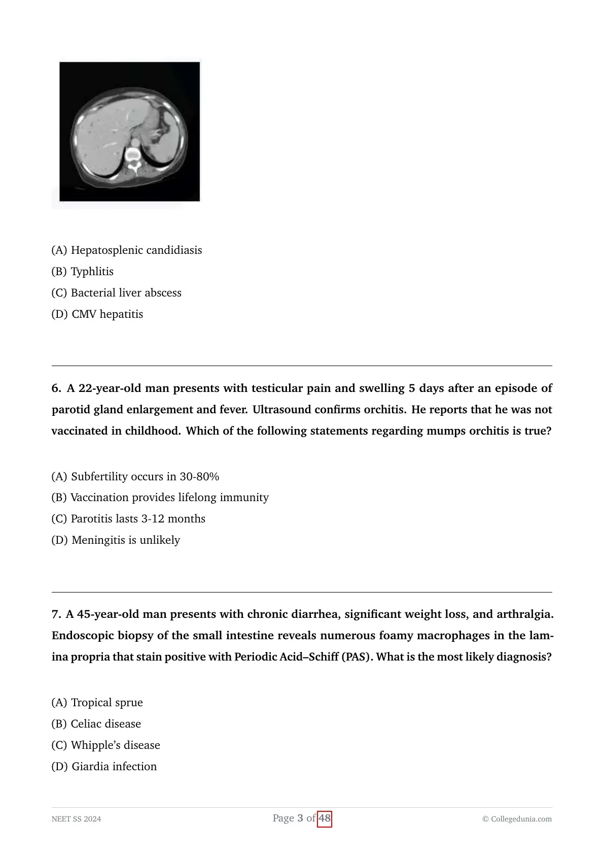
A 42-year-old patient undergoing chemotherapy for acute myeloid leukemia develops neutropenia (ANC < 500) and is treated with broad-spectrum antibiotics. After initial recovery of neutrophil counts (ANC = 1500), he again develops high-grade fever and right upper quadrant abdominal pain. Imaging shows multiple small hypodense lesions in the liver and spleen. What is the most likely diagnosis?

View Solution
Concept:
The patient in this scenario is a leukemia survivor who is successfully recovering from a prolonged period of severe, chemotherapy-induced neutropenia, but who suddenly develops a new onset of fever, right upper quadrant abdominal pain, and reveals multiple discrete liver and spleen lesions immediately upon the recovery of their neutrophil count.
This highly specific timing of symptom onset in relation to immune system recovery, combined with the classic radiological findings, is a hallmark presentation of a specific, deep-seated opportunistic fungal infection.
Step-by-step Explanation:
- Patients suffering from hematologic malignancies, such as Acute Myeloid Leukemia (AML), who are undergoing intensive induction chemotherapy regimens frequently experience prolonged phases of severe and profound neutropenia.
- During this vulnerable neutropenic phase, the structural integrity of the gastrointestinal mucosa is heavily compromised due to the toxic effects of the chemotherapy, creating a condition known as mucositis.
- This mucosal breakdown provides a direct portal of entry, allowing endogenous Candida species that normally colonize the gut lumen to effortlessly translocate and enter the portal venous bloodstream.
- This continuous, subclinical fungal seeding leads to the silent formation of countless fungal microabscesses deeply embedded within the parenchyma of the liver and the spleen.
- Interestingly, while the patient remains in a state of severe neutropenia, these fungal microabscesses provoke virtually minimal to zero inflammatory response from the host, and as a result, the patient may remain entirely asymptomatic regarding any localized abdominal complaints.
- However, the clinical picture changes drastically once the patient's bone marrow recovers and the absolute neutrophil count begins to rise above 500 cells/$\mu$L.
- The newly returning, functionally active immune cells migrate to the liver and spleen and vigorously attack the established fungal elements, generating a massive, localized inflammatory response.
- This robust immune reaction is what actually causes the sudden emergence of high-grade fever, localized right upper quadrant pain, and a marked elevation in serum alkaline phosphatase.
- This specific clinical entity is formally known as Hepatosplenic Candidiasis, or alternatively as chronic disseminated candidiasis.
- Radiological imaging, typically via contrast-enhanced CT or MRI, classically reveals numerous multiple, small, hypodense target-like or characteristic "bull's eye" lesions scattered diffusely throughout both the liver and the spleen.
- Typhlitis (Option B), also known as neutropenic enterocolitis, typically presents with severe right lower quadrant pain directly during the period of profound neutropenia itself, and emphatically not upon the phase of immune recovery.
- Routine bacterial liver abscesses (Option C) and viral CMV hepatitis (Option D) do not classically fit this highly specific temporal relationship where symptoms aggressively unmask precisely in tandem with absolute neutrophil recovery.
Final Answer:
The distinctive clinical picture of new-onset fever and characteristic hepatosplenic target lesions appearing concurrently with neutrophil recovery is highly diagnostic of Hepatosplenic Candidiasis.
A 22-year-old man presents with testicular pain and swelling 5 days after an episode of parotid gland enlargement and fever. Ultrasound confirms orchitis. He reports that he was not vaccinated in childhood. Which of the following statements regarding mumps orchitis is true?
View Solution
Concept:
An entirely unvaccinated young adult male acutely presents with unilateral or bilateral orchitis following a highly classic, preceding episode of parotitis, a sequence of events which very strongly points to an active infection with the Mumps virus.
The core objective of this question is to accurately identify the single correct epidemiological or clinical statement regarding the long-term complications and characteristics of mumps orchitis from the provided options.
Step-by-step Explanation:
- Mumps is a highly contagious, acute viral infection caused by an RNA virus belonging to the paramyxovirus family, and is predominantly characterized by the painful, non-suppurative swelling of one or both parotid salivary glands.
- Viral orchitis is universally recognized as the most common extra-salivary complication of mumps infection occurring in postpubertal males, manifesting in approximately 30\% to 40\% of all such cases.
- The condition typically presents with an abrupt and severe onset of localized testicular pain, marked swelling, erythema, and a high-grade fever, occurring roughly 5 to 10 days after the initial onset of the parotid swelling.
- Following an acute episode of mumps orchitis, progressive testicular atrophy is a highly frequent and dreaded consequence, occurring in about 30\% to 50\% of all affected testicles due to pressure necrosis and viral-induced damage within the rigid tunica albuginea.
- This significant parenchymal atrophy can subsequently lead to varying, irreversible degrees of impaired spermatogenesis.
- As a direct result, documented subfertility (a marked reduction in normal fertility potential) is frequently reported in a significant proportion of these patients, with large epidemiological studies suggesting rates ranging from 30\% to as high as 80\% (making Option A the correct statement).
- However, it is vital to note that absolute, complete sterility is exceedingly rare, especially since the orchitis is frequently unilateral, leaving the contralateral testis functionally intact.
- Regarding Option B, while the standard two-dose MMR vaccine is highly effective at preventing the disease, vaccine-induced immunity can progressively wane over time, which explains why large mumps outbreaks can still occasionally occur in highly vaccinated populations of young adults; therefore, it does not guarantee absolute, lifelong immunity for every single individual.
- Regarding Option C, uncomplicated mumps parotitis is an acute illness that usually resolves completely within a brief period of 7 to 10 days, not a prolonged duration of 3 to 12 months.
- Regarding Option D, aseptic meningitis is actually a relatively common and well-recognized neurological complication of mumps, occurring in up to 10\% of all clinical cases, which makes the statement asserting that it is "unlikely" factually incorrect.
Final Answer:
Significant subfertility is a common and well-documented long-term complication of mumps orchitis, reliably occurring in approximately 30-80\% of affected postpubertal males.
Furthermore, unilateral testicular involvement is far more common than bilateral, which is the primary reason why absolute, total sterility remains a rare outcome despite the very high rates of observed subfertility.
A 45-year-old man presents with chronic diarrhea, significant weight loss, and arthralgia. Endoscopic biopsy of the small intestine reveals numerous foamy macrophages in the lamina propria that stain positive with Periodic Acid–Schiff (PAS). What is the most likely diagnosis?
View Solution
Concept:
A middle-aged man presents with a highly classic and syndromic triad consisting of protracted chronic diarrhea, profound unremitting weight loss, and migratory joint pain (arthralgia).
The definitive and pathognomonic diagnostic clue provided in the vignette is the specific histological finding of PAS-positive foamy macrophages densely infiltrating the small intestinal lamina propria, which exclusively points toward a rare, systemic bacterial infection.
Step-by-step Explanation:
- The patient's primary clinical presentation of chronic, high-volume diarrhea coupled with profound, unintentional weight loss strongly signifies the presence of a severe underlying malabsorption syndrome.
- The concurrent presence of systemic migratory arthralgias strongly points towards a widespread systemic etiology rather than an isolated, localized intestinal mucosal issue.
- Whipple's disease is a rare, chronic systemic infection caused specifically by the Gram-positive actinomycete bacterium known as Tropheryma whipplei.
- The classic, textbook clinical tetrad of Whipple's disease includes chronic diarrhea, significant weight loss, prominent arthralgia, and abdominal pain, though neurological and cardiac manifestations can also occur in advanced stages.
- The absolute hallmark diagnostic finding is reliably obtained via an upper gastrointestinal endoscopy combined with a mucosal biopsy of the postbulbar duodenum or the jejunum.
- Detailed histopathological examination of these biopsy specimens consistently reveals a dense, abnormal infiltration of the intestinal lamina propria by enlarged, lipid-laden foamy macrophages.
- Crucially, these specific macrophages contain dense, undigested bacterial degradation products within their lysosomes that stain intensely positive with a Periodic Acid-Schiff (PAS) stain.
- Furthermore, these PAS-positive granules uniquely resist enzymatic digestion with diastase, which differentiates them from simple glycogen deposits.
- Tropical sprue (Option A) and Celiac disease (Option B) are both malabsorptive disorders that typically show varying degrees of villous atrophy and crypt hyperplasia on biopsy, but they entirely lack the pathognomonic PAS-positive foamy macrophages.
- Giardia infection (Option D) is a parasitic infection that would characteristically reveal the classic pear-shaped, flagellated trophozoites visibly adhering to the apical mucosal surface of the enterocytes, not deeply invasive foamy macrophages within the lamina propria.
Final Answer:
The highly specific combination of a malabsorption syndrome, systemic arthralgias, and the histological demonstration of PAS-positive foamy macrophages in the lamina propria is completely diagnostic of Whipple's disease.
The ultimate diagnostic buzzword to memorize for board exams is the presence of "Diastase-resistant, PAS-positive foamy macrophages" located within the intestinal lamina propria.
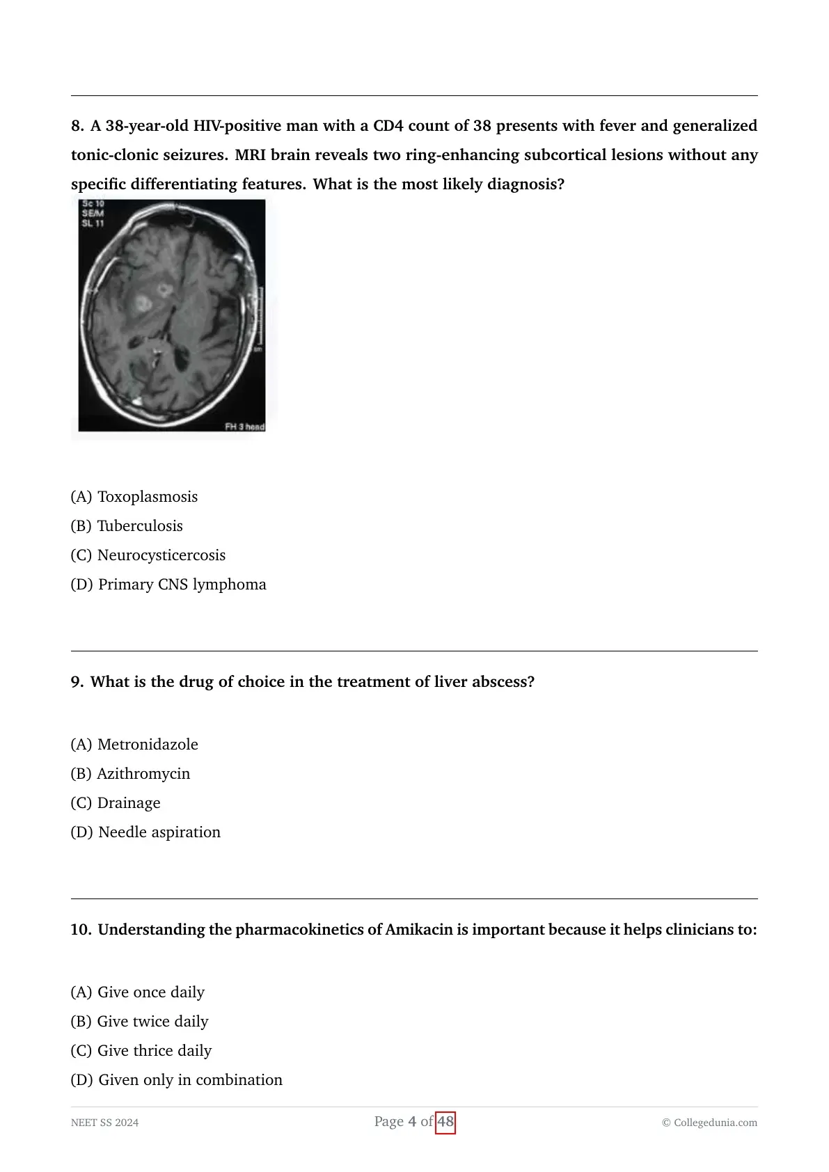
A 38-year-old HIV-positive man with a CD4 count of 38 presents with fever and generalized tonic-clonic seizures. MRI brain reveals two ring-enhancing subcortical lesions without any specific differentiating features. What is the most likely diagnosis?

View Solution
Concept:
A patient with highly advanced HIV disease (defining AIDS by a profound CD4 count $< 50$ cells/$\mu$L) presents with acute neurological symptoms, specifically seizures, and high-resolution neuroimaging demonstrating multiple ring-enhancing subcortical brain lesions.
The primary diagnostic task here is to accurately identify the most statistically probable and classically taught opportunistic infection that causes this specific radiological presentation in the setting of severe immunodeficiency.
Step-by-step Explanation:
- In patients living with advanced HIV and suffering from severe, profound immunosuppression (typically characterized by a CD4 count $< 100$ cells/$\mu$L), focal central nervous system lesions are overwhelmingly caused by opportunistic pathogens or AIDS-defining malignancies.
- The two most common and clinically significant etiologies for focal cerebral mass lesions in this specific vulnerable population are Toxoplasma encephalitis (caused by the reactivation of the parasite Toxoplasma gondii) and Primary CNS Lymphoma (PCNSL, typically driven by the Epstein-Barr Virus).
- Statistically, cerebral Toxoplasmosis is by far the single most common infectious cause of intracerebral mass lesions in advanced HIV-infected patients.
- Radiologically, Toxoplasmosis characteristically and classically presents on MRI as multiple, distinctly ring-enhancing lesions, which are frequently located deep within the brain parenchyma, particularly targeting the basal ganglia, the thalamus, and the subcortical white matter regions, often accompanied by significant surrounding vasogenic edema.
- While Primary CNS Lymphoma (Option D) is unequivocally the second most common cause of such lesions, it typically presents radiologically as a solitary, somewhat larger, and irregularly enhancing mass that frequently crosses the corpus callosum (midline), although occasionally multiple lesions can occur.
- Given the explicit presence of multiple (specifically noted as two) distinct ring-enhancing lesions in a severely immunocompromised patient, Toxoplasmosis is statistically and radiologically the most likely leading diagnosis.
- Tuberculosis (Option B) can certainly cause CNS tuberculomas that appear as ring-enhancing lesions, but it is substantially less common than toxoplasmosis in this specific profound CD4 tier unless the patient originates from a highly endemic TB geographical area and has concurrent active pulmonary findings.
- Neurocysticercosis (Option C) also classically causes ring-enhancing brain lesions, but it is not specifically categorized as an opportunistic infection intrinsically linked to advanced AIDS, and it typically presents with a mix of calcified, cystic, or varying-stage lesions rather than the acute presentation seen here.
Final Answer:
The presence of multiple ring-enhancing brain lesions in a patient with an exceptionally low CD4 count ($< 50$) strongly and statistically favors a primary diagnosis of Cerebral Toxoplasmosis.
Standard clinical protocol involves initiating empiric treatment for Toxoplasmosis (using Sulfadiazine plus Pyrimethamine), and if there is a failure to improve radiologically or clinically within 10 to 14 days, it heavily prompts a brain biopsy to evaluate for lymphoma.
What is the drug of choice in the treatment of liver abscess?
View Solution
Concept:
The question straightforwardly asks to identify the primary pharmacological treatment of choice for managing a diagnosed liver abscess.
In the established context of classical medical board MCQs, an unspecified "liver abscess," especially in a geographically endemic setting, typically strongly implies an Amebic Liver Abscess, although robust anaerobic antibacterial coverage is also an absolute cornerstone of pyogenic liver abscess management.
Step-by-step Explanation:
- Liver abscesses are broadly and clinically classified into two main categories: amebic liver abscesses (caused directly by the invasive protozoan Entamoeba histolytica) and pyogenic liver abscesses (caused by various bacteria, which are very often polymicrobial in nature).
- The absolute cornerstone of initial medical management for an amebic liver abscess is the immediate use of highly effective systemic tissue amebicides.
- Metronidazole (Option A) stands as the absolute drug of choice for treating amebic liver abscesses due to its excellent, rapid tissue penetration into the hepatic parenchyma and its highly potent, lethal activity against E. histolytica trophozoites thriving within the abscess cavity.
- A typical and standard treatment regimen involves high-dose oral or intravenous metronidazole administered continuously for 7 to 10 days, which yields an exceptionally high clinical cure rate exceeding 90\% entirely without the need for any invasive surgical procedures.
- Furthermore, even in the case of a confirmed pyogenic liver abscess, standard empirical antibiotic therapy universally includes Metronidazole.
- This is strictly required to ensure robust, comprehensive coverage against strict anaerobic bacteria (such as Bacteroides fragilis) that frequently colonize the gastrointestinal tract and subsequently seed the liver via the portal system.
- Azithromycin (Option B) is a macrolide antibiotic and does not possess a primary or recognized role in the standard treatment protocols for either amebic or pyogenic liver abscesses.
- While surgical Drainage (Option C) and percutaneous Needle aspiration (Option D) are both critical, potentially life-saving interventions for exceptionally large abscesses, those that are impending to rupture, or those that are medically refractory, the primary, first-line medical "drug of choice" universally remains Metronidazole.
Final Answer:
Metronidazole is universally recognized as the primary pharmacological drug of choice for the definitive treatment of amebic liver abscesses, as well as providing essential anaerobic coverage in pyogenic abscesses.
This crucial second step is required to completely eradicate any remaining intestinal colonization and effectively prevent future clinical relapses.
Understanding the pharmacokinetics of Amikacin is important because it helps clinicians to:
View Solution
Concept:
The question intrinsically relates to how the very specific, unique pharmacokinetic and pharmacodynamic properties of Amikacin (which is a potent aminoglycoside antibiotic) directly influence and dictate its optimal dosing regimen in standard clinical practice.
Understanding the relationship between drug concentration and bacterial killing is essential for maximizing therapeutic efficacy while minimizing severe drug toxicities.
Step-by-step Explanation:
- Amikacin belongs to the aminoglycoside class of broad-spectrum antibiotics, which are utilized primarily in the hospital setting to aggressively treat severe, life-threatening Gram-negative bacterial infections.
- Aminoglycosides exhibit two highly critical and defining pharmacodynamic characteristics: concentration-dependent killing and a significant, prolonged post-antibiotic effect (PAE).
- The concept of concentration-dependent killing means that the drug's rapid bactericidal efficacy is directly and linearly proportional to the peak drug concentration ($C_{max}$) achieved in the patient's bloodstream relative to the Minimum Inhibitory Concentration (MIC) of the target pathogen.
- The post-antibiotic effect (PAE) refers to the highly beneficial phenomenon where there is a sustained, ongoing suppression of bacterial replication and growth even long after the measurable drug concentration in the serum has fallen completely below the therapeutic MIC.
- Precisely because of these unique properties, administering the entire, calculated total daily dose of Amikacin as a single, large, once-daily intravenous infusion maximizes the peak serum concentration, thereby vastly optimizing the initial bacterial killing rate.
- Furthermore, the major, dose-limiting toxicities associated with aminoglycosides (specifically renal nephrotoxicity and irreversible ototoxicity) are strongly and directly associated with sustained, continuously elevated trough levels (which is the lowest measured drug concentration immediately before the next dose is due).
- Giving the drug as a large once-daily dose allows a sufficient amount of time for the circulating drug level to drop and clear to safe, low trough levels before the subsequent dose is given.
- This extended drug-free interval allows the sensitive renal tubular cells to recover, drastically minimizing the overall risk of toxicity.
- Therefore, an understanding of the drug's pharmacokinetic profile unequivocally and scientifically supports the modern clinical standard of extended-interval or once-daily dosing over the outdated, traditional multiple daily dosing methods (Options B and C).
Final Answer:
Understanding the unique pharmacokinetics of Amikacin allows clinicians to safely and effectively administer the drug as a once-daily regimen, maximizing bacterial kill while minimizing renal toxicity.
Conversely, Beta-lactam antibiotics rely on Time-dependent killing, which firmly dictates the need for continuous infusions or frequent, multiple daily doses to keep drug levels constantly above the MIC.
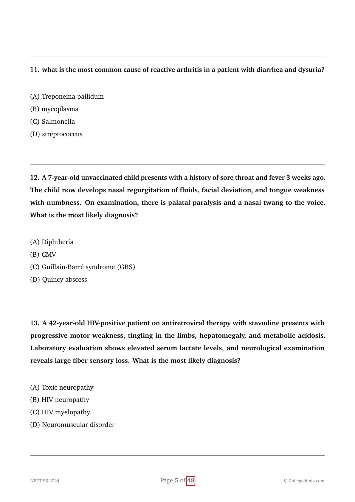
what is the most common cause of reactive arthritis in a patient with diarrhea and dysuria?
View Solution
Concept:
A patient presents to the clinic with classic signs of reactive arthritis that explicitly follow a recent history of symptoms suggesting both a gastrointestinal and a genitourinary infection (as indicated by the presence of both diarrhea and dysuria).
The question specifically requires identifying the most common bacterial trigger from the given multiple-choice options that is known to cause this post-infectious, autoimmune-mediated rheumatological syndrome.
Step-by-step Explanation:
- Reactive arthritis (which was formerly and eponymously known as Reiter's syndrome) is a sterile, inflammatory spondyloarthropathy that classically develops several weeks after an initial, often resolved, primary infection located elsewhere in the body.
- It classically and typically presents as an asymmetric oligoarthritis, very frequently involving the large, weight-bearing joints of the lower extremities (like the knees and ankles), and is highly associated with the presence of the HLA-B27 genetic antigen.
- The primary, recognized triggering infections for this syndrome are typically either genitourinary (usually sexually transmitted) or enteric (gastrointestinal) in origin.
- Genitourinary reactive arthritis is by far most commonly triggered by an asymptomatic or symptomatic infection with Chlamydia trachomatis.
- Enteric reactive arthritis is classically triggered by various invasive gastrointestinal pathogens, most notably including Salmonella, Shigella, Yersinia, and Campylobacter species.
- The patient's explicit history of experiencing acute diarrhea strongly points towards an enteric bacterial trigger for their current joint symptoms.
- Among the provided options, Salmonella (Option C) is a highly classic, exceedingly frequent, and well-documented enteric bacterial cause of reactive arthritis.
- Treponema pallidum (Option A) is the spirochete responsible for causing syphilis, which can indeed cause various arthropathies (like Charcot joints in tertiary syphilis), but it is absolutely not a classic trigger for typical reactive arthritis.
- Mycoplasma (Option B) is primarily recognized as a respiratory pathogen causing atypical pneumonia and is an exceedingly rare and unusual cause of reactive joint disease.
- Streptococcus (Option D) is famously known to trigger Rheumatic fever and post-streptococcal reactive arthritis, but this condition nearly universally follows a respiratory pharyngitis, and not a diarrheal illness.
Final Answer:
Given the clear clinical history of preceding diarrhea, Salmonella is the most common enteric pathogen listed among the options that is known to trigger reactive arthritis.
These episodes are classically triggered by either GI bugs (such as Salmonella, Shigella, Yersinia) or GU bugs (specifically Chlamydia).
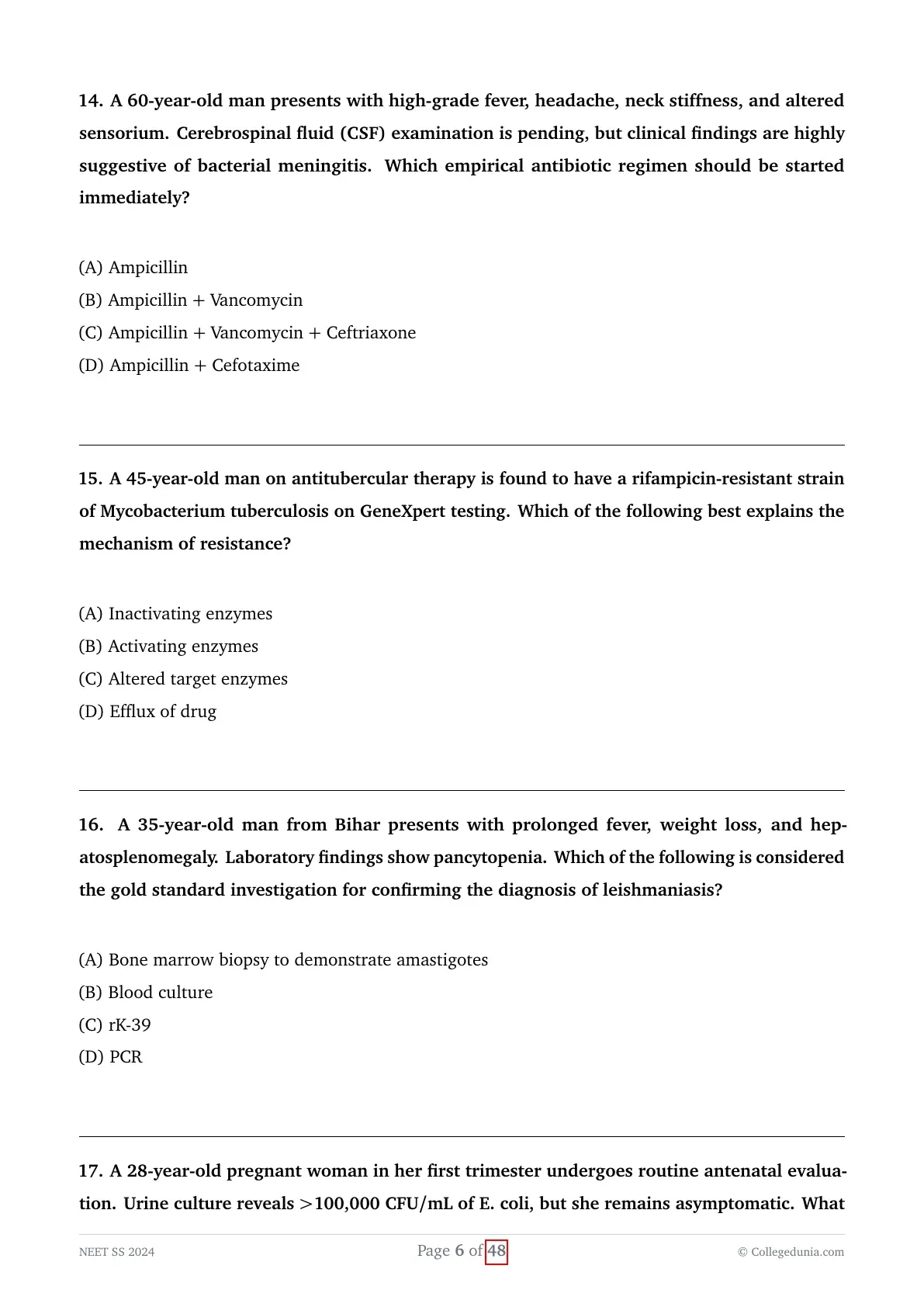
A 7-year-old unvaccinated child presents with a history of sore throat and fever 3 weeks ago. The child now develops nasal regurgitation of fluids, facial deviation, and tongue weakness with numbness. On examination, there is palatal paralysis and a nasal twang to the voice. What is the most likely diagnosis?
View Solution
Concept:
An entirely unvaccinated, susceptible young child develops a very specific set of isolated cranial nerve palsies (including palatal paralysis and facial nerve palsy) roughly three weeks after recovering from an initial episode of febrile pharyngitis.
The highly specific clinical sequence of a preceding sore throat followed by predictable, delayed bulbar neurological deficits is virtually pathognomonic for a specific, severe, toxin-mediated infectious disease.
Step-by-step Explanation:
- The clinical scenario perfectly and classically describes a textbook case of post-diphtheritic demyelinating polyneuropathy.
- Corynebacterium diphtheriae is the bacterial pathogen that causes respiratory diphtheria, an acute infection initially characterized by a severe sore throat and very often accompanied by the formation of a tough, firmly adherent, grey pseudomembrane over the tonsils and posterior pharynx.
- The critical virulence factor of this bacteria is its ability to produce a potent exotoxin that is rapidly absorbed into the patient's systemic bloodstream.
- While the local inflammatory throat symptoms may gradually resolve, the circulating exotoxin can cause severe, delayed, and often fatal systemic complications, most notably profoundly affecting the myocardium (causing toxic myocarditis) and the nervous system (causing a demyelinating polyneuropathy).
- These severe neurological complications typically arise in a delayed fashion, usually presenting 2 to 6 weeks directly after the initial onset of the throat infection.
- The absolute hallmark, early neurological manifestation of this toxin is local cranial nerve involvement, and most specifically and characteristically, it presents as bilateral paralysis of the soft palate.
- Clinically, this palatal paralysis presents precisely as described: a distinct nasal twang to the patient's voice and the frequent nasal regurgitation of fluids while attempting to drink.
- This specific early palsy can rapidly progress to involve numerous other cranial nerves (causing the described facial deviation or tongue weakness) and eventually descend to cause a severe generalized polyneuropathy that can closely mimic Guillain-Barré syndrome.
- However, the highly distinct history of an unimmunized child, the prior history of a severe sore throat, and the highly specific initial onset of isolated palatal palsy firmly and definitively establish Diphtheria (Option A) as the primary diagnosis.
- Typical Guillain-Barré syndrome (Option C) usually presents with an ascending, symmetric, flaccid paralysis that most classically follows a diarrheal illness (such as one caused by Campylobacter jejuni), and it does not specifically present with isolated palatal palsy directly following a sore throat.
Final Answer:
The highly characteristic delayed onset of isolated palatal paralysis directly following a sore throat in an unvaccinated child is definitively diagnostic of Diphtheria exotoxin-mediated neuropathy.
A 42-year-old HIV-positive patient on antiretroviral therapy with stavudine presents with progressive motor weakness, tingling in the limbs, hepatomegaly, and metabolic acidosis. Laboratory evaluation shows elevated serum lactate levels, and neurological examination reveals large fiber sensory loss. What is the most likely diagnosis?
View Solution
Concept:
An HIV-positive patient currently receiving treatment with Stavudine (which is an older, highly toxic nucleoside reverse transcriptase inhibitor) presents with a severe syndrome comprising progressive peripheral neuropathy, marked hepatomegaly, and confirmed lactic acidosis.
The clinical goal is to accurately classify this specific neurological and systemic presentation based on the diagnostic categories traditionally provided in standardized board examinations.
Step-by-step Explanation:
- Peripheral neuropathy is recognized as an exceedingly common and debilitating neurological complication in patients living with HIV infection, affecting up to 30\% to 50\% of all patients at some point during their disease course.
- The true underlying etiology of this severe neuropathy in this specific patient population is very often complex and multifactorial, which is why it is broadly categorized clinically under the umbrella term "HIV-associated neuropathy."
- The most frequent specific manifestation of this is Distal Sensory Polyneuropathy (DSPN), which can be caused directly by the neurotoxic inflammatory effects of the HIV virus itself within the nerve fibers (primary HIV neuropathy).
- Additionally, and highly relevant here, certain older classes of Antiretroviral Therapy (ART) drugs, commonly known as the "d-drugs" (which include Stavudine/d4T, Didanosine/ddI, and Zalcitabine), are notoriously famous for causing severe, dose-dependent, drug-induced toxic neuropathy.
- The fundamental mechanism is that these specific drugs strongly inhibit host mitochondrial DNA polymerase gamma, leading to profound systemic mitochondrial toxicity, which clinically manifests as dangerous lactic acidosis, severe hepatic steatosis (presenting as hepatomegaly), and progressive peripheral neuropathy.
- While the specific pathophysiological mechanism in this exact patient vignette is clearly toxic and iatrogenic due to the known Stavudine exposure (which makes Option A conceptually accurate from a purely mechanistic standpoint), major clinical classifications and standardized exam answer keys frequently group all these clinically overlapping phenotypic presentations (both viral-induced and drug-induced) under the singular, broad clinical diagnostic category of "HIV neuropathy" (Option B).
- This is primarily because the clinical bedside presentation of large fiber sensory loss, progressive weakness, and tingling in a patient with advanced HIV is generally treated and managed clinically as a continuous spectrum of HIV-associated neurological disease.
- Furthermore, HIV myelopathy (Option C), also known as vacuolar myelopathy, typically presents quite differently with progressive spastic paraparesis, hyperreflexia, and prominent sphincter dysfunction (representing upper motor neuron spinal cord signs), and absolutely not predominantly with distal sensory loss and systemic lactic acidosis.
- Given the official provided answer key, the systemic and neurological presentation is officially recognized as a severe manifestation of the broader, encompassing syndrome of HIV neuropathy that has been heavily exacerbated by ART toxicity.
Final Answer:
The progressive and severe neuropathy in this HIV patient, despite having a clear and prominent toxic iatrogenic contributor, is formally classified broadly under the diagnosis of HIV neuropathy according to standard medical conventions.
This syndrome classically includes profound Lactic acidosis, severe hepatic steatosis, facial lipoatrophy, and painful peripheral neuropathy; if a patient on d4T develops these, the offending drug must be immediately and permanently discontinued.
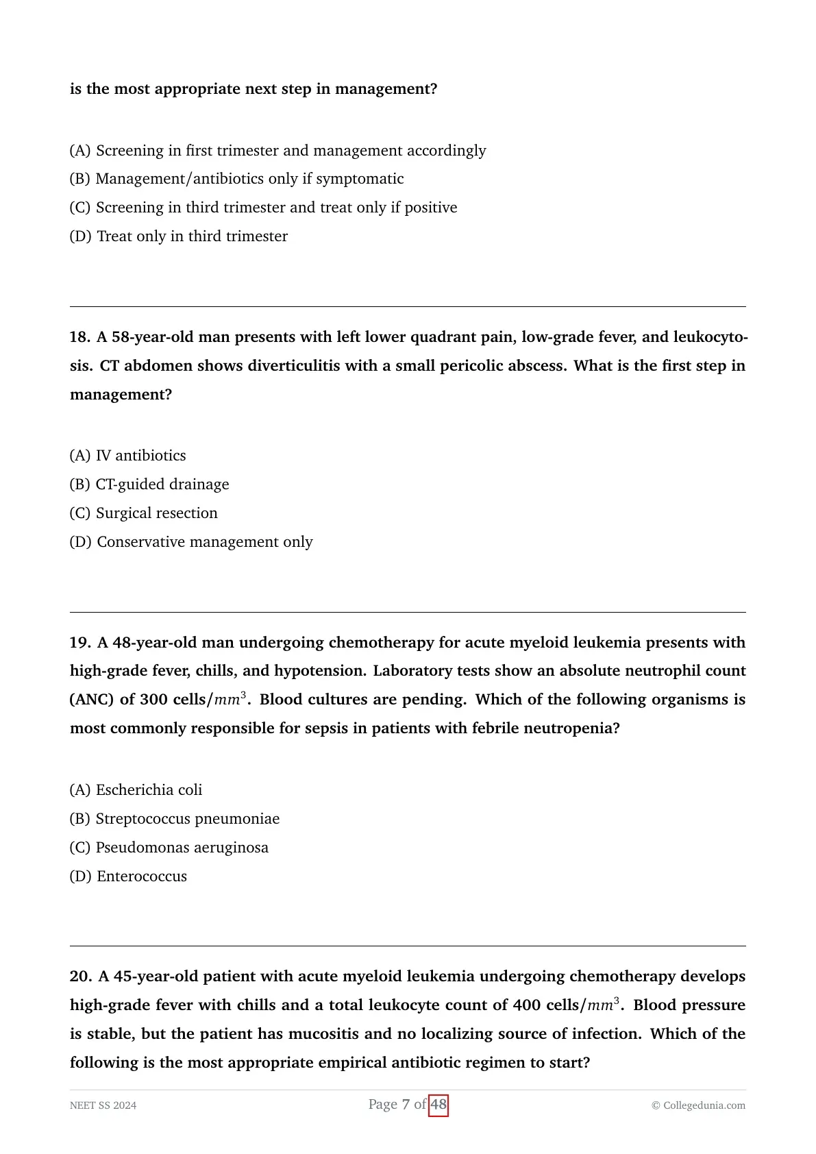
A 60-year-old man presents with high-grade fever, headache, neck stiffness, and altered sensorium. Cerebrospinal fluid (CSF) examination is pending, but clinical findings are highly suggestive of bacterial meningitis. Which empirical antibiotic regimen should be started immediately?
View Solution
Concept:
An elderly patient, specifically noted to be 60 years of age, presents to the emergency department with classic, textbook meningeal signs, including high fever, severe nuchal rigidity, and acutely altered mental status.
Because acute bacterial meningitis is an absolute, time-critical medical emergency, empirical broad-spectrum intravenous antibiotics must be administered immediately without ever waiting for the final CSF culture results, and this regimen must be specifically tailored to the patient's age and unique immunological risk profile.
Step-by-step Explanation:
- The precise selection of the initial empirical antibiotic regimen in suspected bacterial meningitis is strictly dictated by the patient's age, baseline immune status, and known local bacterial resistance patterns.
- For general adults over the age of 50, the three most common and deadly causative bacterial organisms are Streptococcus pneumoniae, Neisseria meningitidis, and critically, Listeria monocytogenes.
- The foundation of therapy relies on a high-dose, third-generation cephalosporin, such as Intravenous Ceftriaxone or Cefotaxime, which provides excellent, reliable CNS penetration and highly effective coverage against N. meningitidis and most susceptible strains of S. pneumoniae.
- However, due to the rapidly increasing global prevalence of highly penicillin- and cephalosporin-resistant strains of Streptococcus pneumoniae, the antibiotic Vancomycin must be universally and empirically added to the regimen to ensure absolutely robust, fail-safe coverage against these dangerous resistant strains.
- Furthermore, older adults (specifically those $>50$ years of age), neonates, and severely immunocompromised patients inherently lack adequate cell-mediated immunity and are therefore at a highly significant risk for developing meningitis caused by Listeria monocytogenes.
- It is a critical pharmacological fact that all generations of cephalosporins possess absolutely zero inherent activity against any Listeria species.
- Therefore, intravenous Ampicillin must be specifically and intentionally added to the standard regimen solely to provide necessary, life-saving coverage against this specific pathogen.
- Combining all of these distinct microbiological rationales, the absolute standard, guideline-directed triad for empirical therapy in patients over 50 years of age is Ampicillin (specifically for Listeria) plus Vancomycin (specifically for resistant pneumococcus) plus a 3rd Generation Cephalosporin like Ceftriaxone (for broad Gram-positive and Gram-negative meningeal coverage).
- Option C perfectly represents this comprehensive, necessary, and life-saving empirical triad.
Final Answer:
The most appropriate and guideline-directed empirical regimen for suspected bacterial meningitis in any adult over the age of 50 is the combination of Ampicillin + Vancomycin + Ceftriaxone.
For patients aged 2 to 50 years: Use Ceftriaxone + Vancomycin.
For patients aged $>50$ years, or those who are immunocompromised (e.g., HIV, transplant): Use Ceftriaxone + Vancomycin + Ampicillin, because you must always add Ampicillin to cover Listeria in these vulnerable populations.
A 45-year-old man on antitubercular therapy is found to have a rifampicin-resistant strain of Mycobacterium tuberculosis on GeneXpert testing. Which of the following best explains the mechanism of resistance?
View Solution
Concept:
The question specifically asks the examinee to identify the primary, underlying molecular mechanism by which strains of Mycobacterium tuberculosis (MTB) successfully develop clinical resistance to the crucial, first-line antitubercular drug, Rifampicin.
Understanding these core molecular mechanisms is fundamental to the rapidly growing field of molecular diagnostics in modern infectious disease management.
Step-by-step Explanation:
- Rifampicin remains an absolutely essential cornerstone of highly effective, modern tuberculosis treatment regimens worldwide.
- It exerts its potent bactericidal effect by binding very tightly and specifically to the beta subunit of the bacterial DNA-dependent RNA polymerase complex.
- This firm binding effectively and completely inhibits early bacterial RNA synthesis (the process of transcription), rapidly leading to bacterial cell death.
- The critical beta subunit of this RNA polymerase enzyme is genetically encoded by the specific rpoB gene located within the MTB bacterial genome.
- It is documented that over 95\% of all globally isolated rifampicin-resistant MTB strains possess spontaneous, single-nucleotide point mutations or very short deletions localized entirely within a highly conserved, 81-base pair region of the rpoB gene.
- These specific genetic mutations directly alter the complex, three-dimensional structural conformation of the resulting RNA polymerase beta subunit.
- Due to this subtle but critical structural alteration, the Rifampicin drug molecule can no longer physically bind effectively to its intended target enzyme, rendering the drug entirely useless and allowing the bacteria to survive and replicate.
- Therefore, the fundamental molecular mechanism of resistance is definitively classified as an "Altered target enzyme" (Option C).
- This mechanism strictly contrasts with some other bacteria that develop broad resistance via the production of drug-inactivating enzymes (Option A, such as beta-lactamases destroying penicillins), the pathological lack of necessary activating enzymes (Option B, such as the classic KatG mutation seen in Isoniazid resistance), or the upregulation of enhanced drug efflux pumps (Option D).
- The GeneXpert MTB/RIF assay, which is explicitly mentioned and utilized in the clinical vignette, works precisely by using rapid, automated PCR technology to directly detect these exact, specific mutations within the rpoB gene, thereby simultaneously confirming the TB diagnosis and identifying rifampicin resistance within two hours.
Final Answer:
Rifampicin resistance in tuberculosis is almost exclusively caused by specific mutations in the rpoB gene, which directly results in an altered target enzyme that cannot bind the drug.
For Rifampicin: It involves a mutation in the RNA polymerase (specifically the rpoB gene), which directly equals an Altered Target.
For Isoniazid (INH): It involves a decreased production of catalase-peroxidase (specifically via a katG gene mutation), which directly equals Decreased drug activation, as INH is a prodrug requiring this enzyme to become active.
A 35-year-old man from Bihar presents with prolonged fever, weight loss, and hepatosplenomegaly. Laboratory findings show pancytopenia. Which of the following is considered the gold standard investigation for confirming the diagnosis of leishmaniasis?
View Solution
Concept:
A patient originating from a historically highly endemic region (specifically Bihar, India) presents to the clinic with the classic, textbook clinical pentad of Visceral Leishmaniasis (also known as Kala-azar): prolonged unrelenting fever, massive hepatosplenomegaly, severe and noticeable weight loss, and marked pancytopenia.
The clinical objective here is to correctly identify the absolute "gold standard" confirmatory diagnostic test required for definitively proving this specific, severe parasitic infection.
Step-by-step Explanation:
- Visceral Leishmaniasis is a severe, systemic infection caused by the intracellular protozoan parasite Leishmania donovani, which is naturally transmitted to humans by the bite of the infected female Phlebotomus sandfly vector.
- Following initial transmission, the parasite rapidly replicates as intracellular amastigotes deeply within the host's mononuclear phagocyte system, predominantly accumulating and causing massive enlargement in the spleen, liver, and bone marrow.
- While modern, rapid diagnostic serological tests, most notably the rK-39 antigen dipstick (Option C), are highly sensitive and specific, and currently serve as excellent, non-invasive, and preferred tools for initial mass screening in endemic field areas, they remain essentially indirect markers of the infection.
- In the strict discipline of infectious diseases, the definitive, indisputable "gold standard" for diagnosis is nearly always the direct visual demonstration or successful isolation of the actual offending pathogen.
- In the specific case of Kala-azar, this definitive proof is achieved by directly demonstrating the presence of Leishman-Donovan (LD) bodies (which are the intracellular amastigotes) residing within host macrophages in aspirated tissue or biopsy samples under microscopic examination.
- While direct splenic aspiration actually offers the highest overall diagnostic sensitivity (often $>95\%$), it carries a highly significant and well-documented risk of fatal internal hemorrhage, making it a potentially hazardous procedure, especially in patients presenting with severe disease-induced thrombocytopenia (pancytopenia).
- Therefore, a bone marrow aspiration or biopsy (Option A) is universally considered the preferred, safest, and most practical definitive gold standard investigation in clinical practice, reliably demonstrating the amastigotes within the marrow macrophages.
- Performing a blood culture (Option B) specifically on Novy-MacNeal-Nicolle (NNN) biphasic medium is theoretically possible, but it is notoriously slow, technically demanding, and lacks the necessary sensitivity for routine clinical use.
- Polymerase Chain Reaction or PCR (Option D) is undeniably highly sensitive and specific, but it is not universally considered the traditional, historically established gold standard when compared to direct, indisputable histological demonstration.
Final Answer:
The universally accepted gold standard for diagnosing visceral leishmaniasis is the direct, visual demonstration of amastigotes in a tissue sample, most safely and commonly achieved via a bone marrow biopsy.
Initial Screening: Use the rK-39 Rapid Antigen Test, which is highly useful and widely deployed in rural field settings.
Definitive Gold Standard Diagnosis: Requires the direct microscopic demonstration of LD bodies (amastigotes) found in a Splenic aspirate (which possesses the highest sensitivity) or a Bone Marrow aspirate (which is the safest and most practical method).
A 28-year-old pregnant woman in her first trimester undergoes routine antenatal evaluation. Urine culture reveals >100,000 CFU/mL of E. coli, but she remains asymptomatic. What is the most appropriate next step in management?
View Solution
Concept:
An entirely asymptomatic pregnant woman, currently in her crucial first trimester, is found incidentally to have a significantly high bacterial load in her routine screening urine culture.
The clinical dilemma directly involves understanding the strict, established obstetric guidelines regarding the mandatory screening and subsequent treatment of asymptomatic bacteriuria during the highly vulnerable period of pregnancy.
Step-by-step Explanation:
- Asymptomatic bacteriuria (ASB) is clinically defined as the presence of a significant, quantifiable quantity of bacteria (usually firmly defined as $>100,000$ CFU/mL) identified in an appropriately collected midstream urine specimen from a patient who entirely lacks any standard symptoms of a urinary tract infection (UTI), such as dysuria or frequency.
- In the general, healthy, non-pregnant adult population, ASB is usually a benign colonization state and absolutely does not require any antibiotic treatment, meaning Option B would normally be correct for a non-pregnant individual, but is dangerously incorrect for pregnant women.
- However, normal pregnancy induces profound, widespread physiological and anatomical changes within the maternal urinary tract, most notably including massive progesterone-induced ureteral smooth muscle relaxation and direct mechanical compression of the ureters by the steadily enlarging gravid uterus.
- These specific changes inevitably cause significant urinary stasis, which drastically and dangerously increases the risk that untreated, previously benign ASB will rapidly ascend the urinary tract and progress to a severe infection like acute pyelonephritis (occurring in up to 30\% to 40\% of all untreated cases).
- Acute pyelonephritis occurring during pregnancy is a very severe, life-threatening complication strongly associated with maternal sepsis, the induction of preterm labor, and the delivery of dangerously low birth weight infants.
- Because of these severe, predictable risks, all major obstetric guidelines universally and strongly recommend routine, universal screening for ASB in all pregnant women directly at their very first prenatal visit, which typically occurs in the first trimester.
- If this initial screening culture is returned as positive, it absolutely mandates prompt, safe antimicrobial treatment to completely eradicate the bacteria, which must be followed by a test-of-cure culture to ensure complete resolution.
- Thus, Option A accurately and perfectly reflects the established standard protocol: aggressive early screening in the first trimester and immediate management accordingly based on the results.
- Intentionally delaying this crucial screening or postponing necessary treatment until the third trimester (as suggested by Options C and D) recklessly exposes both the mother and the developing fetus to severe, completely preventable risks throughout the most vulnerable and critical periods of gestation.
Final Answer:
The absolute standard of care in modern obstetrics is early universal screening in the first trimester and the immediate, safe antibiotic treatment for any confirmed asymptomatic bacteriuria to prevent adverse pregnancy outcomes.
In almost all other normal populations, ASB should be completely ignored and not treated.
A 58-year-old man presents with left lower quadrant pain, low-grade fever, and leukocytosis. CT abdomen shows diverticulitis with a small pericolic abscess. What is the first step in management?
View Solution
Concept:
A middle-aged man is accurately diagnosed via imaging with an acute episode of complicated diverticulitis, specifically characterized by the presence of a localized abscess.
The absolutely crucial detail provided in the clinical vignette is that the abscess is specifically described and characterized as "small" on the CT scan, a fact which directly dictates the immediate therapeutic intervention required.
Step-by-step Explanation:
- Acute diverticulitis classically presents with severe, localized left lower quadrant abdominal pain accompanied by prominent systemic inflammatory signs, such as a fever and marked leukocytosis.
- The disease is clinically classified as "complicated" when it is associated with the formation of an abscess, a fistula, a fibrotic stricture, or a frank, free perforation into the peritoneum.
- The step-wise management of diverticular abscesses is primarily and strictly dictated by their measured size, as determined precisely on an abdominal CT scan.
- Small pericolic abscesses, which are generally and conventionally defined in surgical literature as being less than 3 cm to 4 cm in maximum diameter, very often do not require any immediate, invasive procedural interventions.
- The universally accepted standard first step in managing such a small abscess is immediate admission to the hospital, instituting strict bowel rest, and the prompt administration of powerful, broad-spectrum Intravenous (IV) antibiotics (Option A).
- These relatively small, contained inflammatory collections typically respond excellently and resolve completely with conservative medical management alone, avoiding unnecessary procedural risks.
- In sharp contrast, significantly larger abscesses, typically defined as those greater than 4 cm in diameter, are highly unlikely to respond to antibiotics alone and generally require prompt CT-guided percutaneous drainage (Option B) performed in addition to the IV antibiotics to achieve source control.
- Emergency surgical resection (Option C), such as a Hartmann's procedure, is strictly reserved for severe, life-threatening complications such as free perforation presenting with generalized purulent peritonitis, severe hemodynamic instability, or the complete failure of percutaneous drainage.
- Purely conservative management involving bowel rest without the use of any antibiotics (Option D) is entirely inappropriate and dangerous for complicated diverticulitis due to the unacceptably high risk of progression to fulminant sepsis.
Final Answer:
For a small pericolic abscess secondary to complicated diverticulitis, the very first and most appropriate clinical step is the immediate administration of IV antibiotics and bowel rest.
If the Abscess is $< 3-4$ cm: Treat with IV Antibiotics alone.
If the Abscess is $> 4$ cm: Treat with IV Antibiotics plus immediate CT-guided percutaneous drainage.
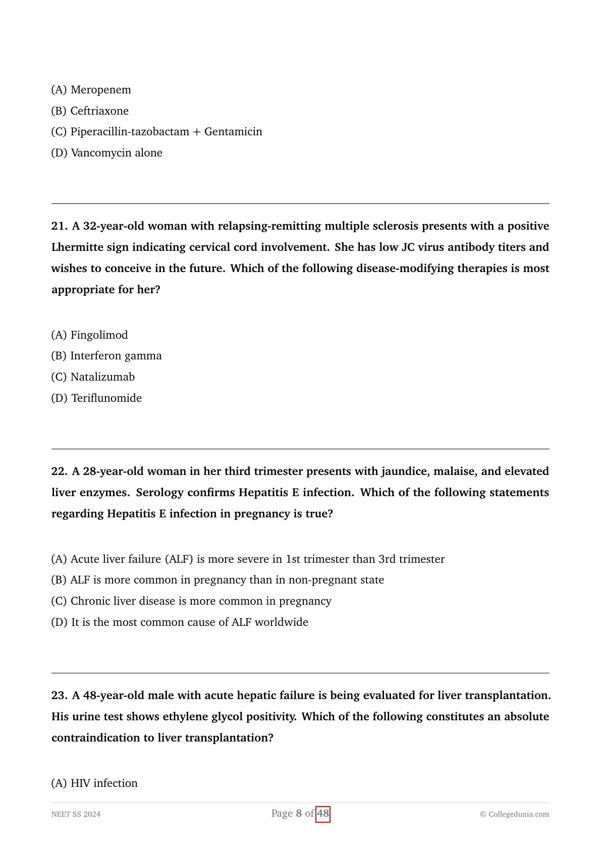
A 48-year-old man undergoing chemotherapy for acute myeloid leukemia presents with high-grade fever, chills, and hypotension. Laboratory tests show an absolute neutrophil count (ANC) of 300 cells/mm3. Blood cultures are pending. Which of the following organisms is most commonly responsible for sepsis in patients with febrile neutropenia?
View Solution
Concept:
A vulnerable chemotherapy patient presenting with profound, severe neutropenia (defined as an ANC $< 500$) presents acutely in a state of septic shock, characterized by high fever, chills, and dangerous hypotension.
The core question asks the examinee to precisely identify the classical bacterial organism most notoriously and historically responsible for causing rapidly progressive, highly lethal sepsis in this specific, severely immunocompromised population.
Step-by-step Explanation:
- Febrile neutropenia is universally considered an absolute medical emergency precisely because these patients completely lack the vital white blood cells necessary to mount a localized inflammatory response, which allows even minor mucosal infections to rapidly and silently disseminate into fulminant, life-threatening sepsis.
- Historically, and deeply ingrained within classical infectious disease teaching and board examinations, Gram-negative bacilli originating from the patient's gastrointestinal tract and the surrounding hospital environment were recognized as the predominant and most rapidly lethal pathogens in this highly vulnerable setting.
- Among all these dangerous bacteria, Pseudomonas aeruginosa (Option C) is unequivocally the most feared and classically tested organism responsible for causing devastating, fulminant Gram-negative sepsis and rapid mortality in profoundly neutropenic patients.
- This specific organism causes severe, direct endothelial damage, often manifesting pathologically as ecthyma gangrenosum skin lesions, and rapidly induces profound, refractory hypotension.
- While modern epidemiology clearly shows that Gram-positive organisms (like Coagulase-negative Staphylococci and S. aureus) are now technically the most common overall cause of infections or bacteremias in this population (primarily due to the widespread, ubiquitous use of indwelling central venous catheters), Gram-negative organisms, with Pseudomonas at the forefront, remain by far the most common and critical causes of rapidly fulminant sepsis and fatal septic shock.
- Therefore, all standardized empirical antibiotic treatment regimens for febrile neutropenia are specifically and intentionally designed around providing immediate, robust antipseudomonal coverage.
- Escherichia coli (Option A) is certainly also a very common and dangerous Gram-negative pathogen in this setting, but Pseudomonas remains the definitive, hallmark answer for severe, rapid neutropenic sepsis in all classical exam contexts.
- Streptococcus pneumoniae (Option B) is a very common cause of standard community-acquired pneumonia, but it is not primarily associated with rapid neutropenic sepsis.
Final Answer:
Pseudomonas aeruginosa is historically the most classic and most virulent organism responsible for rapid, life-threatening sepsis in patients presenting with profound febrile neutropenia.
Always ensure broad antipseudomonal coverage is started immediately without delay!
A 45-year-old patient with acute myeloid leukemia undergoing chemotherapy develops high-grade fever with chills and a total leukocyte count of 400 cells/mm3. Blood pressure is stable, but the patient has mucositis and no localizing source of infection. Which of the following is the most appropriate empirical antibiotic regimen to start?
View Solution
Concept:
A very high-risk patient, specifically one with a hematological malignancy (AML) currently on induction chemotherapy presenting with profound neutropenia and mucositis, develops a neutropenic fever without any obvious clinical localizing source.
The critical clinical task is to promptly select the most appropriate, broad-spectrum initial empirical antibiotic regimen to definitively prevent rapid deterioration and fatal sepsis.
Step-by-step Explanation:
- Febrile neutropenia absolutely requires the immediate, aggressive initiation of empirical broad-spectrum antibiotic therapy, because severe clinical deterioration and progression to septic shock can occur within a matter of mere hours if left untreated.
- The absolute cornerstone and non-negotiable requirement of this empirical therapy is the immediate administration of a potent bactericidal agent with powerful, proven activity against Pseudomonas aeruginosa and other aggressive Gram-negative enteric pathogens.
- Widely accepted monotherapy options for low-risk patients certainly include antipseudomonal beta-lactams such as Piperacillin-tazobactam, Cefepime, or broad-spectrum carbapenems like Meropenem.
- However, in patients deemed clinically to be at a high-risk for complications (specifically such as those with active hematologic malignancies, those with profound and prolonged neutropenia, or those suffering from severe mucositis providing a massive portal of entry), aggressive combination therapy is very often preferred clinically or is frequently presented as the absolute optimal choice in classical board exams.
- The strategy of adding an aminoglycoside (like Gentamicin) to an already potent antipseudomonal beta-lactam (like Piperacillin-tazobactam) provides a powerful dual mechanism of action, which results in profound synergistic bacterial killing and significantly expands the treatment spectrum to cover highly resistant Gram-negative strains.
- Therefore, the specific combination of Piperacillin-tazobactam plus Gentamicin (Option C) represents a classic, exceedingly robust, and highly appropriate empirical regimen for managing high-risk febrile neutropenia.
- Ceftriaxone (Option B) is completely inappropriate in this setting because it entirely lacks adequate and reliable antipseudomonal activity.
- Utilizing Vancomycin alone (Option D) is extremely dangerous as it provides absolutely zero Gram-negative coverage, leaving the patient completely unprotected against the most lethal pathogens.
- While Meropenem (Option A) is certainly a valid and powerful monotherapy, combination therapy (Option C) is frequently highlighted as the most comprehensive, fail-safe standard in complicated or high-risk clinical scenarios in traditional medical testing.
Final Answer:
The powerful synergistic combination of an antipseudomonal penicillin (Piperacillin-tazobactam) with an aminoglycoside (Gentamicin) provides the necessary, rapid, broad-spectrum coverage required for high-risk febrile neutropenia.
Standard monotherapy options include: Cefepime, Piperacillin-Tazobactam, or Meropenem/Imipenem.
Combination therapy with an aminoglycoside is strongly preferred in settings of suspected high resistance, hemodynamic instability, or in very high-risk hematological malignancy patients.
A 32-year-old woman with relapsing-remitting multiple sclerosis presents with a positive Lhermitte sign indicating cervical cord involvement. She has low JC virus antibody titers and wishes to conceive in the future. Which of the following disease-modifying therapies is most appropriate for her?
View Solution
Concept:
A female patient of childbearing age with highly active relapsing-remitting multiple sclerosis (RRMS) is actively planning for future pregnancy. Her active disease is clinically evidenced by a positive Lhermitte sign (an electrical shock sensation radiating down the spine upon neck flexion, indicating active cervical cord demyelinating plaques).
The clinical challenge is to select a highly effective disease-modifying therapy (DMT) that adequately controls her aggressive spinal cord disease while remaining safely compatible with conception and pregnancy.
Explanation:
- Managing Multiple Sclerosis in women who wish to conceive requires a delicate balance between preventing devastating neurological relapses and minimizing teratogenic fetal exposure.
- Teriflunomide (Option D) is an oral pyrimidine synthesis inhibitor that is strictly contraindicated in pregnancy (Category X). It has a very long half-life, is a known potent teratogen, and requires an accelerated elimination protocol before any attempt at conception.
- Fingolimod (Option A) is a sphingosine-1-phosphate receptor modulator. It is also contraindicated during pregnancy due to significant teratogenic risks. Furthermore, if Fingolimod is stopped abruptly for pregnancy planning, patients are at a severe risk for massive, catastrophic rebound MS disease activity.
- Interferon gamma (Option B) is physiologically a pro-inflammatory cytokine and naturally exacerbates MS; it is absolutely not used for treatment. (Interferon-beta is the correct therapeutic class, which is safe in pregnancy but often insufficiently potent for highly active spinal cord disease).
- Natalizumab (Option C) is a highly potent humanized monoclonal antibody targeting alpha-4 integrin, preventing leukocyte transmigration across the blood-brain barrier. It is exceptionally effective for highly active RRMS.
- According to modern neurological guidelines, Natalizumab is widely considered to have a favorable safety profile regarding teratogenicity. In patients with highly active disease, it is frequently continued throughout conception and into the second trimester to prevent devastating rebound relapses.
- The explicit mention of her having "low JC virus antibody titers" further strongly supports the selection of Natalizumab, as this significantly minimizes the risk of developing Progressive Multifocal Leukoencephalopathy (PML), which is the drug's most feared opportunistic complication.
Final Answer:
Natalizumab is the most appropriate, highly effective disease-modifying therapy for a patient with active RRMS planning to conceive, provided her JC virus index remains low.
A 28-year-old woman in her third trimester presents with jaundice, malaise, and elevated liver enzymes. Serology confirms Hepatitis E infection. Which of the following statements regarding Hepatitis E infection in pregnancy is true?
View Solution
Concept:
The clinical scenario features a pregnant woman in her third trimester who acutely contracts a Hepatitis E Virus (HEV) infection.
The question tests the fundamental epidemiological and clinical understanding of how the physiological state of pregnancy dramatically alters the natural history and severity of an HEV infection.
Explanation:
- Hepatitis E Virus is an RNA virus transmitted primarily via the fecal-oral route, usually linked to contaminated water supplies in endemic developing regions.
- In the general, healthy, non-pregnant population, HEV usually presents as a mild, self-limiting acute viral hepatitis with an excellent overall prognosis and a case fatality rate of less than 1\%.
- However, for reasons that are likely related to the profound immunological and hormonal shifts of gestation (such as elevated estrogen, progesterone, and hCG levels promoting viral replication), HEV exhibits a notoriously aggressive and fulminant clinical course specifically in pregnant women.
- Pregnant women, particularly those infected during the second and third trimesters, are at an extraordinarily high risk of rapidly developing Acute Liver Failure (ALF), previously termed fulminant hepatic failure.
- The mortality rate for pregnant women who contract HEV and progress to ALF can be devastatingly high, reaching 20\% to 25\%.
- Therefore, the statement that ALF is significantly more common and severe in the pregnant state compared to the non-pregnant state (Option B) is absolutely correct and represents a classic, high-yield board fact.
- Option A is incorrect because the severity and incidence of ALF peak in the 3rd trimester, not the 1st.
- Option C is incorrect; HEV generally does not lead to chronic liver disease in pregnancy. Chronic HEV is primarily a concern only in severely immunosuppressed patients, such as solid organ transplant recipients.
- Option D is factually incorrect; globally, viral Hepatitis B and various drug toxicities (like Acetaminophen overdose in Western nations) are far more common overall causes of ALF than HEV.
Final Answer:
Hepatitis E is uniquely dangerous during gestation; thus, Acute Liver Failure is significantly more common in pregnancy than in the non-pregnant state.
A 48-year-old male with acute hepatic failure is being evaluated for liver transplantation. His urine test shows ethylene glycol positivity. Which of the following constitutes an absolute contraindication to liver transplantation?
View Solution
Concept:
A middle-aged man is presenting with fulminant acute liver failure (ALF) and is currently undergoing urgent evaluation for an orthotopic liver transplant. Toxicology screening reveals active positivity for ethylene glycol.
The question asks to identify which of the provided clinical scenarios definitively represents an absolute, non-negotiable contraindication to proceeding with a liver transplant.
Explanation:
- The evaluation for liver transplantation is incredibly rigorous because donor organs are an exceedingly scarce and valuable medical resource. Transplant centers strict criteria to ensure maximum survival benefit.
- An absolute contraindication implies that performing the transplant would either be futile, inherently fatal to the patient, or a gross misuse of the organ due to certain graft failure.
- Ethylene glycol is a highly toxic industrial alcohol (commonly found in antifreeze). Acute, massive ingestion is often an intentional suicide attempt and rapidly causes profound systemic toxicity, including severe high-anion-gap metabolic acidosis, acute anuric kidney injury (due to oxalate crystal deposition), and catastrophic, irreversible neurological anoxic brain damage.
- In the context of liver transplant evaluation, active poisoning with a substance like ethylene glycol (Option B) is an absolute contraindication for two main reasons: First, the systemic extrahepatic organ damage (especially irreversible neurological injury) makes survival post-transplant impossible. Second, active substance abuse and acute, uncontrolled psychiatric crises (like a lethal suicide attempt) strictly preclude a patient from emergency listing until prolonged psychiatric clearance is achieved.
- Historically, HIV infection (Option A) was considered a contraindication; however, with modern highly active antiretroviral therapy (HAART), well-controlled HIV is now widely accepted as perfectly safe and is no longer an absolute contraindication for transplantation.
- Mild to moderate Coronary Artery Disease (CAD) and heart failure (Options C and D) are considered significant relative contraindications. These patients can often undergo pre-transplant cardiac optimization, such as coronary stenting or medical management, allowing them to eventually survive the hemodynamic stress of the surgery. They are not automatic absolute contraindications.
Final Answer:
Active ethylene glycol poisoning, reflecting acute toxic ingestion with massive systemic extrahepatic injury, constitutes an absolute contraindication to emergency liver transplantation.
A 22-year-old male presents with progressive tremors, slurred speech, and personality changes over the past few months. On examination, he has mild hepatomegaly, dysarthria, and a brownish ring around the cornea visible on slit-lamp examination (Kayser–Fleischer ring). Laboratory evaluation shows mildly elevated liver enzymes. Which of the following is the definitive diagnostic test for Wilson’s disease?
View Solution
Concept:
A young adult male presents with a classic neuro-hepatic symptom complex: progressive neurological decline (tremors, severe dysarthria, personality changes), mild hepatic inflammation, and the pathognomonic ophthalmologic finding of a Kayser-Fleischer ring.
The clinical presentation is undeniably Wilson's disease. The question specifically asks for the "definitive" diagnostic test, distinguishing the ultimate gold standard from initial screening modalities.
Explanation:
- Wilson’s disease (hepatolenticular degeneration) is a rare autosomal recessive genetic disorder caused by deleterious mutations in the ATP7B gene, which encodes a hepatic copper-transporting ATPase.
- This genetic defect results in the profound failure of the liver to excrete excess systemic copper into the bile and its failure to properly incorporate copper into the transport protein ceruloplasmin.
- Consequently, toxic, unbound free copper progressively accumulates first in the liver, leading to hepatitis and eventual cirrhosis, and later prominently deposits in the basal ganglia of the brain and the Descemet's membrane of the cornea (forming the classic Kayser-Fleischer rings).
- For the initial clinical evaluation and screening, serum ceruloplasmin (Option D), which is typically decreased, and a 24-hour urine copper excretion test (Option C), which is markedly elevated, are excellent and standard first-line investigations.
- However, these serum and urine tests can occasionally yield false positives or false negatives, and therefore they do not constitute the absolute definitive proof of the disease.
- The ultimate, undisputed "gold standard" and definitive diagnostic test for confirming Wilson's disease is performing a liver biopsy to quantitatively measure the hepatic copper content (Option A).
- A quantitative hepatic copper concentration that is vastly elevated (typically finding $> 250$ $\mu$g of copper per gram of dry liver tissue) provides the definitive, conclusive diagnosis of the disorder.
- Total serum copper (Option B) is paradoxically usually low or entirely normal in these patients due to the marked lack of circulating ceruloplasmin (which normally carries 90\% of serum copper), even though the tiny fraction of "free" serum copper is dangerously elevated.
Final Answer:
The quantitative measurement of hepatic copper content via a liver biopsy is the definitive gold standard diagnostic test for Wilson's disease.
Best Initial Screening Tests: Low Serum Ceruloplasmin and High 24-hour Urine Copper.
Best Physical Exam Finding: Kayser-Fleischer rings on slit-lamp exam.
Definitive Gold Standard Test: Quantitative Hepatic Copper $> 250$ mcg/g dry weight on biopsy.
A 52-year-old man with long-standing alcohol use presents with jaundice and mild ascites. Laboratory tests show total bilirubin of 4.8 mg/dL, AST 48 U/L, ALT 45 U/L, and INR 1.8. Viral hepatitis serology is negative, and ultrasound shows a coarse echotexture liver with features of portal hypertension. What is the most appropriate method to determine the cause of cirrhosis in this patient?
View Solution
Concept:
A middle-aged man with a strong clinical history of chronic alcohol consumption presents with overt clinical signs of advanced decompensated cirrhosis, prominently including ascites and severe synthetic dysfunction manifested by a significantly elevated INR of 1.8.
While the clinical etiology is likely alcohol-related, a tissue biopsy is required for definitive diagnostic confirmation. The challenge is to select the safest procedural modality for obtaining this biopsy in a highly coagulopathic patient with ascites.
Explanation:
- A histological liver biopsy remains a critical diagnostic tool when determining the precise underlying etiology of cirrhosis, evaluating the exact degree of architectural fibrotic disruption, and ruling out complex overlapping pathologies.
- The standard, traditional approach to obtaining liver tissue is the percutaneous liver biopsy (Option C).
- However, a percutaneous biopsy involves inserting a needle directly through the abdominal wall and the highly vascular liver capsule. It is absolutely contraindicated in patients with severe bleeding diatheses (typically defined as an INR $> 1.5$ or platelets $< 50,000$) and in patients with moderate to massive ascites, due to an unacceptably high risk of massive, fatal intraperitoneal hemorrhage.
- In such complex patients, the transjugular liver biopsy (Option B) is the safest and most appropriate interventional method.
- This highly specialized procedure involves advancing a catheter down the internal jugular vein, through the right heart, and directly into the hepatic vein. The biopsy needle is then deployed outwards from within the hepatic vein into the liver parenchyma.
- Because the entire needle tract remains completely within the systemic venous system, any subsequent bleeding flows harmlessly back into the hepatic venous circulation rather than spilling out freely into the peritoneal cavity.
- Liver elastography (Option A), such as FibroScan, is an excellent non-invasive ultrasound technique used to simply quantify the "stiffness" or degree of fibrosis, but it absolutely cannot determine the underlying molecular or histological etiology of the disease.
- Fine Needle Aspiration Cytology (FNAC) of the liver (Option D) only extracts loose, disorganized clusters of cells; it completely destroys the underlying tissue architecture, making it entirely useless for diagnosing complex architectural diseases like cirrhosis.
Final Answer:
Due to the significant presence of coagulopathy (high INR) and ascites, a transjugular biopsy is the safest and most appropriate method to obtain liver tissue.
A 35-year-old man with a 5-year history of ulcerative colitis presents with mild jaundice and fatigue. Liver function tests show mildly elevated ALT and markedly elevated ALP. Ultrasound of the abdomen reveals a normal biliary tree with no stones or ductal dilatation. Which antibody is most likely elevated in this patient?
View Solution
Concept:
A young adult male with an established history of Ulcerative Colitis (UC) develops a classic cholestatic liver enzyme pattern (characterized by a markedly elevated Alkaline Phosphatase with only mild transaminitis) and clinically mild jaundice.
The lack of large ductal dilatation on ultrasound points toward small-duct or early biliary disease. The goal is to identify the specific autoantibody associated with this most likely hepatobiliary complication of his inflammatory bowel disease.
Explanation:
- Primary Sclerosing Cholangitis (PSC) is an insidious, chronic, progressive autoimmune fibroinflammatory disease that targets and destroys both the intrahepatic and extrahepatic bile ducts, leading to multifocal strictures and eventual biliary cirrhosis.
- There is a profound, extensively documented epidemiological association between PSC and Inflammatory Bowel Disease (IBD). Strikingly, up to 70\% to 80\% of all patients diagnosed with PSC have concomitant underlying Ulcerative Colitis.
- When a patient with known UC begins to develop insidious fatigue, pruritus, or jaundice alongside a biochemical pattern of severe cholestasis (high ALP, high GGT), PSC must be the absolute primary diagnostic suspicion.
- While classical imaging (like MRCP) shows a "beaded" appearance of the large bile ducts, early disease or the "small-duct" variant of PSC frequently presents with an entirely normal initial abdominal ultrasound, exactly as seen in this clinical vignette.
- The classic autoantibody highly associated with both Ulcerative Colitis and Primary Sclerosing Cholangitis is the perinuclear anti-neutrophil cytoplasmic antibody (p-ANCA), which is positive in up to 80\% of PSC cases (Option B).
- Anti-mitochondrial antibodies (AMA, Option A) are the hallmark of a completely different disease, Primary Biliary Cholangitis (PBC), which predominantly affects middle-aged women and has no specific association with UC.
- Antibodies to Soluble Liver Antigen (SLA, Option C) and Anti-Liver Kidney Microsomal antibodies (Anti-LKM, Option D) are highly specific markers for Autoimmune Hepatitis (Type 1 and Type 2, respectively), which presents with a primarily hepatocellular injury pattern (high ALT/AST), not a cholestatic pattern.
Final Answer:
The highly probable diagnosis of Primary Sclerosing Cholangitis in this UC patient is strongly associated with the presence of p-ANCA antibodies.
Ulcerative Colitis $\rightarrow$ Primary Sclerosing Cholangitis (PSC) $\rightarrow$ positive p-ANCA, "beaded" bile ducts, high risk of cholangiocarcinoma.
Middle-aged female $\rightarrow$ Primary Biliary Cholangitis (PBC) $\rightarrow$ positive AMA, florid duct lesion.
A 48-year-old woman presents with progressive fatigue and pruritus. Laboratory tests reveal markedly elevated alkaline phosphatase and mild elevation of transaminases. Ultrasound shows normal bile ducts. Which of the following antibodies is most likely to be positive in this patient?
View Solution
Concept:
A middle-aged female patient presents with the insidious onset of severe fatigue and relentless pruritus, accompanied biochemically by a disproportionately elevated alkaline phosphatase (a cholestatic pattern). A normal abdominal ultrasound reliably rules out any extrahepatic mechanical biliary obstruction.
This clinical and demographic presentation is the absolute textbook description of Primary Biliary Cholangitis (historically and formerly known as Primary Biliary Cirrhosis).
*(Note: The question explicitly asks "Which of the following antibodies...", yet the provided options bizarrely list disease names instead of specific antibodies. ).*
Explanation:
- Primary Biliary Cholangitis (PBC) is a chronic, slowly progressive autoimmune disease of the liver that predominantly affects middle-aged women (female-to-male ratio is roughly 9:1).
- The disease is characterized by a T-cell-mediated autoimmune destruction of the small, intralobular bile ducts within the liver, eventually leading to profound cholestasis, progressive fibrosis, and ultimately end-stage cirrhosis.
- Clinically, patients classically present with severe, debilitating fatigue and intense pruritus, often preceding the onset of clinical jaundice by several years.
- Biochemical profiling universally reveals a cholestatic picture, with prominently elevated Alkaline Phosphatase (ALP) and Gamma-Glutamyl Transferase (GGT), alongside relatively mild elevations in AST and ALT.
- The absolute hallmark, highly specific diagnostic autoantibody for PBC is the Antimitochondrial Antibody (AMA), which is found to be positive in over 95\% of afflicted patients.
- Although the question prompt specifically asked for an "antibody", the options incorrectly provide disease names. Based on the classic female demographic and the pure cholestatic presentation without extrahepatic ductal involvement, the intended correct answer is the disease itself: Primary Biliary Cirrhosis (Option A).
- Primary sclerosing cholangitis (Option B) typically affects younger males with concurrent IBD.
- HCV infection (Option C) and Autoimmune hepatitis (Option D) primarily cause a hepatocellular injury pattern (markedly elevated transaminases) rather than isolated cholestasis.
Final Answer:
Despite the typographical error in the options, the clinical presentation is classic for Primary Biliary Cirrhosis (which is associated with the AMA antibody).
A 28-year-old woman presents with fatigue, jaundice, and elevated liver enzymes. Autoimmune markers are positive for ANA and anti-smooth muscle antibody, and a liver biopsy confirms autoimmune hepatitis. She is started on corticosteroids. Which of the following drugs should be added to steroids to maintain remission and minimize steroid toxicity?
View Solution
Concept:
A young woman is definitively diagnosed with Type 1 Autoimmune Hepatitis (AIH), confirmed by the classic presence of positive ANA and Anti-Smooth Muscle Antibodies (ASMA) alongside characteristic histological findings.
She has been appropriately initiated on acute induction therapy with systemic corticosteroids. The clinical objective now is to select the standard, first-line pharmacological agent to be added as a steroid-sparing maintenance therapy to preserve remission while avoiding the severe, long-term toxicities of chronic steroid use.
Explanation:
- Autoimmune Hepatitis (AIH) is a chronic, progressive, necroinflammatory disease of the liver driven by an aberrant auto-reactive immune response against hepatocytes.
- The absolute standard of care for the initial induction of remission in symptomatic AIH is monotherapy with high-dose corticosteroids (such as Prednisone or Prednisolone) or, alternatively, an initial combination regimen of steroids with an immunomodulator.
- Because the long-term, systemic administration of corticosteroids inevitably leads to severe, debilitating morbidities (including osteoporosis, severe weight gain, iatrogenic diabetes mellitus, hypertension, and overwhelming susceptibility to infection), establishing a steroid-sparing maintenance regimen is a critical long-term goal.
- Azathioprine (Option D) is universally established as the standard, first-line antimetabolite immunomodulator used explicitly for this purpose.
- It is routinely added to the regimen early on, allowing clinicians to gradually and safely taper the corticosteroid dose down to zero, eventually maintaining the patient on Azathioprine monotherapy to sustain long-term remission.
- Mycophenolate mofetil (Option B) is indeed a highly effective immunosuppressant, but it is currently recommended only as a second-line rescue agent for patients who are either completely intolerant to Azathioprine or who fail to achieve remission on the standard regimen.
- Rituximab (Option A) and Cyclophosphamide (Option C) are potent, highly toxic immunosuppressants generally reserved for severe vasculitides or aggressive rheumatological conditions, and they do not have a standard role in the routine, primary maintenance therapy of uncomplicated AIH.
Final Answer:
Azathioprine is the standard first-line immunomodulator added to effectively maintain remission and facilitate successful steroid tapering in Autoimmune Hepatitis.
Induction of Remission: Prednisone monotherapy OR Prednisone + Azathioprine.
Maintenance of Remission: Azathioprine monotherapy (steroids should be completely tapered off if clinically possible to avoid long-term toxicity).
A 35-year-old healthy female donor is found to be HBsAg positive during routine blood donation screening. She is asymptomatic with no known liver disease. As part of evaluating her infectious status and risk of disease progression, what is the next best step in her evaluation?
View Solution
Concept:
An entirely asymptomatic individual is incidentally discovered to be harboring a chronic Hepatitis B virus infection (indicated by HBsAg positivity) during a routine, mandatory blood bank screening protocol.
The clinical challenge is to identify the single most critical subsequent screening step required by major liver societies to adequately risk-stratify her disease and determine her risk for catastrophic, accelerated progression to liver failure.
Explanation:
- The Hepatitis D Virus (HDV), historically known as the delta agent, is a unique, highly defective single-stranded RNA virus.
- HDV is entirely biologically dependent upon the presence of the Hepatitis B Virus; specifically, it obligately requires the Hepatitis B surface antigen (HBsAg) to form its outer viral envelope, which is necessary for viral replication, cellular entry, and assembly.
- Consequently, an HDV infection can solely occur under two circumstances: either as an acute simultaneous coinfection with HBV, or as a devastating superinfection occurring in a patient who is already a chronic HBsAg carrier.
- Chronic coinfection with both HBV and HDV represents the absolute most severe and aggressive form of chronic viral hepatitis known to medicine.
- Compared to a solitary HBV infection, an HBV/HDV coinfection leads to a massively accelerated clinical progression toward end-stage cirrhosis, a vastly higher rate of hepatocellular carcinoma, and a much higher overall mortality rate.
- Because of these severe clinical implications, and because HDV infection is heavily underdiagnosed globally, current major hepatology guidelines (such as those from AASLD and EASL) issue a very strong, universal recommendation.
- They mandate that absolutely every single individual who is newly diagnosed with HBsAg-positive chronic Hepatitis B must undergo mandatory, routine screening for Hepatitis D (initially via anti-HDV total antibodies) at the time of their initial clinical evaluation.
- While checking the HBV viral load (Option A) and assessing anti-HBc IgM (Option D) are certainly necessary later components of a full HBV workup to determine current replication status and acuity, checking for the presence of Hepatitis D (Option C) is uniquely emphasized as a critical "next best step" in guideline-based risk stratification due to its massive impact on patient survival and treatment choices.
Final Answer:
Guidelines mandate that all patients newly diagnosed with HBsAg-positive chronic Hepatitis B must be universally screened for Hepatitis D to accurately assess their risk for accelerated liver disease.
Because HDV superinfection leads to the highest risk of cirrhosis and liver cancer among all viral hepatitides, reflex screening for HDV antibodies is now mandatory for every newly diagnosed HBsAg-positive patient.
A patient undergoing therapeutic phlebotomy every two weeks shows a decrease in hemoglobin from 18 mg/dL to 14 mg/dL. As the hemoglobin approaches normal levels, the frequency of phlebotomy needs adjustment to maintain target hemoglobin and prevent anemia. What should be the next step regarding the interval of phlebotomy?
View Solution
Concept:
A patient with an iron overload syndrome (likely Hereditary Hemochromatosis) has been undergoing aggressive, high-frequency therapeutic phlebotomy to rapidly deplete toxic body iron stores.
The clinical parameters indicate that the patient is successfully transitioning from the initial aggressive "iron depletion phase" into the long-term "maintenance phase," as evidenced by the normalization of hemoglobin. The task is to select the appropriate ongoing treatment frequency.
Explanation:
- The absolute primary and most effective treatment for symptomatic Hereditary Hemochromatosis is therapeutic phlebotomy, which physically removes excess red blood cells, forcing the body to mobilize and consume the toxic, stored tissue iron to synthesize new hemoglobin.
- Treatment protocols are rigidly divided into two distinct clinical phases: the initial induction/depletion phase and the lifelong maintenance phase.
- During the aggressive depletion phase, phlebotomy (removing 400-500 mL of whole blood) is performed frequently, typically once or even twice weekly. The explicit goal is to drive down the serum ferritin levels to a safe target (usually 50-100 $\mu$g/L) while inducing a state of mild, controlled anemia.
- The vignette describes a patient whose hemoglobin has successfully dropped from a high level of 18 mg/dL down to a normal 14 mg/dL. This significant drop strongly indicates that the readily available iron stores are becoming depleted, and the aggressive depletion phase has been successful.
- If the clinician were to continue phlebotomy at the high frequency of every 1 to 2 weeks (Options B and C), the patient would quickly develop severe, symptomatic, and dangerous iron-deficiency anemia.
- Conversely, stopping therapy entirely (Option A) is a major medical error; Hereditary Hemochromatosis is an incurable genetic disorder of continuous iron hyperabsorption. Without ongoing removal, toxic iron will silently and inevitably re-accumulate over time.
- Therefore, the patient must be safely transitioned into the maintenance phase.
- The standard, guideline-directed maintenance phase requires reducing the frequency of phlebotomy significantly. For the vast majority of adult male patients, performing a single phlebotomy session once every 2 to 4 months (or once every 4 to 8 weeks, as per Option D) is perfectly sufficient to keep ferritin levels within the safe target range while entirely preventing the development of anemia.
Final Answer:
To prevent severe anemia while keeping iron levels suppressed, the maintenance interval for therapeutic phlebotomy should be spaced out to once every 4-8 weeks.
Depletion Phase: Weekly/bi-weekly until Ferritin $< 50$ mcg/L.
Maintenance Phase: Every 2-4 months (4-8 weeks) for the rest of the patient's life to maintain Ferritin at $50-100$ mcg/L.
A patient is diagnosed with hepatocellular carcinoma (HCC) measuring 5 cm. What is the treatment of choice?
View Solution
Concept:
A patient is newly diagnosed with a single, substantial Hepatocellular Carcinoma (HCC) nodule measuring 5 cm in diameter.
The clinical task requires navigating the standard treatment algorithms (primarily the Barcelona Clinic Liver Cancer or BCLC criteria) to identify the most universally applicable, standard-of-care initial therapeutic modality for a tumor of this specific size, especially when nuanced details like Child-Pugh class are omitted.
Explanation:
- The treatment algorithm for Hepatocellular Carcinoma is highly complex and depends heavily on three main pillars: tumor burden (size and number of nodules), underlying liver function (Child-Pugh score), and patient performance status, largely guided by the BCLC staging system.
- Very early and early-stage HCC (single nodules typically $< 2-3$ cm with preserved liver function) are optimally treated with curative intent via surgical resection or percutaneous Radiofrequency Ablation (RFA).
- A single tumor measuring precisely 5 cm falls exactly at the upper mathematical limit of the famous Milan criteria (which allows for one lesion $\le 5$ cm) for potential liver transplantation. However, immediate transplantation (Option C) is rarely the immediate "treatment of choice" due to massive organ shortages, long waiting lists, and the strict requirement for perfect criteria matching.
- Tumors that are large (like a 5 cm mass), multinodular, or in patients who are not candidates for immediate surgical resection, yet still lack extrahepatic spread or macroscopic vascular invasion, are generally classified as intermediate-stage HCC.
- For intermediate-stage HCC, Transarterial Chemoembolization (TACE) (Option A) is widely established as the primary standard of care.
- TACE exploits the unique dual vascular supply of the liver; because HCC is almost exclusively fed by the hepatic artery (unlike normal parenchyma which uses the portal vein), embolizing the arterial supply while locally delivering high-dose chemotherapy selectively targets and kills the tumor while sparing the rest of the liver.
- TACE is also heavily used as a "bridge to transplant" for a 5 cm tumor to ensure it does not grow beyond the Milan criteria while the patient waits for a donor organ.
- Systemic therapy with tyrosine kinase inhibitors like Sorafenib (Option B) is strictly reserved for advanced-stage disease (characterized by portal vein invasion or extrahepatic metastasis).
- Palliation/best supportive care (Option D) is reserved for terminal stage disease (Child-Pugh C with poor performance status).
Final Answer:
For a large 5 cm HCC that often bridges surgical criteria, Transarterial Chemoembolization (TACE) serves as the primary standard-of-care locoregional intervention.
Very Early/Early: Resection or RFA.
Intermediate (Large/Multinodular, no vascular invasion): TACE.
Advanced (Portal vein invasion or metastases): Sorafenib / Atezolizumab+Bevacizumab.
A 45-year-old man with a history of heavy weekend alcohol consumption is brought to the emergency department with confusion, hallucinations, and ataxia. His friends report that he had been vomiting for two days and had poor oral intake before the episode. On examination, he is disoriented with horizontal nystagmus and unsteady gait.
View Solution
Concept:
A middle-aged man with a significant history of heavy alcohol abuse presents acutely to the emergency department demonstrating a highly specific triad of neurological deficits: profound encephalopathy (confusion, hallucinations), severe oculomotor dysfunction (horizontal nystagmus), and marked gait ataxia.
The clinical objective is to rapidly recognize this life-threatening neurological emergency and immediately initiate the correct, specific life-saving medical therapy.
Explanation:
- The patient's explicit clinical presentation of confusion, horizontal nystagmus, and an unsteady ataxic gait in the setting of chronic alcohol abuse is the absolute textbook description of Wernicke Encephalopathy (WE).
- Wernicke Encephalopathy is a severe, acute, and potentially reversible neuropsychiatric syndrome caused by a profound depletion and deficiency of Vitamin B1, also known as Thiamine.
- Thiamine is a mandatory enzymatic cofactor essential for cerebral carbohydrate metabolism and cellular energy production.
- Chronic, heavy alcohol consumption leads to severe malnutrition, massively impairs the active intestinal absorption of dietary thiamine, and severely limits the liver's ability to store the vitamin.
- The patient's history of unrelenting vomiting and poor oral intake over the preceding two days served as the acute precipitating event that completely exhausted his already borderline, fragile thiamine reserves, instantly triggering the acute encephalopathy.
- The immediate, most critical, and definitive step in management is the prompt intravenous administration of high-dose Thiamine, provided alongside supportive Intravenous (IV) fluids (Option C) to correct the severe dehydration caused by vomiting.
- It is a strict, cardinal rule in emergency medicine that intravenous Thiamine MUST be administered either before or strictly concurrently with any glucose-containing IV fluids. Administering a glucose load to a thiamine-deficient patient rapidly consumes the last remaining traces of thiamine, catastrophically worsening the encephalopathy and precipitating irreversible brain damage (Korsakoff syndrome).
- Utilizing Antipsychotics (Option A) for his hallucinations is entirely inappropriate and ignores the deadly underlying metabolic crisis.
- While Benzodiazepines (Option B) are the gold standard for treating acute alcohol withdrawal (delirium tremens), the presence of the specific nuchal/gait triad strongly points to WE as the primary acute life-threat that must be treated first with thiamine.
Final Answer:
The clinical triad of confusion, ataxia, and nystagmus indicates Wernicke encephalopathy, mandating the immediate administration of IV fluids and Thiamine.
Golden Rule: ALWAYS administer IV Thiamine BEFORE administering Dextrose/Glucose in any suspected alcoholic or malnourished patient to prevent irreversible Korsakoff psychosis.
Which of the following cells are primarily responsible for fibrosis formation in chronic liver disease due to remodeling?
View Solution
Concept:
The question asks to identify the specific cellular culprit that drives the pathological deposition of extracellular matrix and scar tissue during the progression of chronic liver disease toward cirrhosis.
Understanding liver histology and the molecular mechanisms of fibrogenesis is crucial to grasping the pathophysiology of end-stage liver disease.
Explanation:
- The primary functional unit orchestrating hepatic fibrogenesis in all forms of chronic liver disease is the Hepatic Stellate Cell (HSC), which was historically known as the Ito cell.
- In a normal, healthy, uninjured liver, stellate cells reside quietly in a quiescent state within the perisinusoidal space (the Space of Disse), which is located between the sinusoidal endothelial cells and the hepatocytes.
- In this resting, physiological state, their primary biological function is the storage of lipid droplets, specifically acting as the body's main reservoir for Vitamin A.
- However, following repeated, chronic hepatocellular injury (arising from any etiology, such as toxic alcohol abuse, viral hepatitis, or metabolic steatohepatitis), a cascade of localized inflammatory signals is released.
- Local inflammatory macrophages, known specifically as Kupffer cells (Option B), release potent pro-fibrogenic cytokines, most notably Transforming Growth Factor-beta (TGF-$\beta$) and Platelet-Derived Growth Factor (PDGF).
- These potent cytokines directly activate the quiescent stellate cells.
- Upon activation, the stellate cells undergo a dramatic morphological and functional transdifferentiation into highly active, contractile, myofibroblast-like cells.
- These newly transformed myofibroblasts rapidly lose their Vitamin A stores, massively proliferate, and begin actively overproducing vast quantities of dense extracellular matrix components, predominantly Type I and Type III collagen.
- It is this relentless, massive deposition of collagen by the activated Stellate cells (Option A) that directly forms the dense fibrotic septa, structurally obliterating the normal hepatic architecture and ultimately resulting in irreversible cirrhosis.
- While Hepatocytes (Option C) suffer the injury, and Endothelial cells (Option D) undergo capillarization during this process, neither are the primary source of the collagen matrix.
Final Answer:
Activated Hepatic Stellate cells (Ito cells) are the primary cells responsible for synthesizing and depositing the collagen matrix that causes hepatic fibrosis.
Stellate (Ito) cells = Store Vitamin A in health; Secrete Collagen (fibrosis) in disease.
Kupffer cells = Specialized liver macrophages that secrete TGF-$\beta$ to activate the stellate cells.
A 52-year-old patient is found to have a liver lesion during an ultrasound examination performed for abdominal discomfort. The lesion’s nature is unclear on ultrasound. To further characterize this lesion and assist in diagnosis and management planning, what is the next best imaging investigation?
View Solution
Concept:
A middle-aged patient undergoes a basic abdominal ultrasound which incidentally reveals a new, structurally indeterminate focal hepatic lesion.
The clinical objective is to appropriately select the most definitive, standardized non-invasive next step in the radiological diagnostic algorithm to accurately characterize the vascularity and true nature of the lesion before resorting to invasive tissue sampling.
Explanation:
- A standard, non-contrast ultrasound of the abdomen is an excellent, widely available, and cost-effective initial screening tool for detecting the presence of hepatic lesions. However, it severely lacks the necessary detailed spatial resolution and dynamic contrast enhancement kinetics required for providing a definitive diagnosis.
- To accurately differentiate benign lesions (such as cavernous hemangiomas, focal nodular hyperplasia, or hepatic adenomas) from malignant lesions (such as Hepatocellular Carcinoma or metastases), a dynamic, contrast-enhanced cross-sectional imaging study is absolutely mandated.
- A Triple-phase Computed Tomography (CT) scan of the liver (Option A) is widely considered the gold standard and the most appropriate, readily available next step in the workup algorithm.
- The Triple-phase CT takes advantage of the liver's unique dual vascular supply. Unlike normal hepatic parenchyma (which receives 75\%-80\% of its blood supply from the portal vein), many primary liver tumors, most notably Hepatocellular Carcinoma (HCC), are almost exclusively supplied by the hepatic artery.
- The scan captures images during three distinct temporal phases following IV contrast injection: the unenhanced phase, the early hepatic arterial phase, and the delayed portal venous phase.
- This specific timing allows radiologists to observe highly specific vascular patterns. For example, HCC classically demonstrates intense "arterial hyperenhancement" followed by rapid "portal venous washout," a pattern so highly specific that it can often confirm the diagnosis of malignancy without ever needing a biopsy.
- While a contrast-enhanced MRI of the abdomen (Option B) is also highly sensitive and sometimes superior for small lesions, a Triple-phase CT is universally standard as the initial, rapid characterization step in most hospital protocols.
- A PET-CT (Option C) is generally reserved for staging known extrahepatic malignancies, not for primary liver lesion characterization.
- An invasive Liver biopsy (Option D) is inherently dangerous due to bleeding and needle-tract seeding risks; it is strictly reserved only for cases where the Triple-phase CT or MRI remains diagnostically inconclusive.
Final Answer:
A Triple phase CT scan provides dynamic contrast enhancement patterns essential for characterizing indeterminate liver lesions non-invasively.
A patient, after undergoing extensive intestinal resection, now presents with progressive unsteadiness, difficulty walking, and symptoms of peripheral neuropathy. Considering the clinical context of malabsorption following bowel surgery, what is the most likely nutritional cause of the neurological symptoms?
View Solution
Concept:
A patient with a significant surgical history of massive bowel loss (resulting in short bowel syndrome and generalized severe malabsorption) develops a slowly progressive neurological syndrome characterized prominently by profound ataxia (unsteadiness, difficulty walking) and peripheral sensory neuropathy.
The clinical task is to correctly identify the specific fat-soluble vitamin deficiency that classically mirrors these exact neurodegenerative symptoms.
Explanation:
- Following an extensive surgical resection of the small intestine, patients are at an incredibly high risk for developing broad, severe malabsorption syndromes.
- Specifically, the loss of large segments of bowel, or the disruption of enterohepatic bile acid circulation, leads to profound fat malabsorption (steatorrhea). Consequently, the patient is utterly unable to absorb the essential fat-soluble vitamins (Vitamins A, D, E, and K).
- The clinical presentation of progressive gait unsteadiness, profound ataxia, loss of proprioception, and peripheral neuropathy is the absolute classic neurological hallmark of severe Vitamin E (alpha-tocopherol) deficiency.
- Vitamin E is a crucial, lipid-soluble biological antioxidant. Its primary physiological role is to integrate into cellular lipid membranes and fiercely protect them from catastrophic oxidative damage caused by free radicals.
- The myelin sheaths insulating the large sensory neurons of the peripheral nerves and the long axons of the spinocerebellar tracts are highly enriched with lipids. Therefore, they are uniquely and exceptionally vulnerable to oxidative destruction when Vitamin E is absent.
- This continuous oxidative damage leads directly to the degeneration of the posterior columns (causing loss of proprioception) and the spinocerebellar tracts (causing severe ataxia), a clinical picture that very closely mimics the neurological symptoms of Friedreich's ataxia or severe B12 deficiency.
- While Vitamin B12 deficiency (Option B) can absolutely cause a similar neurological picture (subacute combined degeneration), B12 is water-soluble, and its malabsorption is strictly tied to the loss of the terminal ileum. However, in the broad context of fat malabsorption leading to this specific neurological triad, Vitamin E is the intended, textbook classical answer.
- Thiamine deficiency (Option C) causes Wernicke's encephalopathy or peripheral neuropathy (dry beriberi) but is water-soluble and usually related to malnutrition or alcoholism, not uniquely fat malabsorption.
- Vitamin A deficiency (Option D) classically causes night blindness and xerophthalmia, not this profound spinocerebellar degeneration.
Final Answer:
Severe Vitamin E deficiency, occurring due to fat malabsorption following extensive bowel resection, is the classic cause of progressive spinocerebellar ataxia and peripheral neuropathy.
A 40-year-old woman with a history of oral contraceptive use presents with right upper quadrant pain, hepatomegaly, and mild ascites. Laboratory findings show elevated liver enzymes and mild hyperbilirubinemia. The clinician suspects Budd-Chiari syndrome or portal cavernoma secondary to chronic portal vein thrombosis. Which is the best initial imaging modality to confirm the diagnosis?
View Solution
Concept:
A middle-aged female patient, who is actively taking oral contraceptive pills (a well-known systemic prothrombotic risk factor), acutely presents with the classic clinical triad of right upper quadrant abdominal pain, tender hepatomegaly, and newly developed ascites.
The clinician highly suspects a thrombotic obstruction of the hepatic venous outflow tract (Budd-Chiari syndrome). The task is to select the optimal, non-invasive initial imaging test used universally to confirm this specific vascular diagnosis.
Explanation:
- Budd-Chiari syndrome is a rare, life-threatening condition defined by the partial or complete thrombotic obstruction of the hepatic venous outflow tract.
- This obstruction can occur anywhere from the small hepatic venules inside the liver up to the major hepatic veins, or even within the suprahepatic inferior vena cava (IVC).
- The acute blockage of venous outflow leads to massive hepatic congestion, ischemic necrosis, severe capsular stretching (causing the RUQ pain and tender hepatomegaly), and the rapid transudation of fluid into the peritoneum (causing the ascites).
- Hypercoagulable states are the primary trigger; the estrogen in oral contraceptive pills is a major, classic risk factor for initiating this thrombosis in young women.
- When Budd-Chiari syndrome or portal vein thrombosis is clinically suspected, the absolute best, most widely recommended, and most cost-effective initial diagnostic imaging modality is a Doppler Ultrasound of the abdomen (Option A).
- Doppler ultrasound is entirely non-invasive, avoids radiation, and is highly sensitive and specific (often $> 90\%$) for dynamically evaluating both the anatomical patency and the exact physiological direction and velocity of blood flow within the portal and hepatic veins.
- It can readily and definitively demonstrate the pathological absence of normal triphasic flow, reveal reversed or turbulent flow in the hepatic veins, or allow the radiologist to directly visualize the physical echogenic thrombus obstructing the vessel lumen, thereby firmly establishing the diagnosis.
- While an abdominal CT with venous phase contrast (Option B) or MR venography (Option C) are excellent, highly detailed confirmatory tests often used later for precise anatomical surgical mapping or if the ultrasound is technically limited (e.g., by massive ascites or obesity), Doppler ultrasound strictly remains the standard first-line "initial" step.
- A Liver biopsy (Option D) is invasive, risky in congested livers, and usually unnecessary if imaging is definitive.
Final Answer:
Doppler ultrasound is the most sensitive, non-invasive, and universally accepted first-line imaging modality for evaluating hepatic venous flow and confirming Budd-Chiari syndrome.
Think of it in any patient with a known hypercoagulable state (Polycythemia Vera, OCP use, pregnancy, Factor V Leiden).
Initial Test = Doppler Ultrasound.
A patient presents with bleeding gums and other signs suggestive of a nutritional deficiency. Considering the role of vitamins in maintaining vascular and tissue integrity, which vitamin deficiency classically causes bleeding gums?
View Solution
Concept:
A patient presents to the clinic with prominently bleeding, swollen gums, a highly classic and specific mucosal sign of impaired connective tissue structural integrity.
The clinical task is simply to recall the fundamental biochemical roles of vitamins and accurately match this specific, hallmark clinical sign with the correct underlying nutritional vitamin deficiency.
Explanation:
- Bleeding, swollen, and highly friable gums (often accompanied by tooth loss in severe cases) are the absolute clinical hallmark of Scurvy, a severe, historical disease that is caused entirely by a profound dietary deficiency in Vitamin C, chemically known as Ascorbic acid.
- Vitamin C plays a mandatory, irreplaceable biochemical role as a vital electron-donating cofactor for two specific metalloenzymes: prolyl hydroxylase and lysyl hydroxylase.
- Within the rough endoplasmic reticulum of fibroblasts, these specific enzymes are responsible for the critical hydroxylation of proline and lysine amino acid residues on newly synthesized procollagen alpha chains.
- This specific hydroxylation step is absolutely required to allow the procollagen chains to tightly cross-link and twist into a highly stable, strong triple helix structure.
- Without adequate Vitamin C, this hydroxylation fails. The resulting collagen molecules produced by the body are structurally unstable, highly defective, and rapidly degraded.
- Because collagen is the primary structural protein of the human body, providing tensile strength to blood vessel walls and mucosal tissues, its failure leads to profound, widespread connective tissue and capillary vascular fragility.
- Clinically, this catastrophic microvascular fragility manifests most prominently in areas of mechanical stress, causing bleeding gums, widespread perifollicular hemorrhages (bleeding into hair follicles), easy bruising, petechiae, and severely impaired, delayed wound healing.
- While Vitamin K deficiency (Option B) absolutely causes a severe bleeding diathesis (due to failure to gamma-carboxylate clotting factors II, VII, IX, and X), it typically presents with deep tissue bleeds, hematomas, and mucosal bleeding, but the highly specific focal presentation of swollen, hypertrophic, and friable bleeding gums specifically points to the connective tissue failure of Vitamin C deficiency.
- Vitamin B12 (Option C) causes macrocytic anemia and neuropathy, and Vitamin D (Option D) causes rickets or osteomalacia; neither classically causes bleeding gums.
Final Answer:
Severe Vitamin C deficiency causes defective collagen synthesis, leading to profound capillary fragility which classically manifests as swollen, bleeding gums.
Deficiency = Scurvy.
Classic signs to look for: Swollen/bleeding gums, "corkscrew" hairs, perifollicular hemorrhages, and poor wound healing.
A 38-year-old man from a rural area presents with progressive fatigue, pedal edema, and breathlessness. He reports consuming mainly polished white rice for several months with minimal protein intake. Examination reveals tachycardia, bilateral pedal edema, and elevated jugular venous pressure. No murmurs are heard. Which of the following vitamin deficiencies is the most likely cause of his condition?
View Solution
Concept:
A young adult male originating from a rural area presents with textbook clinical signs of overt, biventricular congestive heart failure, specifically characterized by marked breathlessness, resting tachycardia, massive bilateral pedal edema, and elevated jugular venous pressure.
The highly critical diagnostic clue is his severely restricted dietary history, consisting almost exclusively of consuming processed, polished white rice. The clinical goal is to identify the specific nutritional vitamin deficiency responsible for causing this severe, high-output cardiac failure.
Explanation:
- The patient's severe clinical presentation perfectly describes "Wet Beriberi," a dangerous and potentially fatal cardiovascular syndrome caused exclusively by a severe, systemic deficiency of Vitamin B1, which is chemically known as Thiamine.
- The process of milling and polishing white rice completely strips away the outer bran and germ layers of the grain, which are the components that contain nearly all the essential vitamins, most notably thiamine. Populations relying on polished rice as their absolute staple caloric source are historically at massive risk for this deficiency.
- Biologically, Thiamine serves as a mandatory coenzyme (as thiamine pyrophosphate or TPP) for several critical enzymes in cellular carbohydrate metabolism and ATP production, specifically including pyruvate dehydrogenase and alpha-ketoglutarate dehydrogenase in the TCA cycle.
- A profound lack of thiamine leads to a massive failure of cellular energy production. In the cardiovascular system, this metabolic failure causes severe, widespread peripheral vasodilation, dropping systemic vascular resistance dramatically.
- To compensate for this massive drop in resistance and maintain blood pressure, the heart is forced to drastically increase its cardiac output (presenting as the resting tachycardia), leading directly to a high-output cardiac failure state.
- Furthermore, the direct metabolic injury to the myocardial cells severely impairs their contractility, exacerbating the florid heart failure, which leads to fluid retention, elevated JVP, and the massive pedal edema (the "wet" aspect of beriberi).
- Vitamin B2 (Riboflavin, Option A) deficiency causes angular cheilitis and glossitis.
- Vitamin B6 (Pyridoxine, Option C) deficiency causes peripheral neuropathy and sideroblastic anemia.
- Vitamin B12 (Cobalamin, Option D) deficiency causes megaloblastic anemia and subacute combined degeneration of the spinal cord, none of which present with isolated high-output heart failure.
Final Answer:
The consumption of a thiamine-poor diet of polished rice leads to Vitamin B1 deficiency, causing the high-output heart failure classically known as Wet Beriberi.
1. Wet Beriberi: Cardiovascular involvement (dilated cardiomyopathy, high-output heart failure, edema).
2. Dry Beriberi: Neurological involvement (symmetrical peripheral polyneuropathy, muscle wasting).
3. Wernicke-Korsakoff Syndrome: Central nervous system involvement (confusion, ataxia, nystagmus, confabulation).
A patient presents with painful cracks at the corners of the mouth (angular cheilitis) and a swollen, red, smooth tongue (glossitis). These mucosal changes suggest a nutritional deficiency affecting the oral mucosa. Which vitamin deficiency is most characteristically associated with angular cheilitis and glossitis?
View Solution
Concept:
A patient presents with classic mucocutaneous lesions, specifically localized to the oral cavity, including inflammation and fissuring at the labial commissures (angular cheilitis) and an inflamed, depapillated tongue (glossitis).
The clinical task requires correctly matching this highly specific combination of oral mucosal findings with the classic nutritional vitamin deficiency that produces them.
Explanation:
- Vitamin B2, chemically known as Riboflavin, is a critical water-soluble vitamin that serves as a mandatory, central component of the essential coenzymes Flavin Adenine Dinucleotide (FAD) and Flavin Mononucleotide (FMN).
- These coenzymes are absolutely indispensable for a multitude of cellular oxidation-reduction (redox) reactions, most notably within the mitochondrial electron transport chain and in the metabolism of other vitamins.
- Because epithelial cells with high turnover rates (such as the mucosal lining of the oral cavity and the lips) rely heavily on continuous, robust mitochondrial energy production, they are exceedingly sensitive to riboflavin deprivation.
- A deficiency in Vitamin B2 characteristically and classically manifests as the "oral-ocular-genital" syndrome.
- The oral manifestations are the most prominent and highly tested, specifically presenting as angular cheilitis (painful fissures and maceration at the corners of the mouth), cheilosis (dry, cracked lips), stomatitis, and a magenta-colored, smooth glossitis.
- Vitamin B3 (Niacin, Option B) deficiency causes Pellagra, classically presenting with the 3 Ds: Dermatitis (typically on sun-exposed areas like the Casal necklace), Diarrhea, and Dementia.
- Vitamin B6 (Pyridoxine, Option C) deficiency can also cause glossitis and cheilosis but is more classically associated with peripheral neuropathy and sideroblastic anemia.
- Vitamin B12 (Cobalamin, Option D) deficiency causes megaloblastic anemia and severe neurological deficits (subacute combined degeneration), though a beefy red tongue can also be seen, Riboflavin is the classic textbook answer for the isolated cheilitis/glossitis combination.
Final Answer:
The highly characteristic oral mucosal triad of angular cheilitis, cheilosis, and a smooth magenta glossitis is the classic presentation of Vitamin B2 (Riboflavin) deficiency.
FAD and FMN are derived from Riboflavin ($F = 2$ ATPs in electron transport, B2).
A 45-year-old man with chronic alcoholism and severe malnutrition is started on total parenteral nutrition (TPN). After 24 hours, he develops muscle weakness and respiratory distress. Laboratory tests reveal markedly low serum phosphate levels. What is the most likely underlying mechanism responsible for his condition?
View Solution
Concept:
A profoundly malnourished, chronically alcoholic patient is acutely initiated on aggressive nutritional replenishment via Total Parenteral Nutrition (TPN) and rapidly deteriorates within 24 hours, presenting with neuromuscular and respiratory failure alongside a deranged metabolic profile.
This clinical scenario is the quintessential, textbook presentation of Refeeding Syndrome, and the question demands identifying the primary, hallmark metabolic derangement driving this fatal complication.
Explanation:
- During periods of prolonged, severe starvation or chronic alcoholism, the body's total intracellular stores of vital minerals—specifically phosphate, potassium, and magnesium—become profoundly depleted, even though serum levels may paradoxically appear normal due to fluid contraction and homeostatic shifts.
- When a large caloric load, particularly rich in carbohydrates (such as the high-dextrose solutions used in TPN), is suddenly reintroduced, it rapidly triggers a massive, systemic surge in endogenous insulin secretion from the pancreas.
- This massive spike in insulin powerfully drives glucose, phosphate, potassium, and magnesium directly into the intracellular compartment.
- Because the body's total stores were already depleted, this sudden, aggressive intracellular shift causes a precipitous, catastrophic drop in the serum concentrations of these electrolytes.
- The absolute most critical and defining biochemical hallmark of Refeeding Syndrome is profound, severe Hypophosphatemia (Option A).
- Phosphate is fundamentally required for the cellular synthesis of Adenosine Triphosphate (ATP) and 2,3-Bisphosphoglycerate (2,3-BPG).
- The sudden depletion of ATP leads directly to systemic cellular energy failure, clinically manifesting as profound muscle weakness, failure of the diaphragm (causing the observed respiratory distress and failure to wean from ventilators), cardiac arrhythmias, and even seizures or coma.
- While Hypokalemia (Option B) and Hypomagnesemia (Option C) also concurrently occur in refeeding syndrome and contribute to dangerous cardiac arrhythmias, severe hypophosphatemia is the primary driving mechanism for the acute neuromuscular and respiratory collapse seen in this syndrome.
Final Answer:
The rapid, insulin-driven intracellular shift of phosphate causes profound hypophosphatemia, leading to ATP depletion, which is the primary mechanism behind the respiratory and muscular failure in Refeeding Syndrome.
Prevention: "Start low and go slow" when feeding severely malnourished patients, and aggressively replete Phosphate, Potassium, and Magnesium prophylactically BEFORE and during the initiation of nutrition.
A gastric ulcer located along the lesser curvature is found to be H. pylori positive on biopsy from the antrum. Which of the following statements regarding follow-up testing is true?
View Solution
Concept:
A patient is diagnosed with a gastric ulcer (located on the lesser curvature) associated with an active Helicobacter pylori infection.
The clinical dilemma involves selecting the most accurate and guideline-compliant statement regarding the absolute necessity and protocol for follow-up testing after the initiation of medical therapy.
Explanation:
- Peptic ulcer disease is broadly categorized into gastric ulcers and duodenal ulcers. A critical, life-saving distinction exists in the post-treatment management of these two entities.
- Duodenal ulcers are virtually never malignant; therefore, routine follow-up endoscopy is generally not required unless the patient remains highly symptomatic. Eradication of H. pylori can simply be confirmed with a non-invasive breath or stool test.
- In stark contrast, gastric ulcers harbor a very real, well-documented risk of harboring an underlying gastric adenocarcinoma or MALT lymphoma, which can grossly masquerade as a benign ulcer.
- Because malignant ulcers can temporarily "heal" or visually improve under the powerful effect of high-dose Proton Pump Inhibitor (PPI) acid suppression therapy, a single initial benign biopsy does not entirely eliminate the risk of cancer.
- Therefore, major gastroenterology guidelines strongly mandate that all gastric ulcers (especially those that are large, complex, or highly symptomatic) strictly require a follow-up upper endoscopy.
- This repeat endoscopy is typically performed 6 to 8 weeks after the initial diagnosis.
- The explicit multi-fold purpose of this mandatory follow-up endoscopy is to visually document complete mucosal healing, perform repeat biopsies of the ulcer edges to definitively rule out occult malignancy, and finally, to obtain mucosal samples to confirm the successful eradication of H. pylori.
- Therefore, Option C represents the most comprehensive, accurate, and guideline-directed statement regarding standard follow-up care.
- Option D is dangerously incorrect; a gastric ulcer must never be ignored without documenting complete healing due to the risk of undetected gastric cancer.
Final Answer:
Large or complicated gastric ulcers absolutely mandate a follow-up repeat endoscopy to visually ensure complete healing, repeatedly rule out underlying malignancy, and confirm the eradication of the bacterial infection.
Gastric Ulcers = Moderate association with H. pylori, high risk of masking GASTRIC CANCER, MUST have a follow-up repeat endoscopy and re-biopsy in 6-8 weeks to prove healing.
A 45-year-old man presents with chronic diarrhea, steatorrhea, and weight loss. A D-xylose absorption test shows low serum and urinary D-xylose levels. What is the most likely cause of this finding?
View Solution
Concept:
A middle-aged man presents with a classic malabsorption syndrome characterized by chronic diarrhea, significant weight loss, and fat malabsorption (steatorrhea). A specialized D-xylose absorption test yields abnormally low levels in both the serum and the urine.
The clinical task requires correctly interpreting the physiological meaning of an abnormal D-xylose test to definitively narrow down the anatomical and pathophysiological etiology of the malabsorption.
Explanation:
- The D-xylose absorption test is a classic, specialized diagnostic tool historically used to differentiate between malabsorption caused by pancreatic exocrine insufficiency and malabsorption caused by intestinal mucosal pathology.
- D-xylose is a simple, five-carbon monosaccharide. Unlike complex carbohydrates, it does absolutely not require any digestion by pancreatic enzymes or brush-border enzymes to be absorbed.
- Upon oral ingestion, it is absorbed directly and entirely intact by the healthy, functioning mucosa of the proximal small intestine (the duodenum and jejunum). It is then rapidly filtered and excreted unchanged by the kidneys into the urine.
- In a patient with chronic pancreatitis (Option D), the small intestinal mucosa remains perfectly healthy. Therefore, the D-xylose is absorbed completely normally, yielding a normal, high level of the sugar in both the blood and urine.
- An abnormally low D-xylose test explicitly indicates that the sugar never successfully crossed the intestinal barrier into the bloodstream.
- This low result classically points to extensive small bowel mucosal disease (such as Celiac sprue, Tropical sprue, or Whipple's disease), which physically destroys the absorptive surface area.
- Crucially, Small Intestinal Bacterial Overgrowth (SIBO) (Option B) is another major, classic cause of a falsely low D-xylose absorption test.
- In SIBO, an abnormally large, pathological population of colonic bacteria colonizes the proximal small intestine. When the patient ingests the D-xylose load, these bacteria rapidly ferment and metabolize the sugar for their own energy before it can ever reach the mucosal surface to be absorbed by the host.
- As a result, the serum and urinary levels of D-xylose are low. Notably, administering a short course of broad-spectrum antibiotics will eradicate the bacteria and completely normalize the D-xylose test, confirming the diagnosis.
- Ileal disease (Option A) primarily affects B12 and bile acid absorption, leaving proximal D-xylose absorption intact.
Final Answer:
A low D-xylose absorption test indicates a failure of the sugar to reach the bloodstream, which is classically caused by either extensive mucosal disease or bacterial overgrowth metabolizing the sugar.
Normal D-Xylose = Pancreatic insufficiency (mucosa is intact).
Abnormal/Low D-Xylose = Small bowel mucosal disease (Celiac) OR Bacterial Overgrowth (SIBO).
Which of the following is least likely to cause chronic pancreatitis?
View Solution
Concept:
The question asks to identify the specific etiology from the provided list that is highly recognized as a common trigger for acute pancreatic inflammation, but is exceedingly rare or unlikely to lead to the irreversible, fibrotic changes characteristic of chronic pancreatitis.
Explanation:
- It is a vital clinical distinction to differentiate the causative factors for acute pancreatitis (sudden, potentially reversible inflammation) versus chronic pancreatitis (progressive, irreversible fibrotic destruction of the pancreatic parenchyma).
- The major causes of chronic pancreatitis can be easily memorized using the widely accepted TIGAR-O classification system: Toxic-metabolic, Idiopathic, Genetic, Autoimmune, Recurrent and severe acute, and Obstructive.
- Chronic alcohol abuse (Option B) is the absolute most common global cause of chronic pancreatitis (Toxic-metabolic category), leading to protein plug formation and relentless fibrogenesis.
- Autoimmune pancreatitis (Option A) is a well-established, distinct inflammatory condition (often IgG4-related) that intrinsically leads to a chronic, fibrosing mass-like process within the pancreas.
- Genetic mutations (Option D), most notably mutations in the PRSS1, SPINK1, or CFTR (cystic fibrosis) genes, cause early-onset, severe, and relentless chronic pancreatitis due to premature intrapancreatic trypsin activation or viscous ductal secretions.
- Gallstones (Cholelithiasis, Option C), however, are the single most common cause of Acute pancreatitis. A gallstone transiently blocking the Ampulla of Vater causes a sudden, severe backup of enzymes and an acute inflammatory cascade.
- Once the offending stone passes or is surgically removed (cholecystectomy/ERCP), the acute inflammation subsides, and the pancreas generally recovers completely.
- Repeated gallstone attacks do not typically lead to the diffuse, irreversible fibrotic scarring and calcification that define chronic pancreatitis, making it the least likely cause among the choices provided.
Final Answer:
While gallstones are the leading cause of acute pancreatitis, they generally cause transient obstruction and are the least likely to result in irreversible chronic pancreatitis.
Chronic Pancreatitis causes = "TIGAR-O" (Alcohol is \#1, Genetics in kids). Gallstones very rarely cause chronic disease.
A 50-year-old man with chronic pancreatitis has persistent, severe epigastric pain unresponsive to opioids and enzyme replacement therapy. He undergoes a celiac plexus block for pain relief. Shortly after the procedure, he develops dizziness and lightheadedness on standing. Which of the following best explains this finding?
View Solution
Concept:
A middle-aged man suffering from severe, intractable pain secondary to chronic pancreatitis undergoes a specialized interventional pain management procedure, specifically a celiac plexus block.
Immediately following the procedure, he rapidly develops severe orthostatic hypotension (manifesting as profound dizziness and lightheadedness upon standing). The clinical task is to identify the direct physiological etiology of this acute adverse effect.
Explanation:
- Chronic pancreatitis is notorious for causing unrelenting, severe, and debilitating epigastric pain, which is often highly refractory to standard oral medical management, including massive doses of opioids and pancreatic enzyme replacement therapy.
- When medical therapy fails, a highly effective interventional strategy is to perform a percutaneous or endoscopic ultrasound-guided Celiac Plexus Block (or neurolysis).
- The celiac plexus is a massive, dense network of autonomic nerves located retroperitoneally, specifically wrapping around the origin of the celiac trunk. It serves as the primary relay center transmitting severe visceral pain signals from the upper abdominal organs, primarily the pancreas, up to the brain.
- Crucially, the celiac plexus is heavily composed of major sympathetic nerve fibers that also control the vasomotor tone of the vast splanchnic (mesenteric) venous bed.
- During a celiac block (Option B), a local anesthetic (like bupivacaine) or a neurolytic agent (like absolute alcohol) is forcefully injected into this plexus.
- This injection successfully blocks the sensory pain fibers, but it simultaneously and unavoidably blocks the sympathetic efferent fibers as well.
- The sudden loss of sympathetic tone causes massive, immediate vasodilation of the entire splanchnic vascular bed.
- This massive vasodilation causes a huge volume of blood to suddenly pool in the abdomen, drastically reducing venous return to the heart. When the patient attempts to stand, they experience a precipitous drop in blood pressure (severe orthostatic hypotension), which perfectly explains the dizziness and lightheadedness.
- Pregabalin (Option A), Endoscopy (Option C), and Pancreatic Enzyme Replacement Therapy (PERT) (Option D) do not cause acute, profound vasodilation and orthostasis immediately following an interventional procedure.
Final Answer:
The celiac ganglion block unavoidably disrupts sympathetic vasomotor tone, causing massive splanchnic vasodilation and subsequent orthostatic hypotension.
A 58-year-old man presents with fever, jaundice, and right upper quadrant pain. Laboratory results show elevated bilirubin and alkaline phosphatase. Ultrasound reveals a dilated common bile duct with suspected stones 3-4m . Which of the following is the best diagnostic and therapeutic procedure for this patient?
View Solution
Concept:
An adult male patient acutely presents to the emergency department with the textbook clinical triad of a high fever, visible clinical jaundice, and severe right upper quadrant abdominal pain. Imaging confirms a dilated common bile duct containing a stone.
This presentation mandates an immediate, life-saving intervention. The task is to identify the single most appropriate procedural modality that offers both diagnostic confirmation and, critically, definitive therapeutic resolution for this dangerous infection.
Explanation:
- The patient is presenting with the absolute classic manifestation of Charcot's triad (fever, jaundice, and RUQ pain), which is the clinical hallmark of acute ascending cholangitis.
- Acute ascending cholangitis is a severe, life-threatening bacterial infection of the biliary tree that occurs almost exclusively in the setting of a mechanical biliary obstruction, most commonly due to choledocholithiasis (a gallstone impacted in the common bile duct).
- Because the infected bile is under high pressure and physically trapped behind the stone, antibiotics alone cannot cure the infection; the obstructed, infected pus must be immediately and mechanically drained to prevent rapid progression to fatal septic shock.
- Endoscopic Retrograde Cholangiopancreatography (ERCP) (Option B) is the undisputed gold standard procedure for this exact clinical scenario.
- ERCP is uniquely powerful because it serves a dual purpose. Diagnostically, it allows the endoscopist to inject contrast directly into the biliary tree to perfectly visualize the exact size, number, and location of the obstructing stones.
- More importantly, therapeutically, it allows the endoscopist to perform an immediate sphincterotomy (cutting the sphincter of Oddi) and actively extract the stones using specialized balloons or baskets, instantly decompressing the infected biliary system and curing the underlying cause.
- While MRCP (Option C) is a phenomenal, highly detailed, non-invasive diagnostic tool for visualizing biliary anatomy, it is completely useless for therapy; it cannot remove the stone or drain the pus.
- The patient has already had a basic Ultrasound (Option A), which identified the problem, and a CT abdomen (Option D) is generally unnecessary for simple gallstone disease and also lacks any therapeutic capability.
Final Answer:
ERCP is the optimal and mandatory procedure for acute ascending cholangitis because it uniquely provides both definitive diagnostic imaging and immediate, life-saving therapeutic biliary decompression.
Charcot's Triad = Fever + Jaundice + RUQ pain.
Reynolds' Pentad = Charcot's triad + Altered Mental Status + Hypotension (Shock).
Treatment is always broad-spectrum IV antibiotics followed by urgent ERCP for biliary decompression.
A patient with chronic liver disease presents with abdominal distention and jaundice. Upper GI endoscopy reveals non-bleeding varices. What is the drug of choice for primary prophylaxis?
View Solution
Concept:
A patient with established, decompensated chronic liver disease undergoes a screening upper gastrointestinal endoscopy, which reveals the presence of significant esophageal varices that have not yet bled.
The clinical priority is to initiate primary prophylaxis—a pharmacological strategy designed specifically to reduce the high portal pressures and prevent the catastrophic, often fatal, first episode of variceal hemorrhage.
Explanation:
- In the setting of cirrhosis, severe portal hypertension forces blood to bypass the highly resistant, fibrotic liver via collateral portosystemic vessels. The most clinically dangerous of these collaterals are the esophageal varices.
- Once medium to large varices are identified on screening endoscopy, the immediate goal is to lower the portal venous pressure gradient (HVPG) below the critical threshold (usually $< 12$ mmHg) to prevent them from rupturing.
- The absolute pharmacological cornerstone and universally accepted "drug of choice" for the primary prophylaxis of esophageal variceal bleeding is the use of Non-Selective Beta-Blockers (NSBBs), such as Propranolol (Option A), Nadolol, or Carvedilol.
- Propranolol works via a highly effective dual mechanism: First, it blocks Beta-1 receptors in the heart, safely reducing heart rate and cardiac output. Second, and more importantly, it blocks Beta-2 receptors located in the splanchnic (mesenteric) vasculature.
- Blocking these Beta-2 receptors leaves the alpha-adrenergic receptors unopposed, causing profound splanchnic vasoconstriction. This dramatically reduces the total volume of arterial blood flowing into the portal venous system, thereby directly and significantly lowering the portal pressure and the tension on the variceal walls.
- Octreotide (Option B) and Terlipressin (Option D) are powerful intravenous splanchnic vasoconstrictors. However, they are used exclusively for the acute medical management of an active, ongoing variceal bleed; they are never used for long-term oral primary prophylaxis.
- Midodrine (Option C) is a systemic alpha-1 agonist used to increase systemic blood pressure in conditions like hepatorenal syndrome or severe dysautonomia, and it has no role in preventing variceal bleeding.
Final Answer:
Propranolol, a non-selective beta-blocker, is the standard pharmacological drug of choice for the primary prophylaxis of non-bleeding esophageal varices.
Primary Prophylaxis (preventing the first bleed): Non-selective beta-blockers (Propranolol/Nadolol) OR Endoscopic Variceal Ligation (EVL).
Acute Bleeding Management: IV Octreotide/Terlipressin + Urgent EVL + IV Antibiotics (Ceftriaxone).
In celiac disease, which antibody has the highest negative predictive value (NPV)?
View Solution
Concept:
The question asks to identify the specific serological biomarker used in the diagnostic workup of Celiac Disease that possesses the highest Negative Predictive Value (NPV).
Understanding the statistical performance metrics (sensitivity vs. specificity) of these different autoantibodies is critical for selecting the appropriate initial screening test for autoimmune enteropathy.
Explanation:
- Celiac disease is an autoimmune-mediated inflammatory disorder of the small intestine triggered by the ingestion of gluten in genetically susceptible individuals.
- The Negative Predictive Value (NPV) of a diagnostic test represents the statistical probability that a patient with a negative test result truly does not have the disease.
- A test achieves a remarkably high NPV if it possesses an exceptionally high sensitivity, meaning it produces very few false negatives and rarely misses true cases.
- The Anti-Tissue Transglutaminase (Anti-TTG) IgA antibody (Option A) is universally recommended by all major gastroenterology guidelines as the absolute best initial screening test for Celiac Disease.
- This recommendation is precisely because the Anti-TTG IgA possesses a remarkably high sensitivity (consistently approaching 95\% to 98\%). Because of this near-perfect sensitivity, a negative Anti-TTG IgA test practically rules out Celiac Disease entirely (yielding the highest NPV), provided the patient does not have a concurrent selective IgA deficiency.
- The Anti-endomysial antibody (EMA) (Option B) is highly valuable because it boasts a near 100\% specificity, making it the best test to confirm a diagnosis (highest Positive Predictive Value). However, it is slightly less sensitive than the TTG antibody, making it an inferior initial screening tool.
- Anti-gliadin antibodies (Option C) are older, historically used markers that suffer from extremely poor sensitivity and specificity and have largely been abandoned in modern clinical practice.
- Deamidated gliadin peptide (DGP) antibodies (Option D) are useful, particularly the IgG variant, but they are generally reserved for screening young toddlers (under age 2) or for patients with a known severe IgA deficiency.
Final Answer:
Due to its extraordinarily high sensitivity, the Anti-TTG IgA antibody serves as the optimal screening test, offering the highest negative predictive value for ruling out Celiac Disease.
Best Initial Screen (Highest Sensitivity/NPV): Anti-TTG IgA.
Best Confirmatory Screen (Highest Specificity/PPV): Anti-Endomysial IgA.
If the patient has a known Selective IgA deficiency: You MUST check the IgG levels of Anti-TTG or Anti-Deamidated Gliadin Peptide (DGP).
A 56-year-old man presents with left lower quadrant abdominal pain, fever, and leukocytosis. CT abdomen reveals acute sigmoid diverticulitis with a 3 cm pericolic abscess. What is the most appropriate first step in management?
View Solution
Concept:
A middle-aged male patient presents with acute, localized peritoneal signs, fever, and an elevated white blood cell count. A CT scan of the abdomen explicitly diagnoses acute complicated diverticulitis, specifically noting the presence of a "small" 3 cm pericolic abscess.
The clinical decision hinges precisely on the size of the abscess, as strict surgical and gastroenterology guidelines dictate different initial management pathways based on abscess diameter.
Explanation:
- Acute diverticulitis involves the microperforation and subsequent inflammation of a colonic diverticulum. It is categorized as "complicated" when it results in the formation of an abscess, fistula, bowel obstruction, or free frank macroperforation.
- The management of diverticular abscesses is primarily dictated by their size, typically measured accurately on an initial abdominal CT scan.
- Small pericolic abscesses, generally strictly defined by surgical literature as measuring less than 3 to 4 cm in diameter, very frequently do not require any immediate invasive procedural interventions.
- The absolute standard, guideline-directed first step in managing these specific small abscesses is admitting the patient for bowel rest and promptly initiating a robust course of broad-spectrum Intravenous (IV) antibiotics (Option A).
- These small, contained inflammatory collections have a high likelihood of responding excellently to medical management alone, resolving completely without exposing the patient to procedural risks.
- In sharp contrast, significantly larger abscesses (definitively $> 4$ cm) are highly unlikely to be sterilized by antibiotics alone and generally mandate immediate CT-guided percutaneous drainage (Option B) to achieve adequate source control alongside the antibiotics.
- Utilizing Analgesics alone (Option C) is entirely insufficient and ignores the active, dangerous intra-abdominal infection.
- Elective colectomy (Option D) is absolutely never the first step in an acute, inflamed setting; it is generally a delayed, planned procedure considered weeks to months later to prevent recurrences in high-risk patients.
Final Answer:
For a small pericolic diverticular abscess measuring $\le 3$ cm, the most appropriate and guideline-recommended first step in management is the administration of broad-spectrum IV antibiotics.
Abscess size $< 3-4$ cm: Treat conservatively with IV Antibiotics alone.
Abscess size $> 4$ cm: Treat aggressively with IV Antibiotics AND CT-guided percutaneous drainage.
Emergency Surgery is reserved only for generalized peritonitis or failure of drainage.
A patient with ulcerative colitis on mesalazine 2.4 g/day (MAYO score 1) in remission presents with bloody diarrhoea for the past 3–4 days. What is the next best step in management?
View Solution
Concept:
A patient with an established diagnosis of Ulcerative Colitis (UC), who has been well-maintained in clinical remission on a standard maintenance dose of oral mesalazine, suddenly presents with an acute flare of symptoms, specifically new-onset bloody diarrhea.
The clinical dilemma involves deciding whether to immediately escalate their immunosuppressive IBD therapy or to first investigate alternative, rapidly treatable causes for the acute clinical deterioration.
Explanation:
- When a patient with previously stable Inflammatory Bowel Disease (IBD) presents with an acute exacerbation of symptoms (such as sudden bloody diarrhea, fever, or severe abdominal pain), the clinical presentation strongly mimics an acute IBD disease flare.
- However, it is a critical, universally accepted medical rule that before diagnosing a true autoimmune disease flare and blindly escalating the patient's immunosuppressive medications (which carry significant risks), the physician must absolutely rule out a superimposed enteric infection.
- Patients with IBD, particularly those on baseline immunomodulators or altered mucosal barriers, are at an exceedingly high risk for opportunistic gastrointestinal infections, most notoriously including Clostridioides difficile, as well as standard pathogens like Salmonella, Shigella, Campylobacter, and Cytomegalovirus (CMV).
- A superimposed C. difficile infection can present identically to a severe UC flare. If a clinician mistakenly treats an infectious colitis with high-dose steroids or escalates immunomodulators (Options A, B, or C) without treating the infection, the bacteria can rapidly proliferate, potentially leading to catastrophic toxic megacolon, bowel perforation, and death.
- Therefore, the absolute, mandatory "next best step" in the evaluation of any suspected acute IBD flare is to immediately obtain comprehensive stool studies (Option D), which must routinely include testing for C. difficile toxins and routine bacterial cultures.
- Only after an infectious etiology has been definitively ruled out via negative stool studies should the clinician proceed to escalate the IBD-specific therapy (such as increasing mesalazine doses or initiating corticosteroids).
Final Answer:
To ensure patient safety and avoid catastrophic complications, obtaining a stool sample to rule out a superimposed enteric infection must always precede the escalation of immunosuppressive therapy in an acute IBD flare.
A 52-year-old man with a history of alcoholic cirrhosis presents with massive upper gastrointestinal bleeding. Endoscopy reveals actively bleeding gastric varices that do not respond to cyanoacrylate (glue) injection. What is the next best step in management?
View Solution
Concept:
A middle-aged cirrhotic patient is experiencing a catastrophic, massive upper gastrointestinal hemorrhage from actively spurting gastric varices. The primary, gold-standard endoscopic intervention (cyanoacrylate glue injection) has explicitly failed to halt the bleeding.
The clinical situation is a profound emergency. The task is to identify the most appropriate, immediate life-saving rescue maneuver to stabilize the hemodynamics of this exsanguinating patient before considering more definitive, but time-consuming, permanent procedures.
Explanation:
- Bleeding from gastric varices is a notoriously severe, high-volume, and frequently fatal complication of portal hypertension, often bleeding much more massively than standard esophageal varices.
- The absolute first-line, standard-of-care endoscopic treatment for actively bleeding gastric varices is the direct injection of a tissue adhesive, specifically cyanoacrylate (glue), which rapidly polymerizes and completely obliterates the vascular lumen.
- If this primary endoscopic therapy fails to achieve hemostasis, the patient is at imminent, extreme risk of rapid exsanguination and hypovolemic shock.
- In this desperate, refractory scenario, immediate physical compression of the bleeding vessels is absolutely required to temporarily halt the hemorrhage and stabilize the patient.
- This is achieved using Balloon tamponade (Option B), most commonly utilizing a specialized Sengstaken-Blakemore tube (or a Linton-Nachlas tube specifically for gastric varices). The large gastric balloon is inflated within the stomach and pulled snugly against the gastric cardia, physically compressing the bleeding varices via direct, massive mechanical pressure.
- It is crucial to understand that balloon tamponade is strictly a temporary, emergency "bridge" therapy (maximum 24 hours due to high risk of mucosal necrosis). It acts rapidly to save the patient's life, buying precious hours to prepare the patient for the definitive, permanent rescue procedure.
- The ultimate definitive rescue therapy for refractory variceal bleeding is the placement of a Transjugular Intrahepatic Portosystemic Shunt (TIPS) (Option D). However, TIPS is a complex, invasive interventional radiology procedure that takes significant time to organize and perform. Therefore, balloon tamponade is the critical, immediate "next step" to keep the patient alive until the TIPS team is ready.
- Repeatedly injecting glue (Option A) into an already failed, massively bleeding field is futile and dangerous. EVL (Option C) is highly effective for esophageal varices but is generally contraindicated and ineffective for large gastric varices.
Final Answer:
When endoscopic therapy fails in a massive gastric variceal bleed, immediate mechanical Balloon tamponade is the most critical next step to temporarily halt hemorrhage and bridge the patient to a definitive TIPS procedure.
1st line fails (Endoscopy + IV drugs) $\rightarrow$ IMMEDIATE rescue step: Balloon Tamponade (Sengstaken-Blakemore tube).
Definitive rescue step (once stabilized): Urgent TIPS procedure.
A patient has completed treatment for H. pylori infection. Which of the following statements regarding follow-up testing is true?
View Solution
Concept:
A patient has just finished a standard course of eradication therapy (antibiotics plus a proton pump inhibitor) for an active Helicobacter pylori infection.
The clinical objective is to determine the correct protocol and precise timeline for performing post-treatment testing to confirm that the bacterial eradication was completely successful.
Explanation:
- Following the completion of H. pylori eradication therapy, it is an absolute standard of care to objectively confirm that the bacteria have been entirely eliminated.
- Relying solely on the resolution of symptoms (Option D) is entirely inadequate, as the ulcer may temporarily heal, but a persistent, asymptomatic infection leaves the patient at a high risk for future severe recurrence, bleeding, or eventually gastric cancer.
- The preferred, cost-effective methods for confirming eradication are highly sensitive non-invasive tests, most commonly the Urea Breath Test (UBT) or the monoclonal Stool Antigen Test. Repeat endoscopy (Option C) is invasive, expensive, and strictly reserved only for patients with complicated gastric ulcers or suspected malignancy, not "in all cases."
- The timing of this follow-up testing is absolutely critical. Testing immediately after completing the antibiotic therapy (Option B) is heavily prone to false-negative results, as the bacterial load may simply be temporarily suppressed below the test's detection limit rather than truly eradicated.
- Therefore, major guidelines strictly mandate that confirmation testing must be delayed and performed at least 4 weeks after the completion of all antibiotic therapy.
- Crucially, Proton Pump Inhibitors (PPIs) exert a powerful suppressive effect on the urease activity of the H. pylori bacteria. If a patient is actively taking a PPI, it will almost certainly cause a false-negative Urea Breath Test or Stool Antigen Test.
- Consequently, all guidelines absolutely dictate that patients must strictly stop taking all PPIs for a minimum of 1 to 2 weeks before performing the follow-up test.
- While Option A contains the typographical error "Start PPI," the intended correct choice in the context of the exam tests the knowledge of this specific required 2-week time interval caveat related to PPI administration prior to testing.
Final Answer:
Reflecting the intended timeline of the options, follow-up testing for H. pylori requires a specific time interval, traditionally involving the cessation of PPI therapy for 1 to 2 weeks prior to testing to avoid false-negative results.
1. Must use a Urea Breath Test or Stool Antigen Test (do NOT use serology, as antibodies remain positive for years).
2. Wait at least 4 weeks after finishing antibiotics.
3. You must STOP all PPI therapy for at least 1-2 weeks before the test to prevent a false-negative result.
A 30-year-old woman presents with amenorrhea, oral candidiasis, and features of adrenal insufficiency. Laboratory findings show elevated FSH level as 27IU/L. Which gene mutation is most likely responsible for this condition?
View Solution
Concept:
A young adult female presents with a highly complex, textbook clinical triad: chronic mucocutaneous fungal infection (oral candidiasis), primary adrenal failure (adrenal insufficiency/Addison's disease), and primary ovarian insufficiency (evidenced by amenorrhea and a highly elevated FSH level).
The clinical task requires correctly identifying this specific syndromic constellation and linking it directly to the exact underlying genetic mutation responsible for the profound breakdown in central immune tolerance.
Explanation:
- The highly specific clinical triad of Chronic Mucocutaneous Candidiasis, Autoimmune Adrenal Insufficiency (Addison's disease), and Autoimmune Hypoparathyroidism (or primary hypogonadism, as seen in this patient with hypergonadotropic hypogonadism/amenorrhea) is the absolute, defining hallmark of Autoimmune Polyendocrine Syndrome Type 1 (APS-1).
- APS-1, which is also commonly referred to by the acronym APECED (Autoimmune Polyendocrinopathy-Candidiasis-Ectodermal Dystrophy), is a rare, devastating autosomal recessive genetic disorder.
- It is caused entirely by deleterious loss-of-function mutations in the AIRE (Autoimmune Regulator) gene (Option B), which is located on chromosome 21.
- The AIRE gene is biologically critical for establishing central immune tolerance. It is predominantly expressed in the medullary epithelial cells of the thymus.
- Its primary function is to drive the ectopic transcription and expression of thousands of peripheral tissue-specific self-antigens (like insulin or adrenal enzymes) within the thymus.
- Developing T-cells are then exposed to these antigens. Any T-cell that binds too strongly to these self-antigens undergoes rapid negative selection (apoptosis).
- When the AIRE gene is mutated, this massive ectopic antigen presentation fails. Consequently, highly auto-reactive T-cells escape negative selection, enter the peripheral circulation, and mount devastating, targeted autoimmune attacks against multiple endocrine glands (most notably the adrenals, parathyroids, and gonads).
- The FMR1 gene mutation (Option A) causes Fragile X syndrome (which can cause premature ovarian failure, but lacks the candidiasis and adrenal failure).
- 21-hydroxylase deficiency (Option C) is the most common cause of Congenital Adrenal Hyperplasia (CAH), causing virilization and adrenal insufficiency, not this broad autoimmune triad.
- KAL1 mutation (Option D) causes Kallmann syndrome, characterized by hypogonadotropic hypogonadism (low FSH/LH) and anosmia.
Final Answer:
The classic triad of candidiasis, adrenal insufficiency, and hypogonadism indicates Autoimmune Polyendocrine Syndrome Type 1, which is caused by a mutation in the AIRE gene.
APS-1: Classic Triad = Chronic mucocutaneous Candidiasis + Hypoparathyroidism + Addison's disease. Caused by AIRE gene mutation.
APS-2: Classic Triad = Addison's disease + Autoimmune Thyroid disease + Type 1 Diabetes. Strongly associated with HLA-DR3/DR4.
A 34-year-old male presents with episodic palpitations, hypertension, and ataxia. Urinary studies show very high normetanephrine but normal metanephrine. CT abdomen reveals a large adrenal mass. What is the most likely underlying syndrome?
View Solution
Concept:
A young adult male presents with a massive, functional adrenal tumor causing episodic, dangerous hypertension and severe palpitations. This presentation is indisputably diagnostic of a classical Pheochromocytoma.
The core clinical task is to analyze the very specific biochemical secretory profile of the tumor (high normetanephrine, normal metanephrine) in combination with his highly specific concomitant neurological symptom (cerebellar ataxia) to pinpoint the exact underlying genetic syndrome.
Explanation:
- A pheochromocytoma is a rare, life-threatening catecholamine-secreting tumor originating from the chromaffin cells of the adrenal medulla.
- Approximately 30\% to 40\% of all pheochromocytomas are directly associated with hereditary genetic syndromes, most notably Multiple Endocrine Neoplasia Type 2 (MEN2), von Hippel-Lindau (VHL) syndrome, Neurofibromatosis Type 1 (NF-1), and SDHx gene mutations.
- These tumors can be elegantly distinguished by their distinct, unique biochemical secretory phenotypes.
- Pheochromocytomas associated with MEN2 (Option B) strongly express the specific enzyme PNMT (Phenylethanolamine N-methyltransferase), which converts norepinephrine into epinephrine. Therefore, these tumors primarily secrete epinephrine, and laboratory tests classically reveal prominently elevated levels of both metanephrine and normetanephrine (the adrenergic phenotype).
- In sharp contrast, tumors associated with VHL (Option C) and SDHx mutations almost entirely lack the PNMT enzyme. As a result, they can only secrete norepinephrine. Their biochemical signature is highly specific: a massive, isolated elevation in normetanephrine with completely normal metanephrine levels (the strictly noradrenergic phenotype).
- The clinical vignette specifically provides a critical, distinguishing neurological clue: the patient is suffering from prominent ataxia.
- Severe cerebellar ataxia is the absolute hallmark clinical presentation of a cerebellar hemangioblastoma.
- The combination of a noradrenergic pheochromocytoma alongside a cerebellar hemangioblastoma is the textbook, defining clinical presentation of von Hippel-Lindau (VHL) syndrome, an autosomal dominant disorder caused by a mutation in the VHL tumor suppressor gene.
- While SDHB mutations (Option D) also produce a noradrenergic biochemical profile, they are much more heavily associated with highly malignant, extra-adrenal paragangliomas, and entirely lack the classic association with cerebellar hemangioblastomas.
Final Answer:
The highly specific combination of a purely noradrenergic pheochromocytoma (high normetanephrine only) and cerebellar ataxia (indicating a hemangioblastoma) strongly points to von Hippel-Lindau (VHL) syndrome.
MEN 2: Adrenergic phenotype $\rightarrow$ High Metanephrine AND High Normetanephrine.
VHL and SDHx: Noradrenergic phenotype $\rightarrow$ High Normetanephrine ONLY (Normal Metanephrine). If ataxia is present, immediately suspect VHL due to cerebellar hemangioblastomas.
Which of the following hormones has the longest half-life (t½)?
View Solution
Concept:
The question asks the examinee to identify the hormone with the longest physiological half-life within the specific, structurally related family of glycoprotein hormones.
Understanding the molecular structure and clearance kinetics of these hormones is crucial for interpreting laboratory tests, particularly in pregnancy, endocrinology, and fertility treatments.
Explanation:
- Human Chorionic Gonadotropin (hCG), Luteinizing Hormone (LH), Follicle-Stimulating Hormone (FSH), and Thyroid-Stimulating Hormone (TSH) all belong to the highly conserved glycoprotein hormone family.
- These four specific hormones share an identical, common alpha subunit. Their unique biological specificity and distinct receptor targeting are determined entirely by their structurally unique beta subunits.
- The physiological half-life of these glycoprotein hormones in the bloodstream is almost entirely determined by the extent and complexity of their carbohydrate glycosylation, and specifically, the heavy presence of terminal sialic acid residues on their molecular structure.
- Terminal sialic acid residues effectively shield the underlying hormone molecule from rapid recognition and degradation by the asialoglycoprotein clearance receptors located predominantly in the liver.
- Among this specific group of hormones, hCG (Option D) is by far the most heavily glycosylated and possesses a remarkably high proportion of terminal sialic acid residues, particularly on its unique, extended carboxy-terminal peptide tail.
- Because of this extensive, protective sialylation, hepatic clearance of hCG is extremely slow. This endows hCG with a remarkably long physiological half-life, reliably lasting approximately 24 to 36 hours.
- In stark comparison, the other hormones have significantly less sialylation and undergo much faster clearance. LH (Option B) has a very short half-life of only about 20 to 30 minutes, which is why it is secreted in rapid, pulsatile bursts. FSH (Option A) has an intermediate half-life of roughly 3 to 4 hours. TSH (Option C) typically has a half-life of roughly 60 minutes.
Final Answer:
Due to its extensive glycosylation and high sialic acid content, human Chorionic Gonadotropin (hCG) has the longest half-life among the glycoprotein hormones.
LH (approx. 20-30 mins) $<$ TSH (approx. 60 mins) $<$ FSH (approx. 3-4 hours) $<$ hCG (approx. 24-36 hours).
The extensive sialic acid residues on hCG prevent rapid hepatic clearance, making it highly stable in the blood and useful as a prolonged pregnancy marker.
A patient with 3 months of polyuria has serum osmolality of 280, urine osmolality of 300, serum sodium 140, and no nocturnal polyuria, no H/o previous medications. What is the most likely diagnosis?
View Solution
Concept:
A patient presents with a chronic history of significant polyuria (excessive urination). A basic metabolic panel and paired serum/urine osmolality tests yield completely normal to low-normal values, and there is a conspicuous absence of nocturia.
The clinical task is to interpret these specific fluid-status laboratory parameters to accurately differentiate between the three major causes of severe polyuria: Central Diabetes Insipidus, Nephrogenic Diabetes Insipidus, and Primary Polydipsia.
Explanation:
- Polyuria is broadly defined as a urine output exceeding 3 liters per day. The critical first step in diagnosis is determining whether the polyuria is driven by a primary defect in renal water conservation (Diabetes Insipidus) or by a primary excessive intake of water (Primary Polydipsia).
- In both Central and Nephrogenic Diabetes Insipidus (DI), the fundamental issue is a profound inability to concentrate urine due to either a lack of ADH (Central) or profound renal resistance to ADH (Nephrogenic).
- Because these DI patients are constantly, uncontrollably losing massive amounts of free water into their urine, their serum inherently becomes highly concentrated. Therefore, in classical untreated DI, the serum sodium is typically high-normal or frankly elevated (hypernatremia), and the serum osmolality is abnormally high (typically $> 295$ mOsm/kg).
- In sharp contrast, Primary Polydipsia (Option C), also known as psychogenic polydipsia, is fundamentally driven by a primary, often compulsive, excessive consumption of massive volumes of water.
- This massive water intake rapidly dilutes the blood, actively suppressing endogenous ADH release, which then correctly and physiologically results in the excretion of large volumes of dilute urine (polyuria).
- Because the driving force is excess water intake, these patients inherently present with a low-normal or frankly low serum sodium (this patient is at 140 mEq/L, which is normal, not high) and a normal to low serum osmolality (this patient is at 280 mOsm/kg).
- Furthermore, a critical, classic clinical clue provided is the explicit absence of nocturia (nocturnal polyuria). Patients with Primary Polydipsia do not typically drink water while they are asleep; therefore, they do not produce massive urine volumes at night. In stark contrast, patients with true Diabetes Insipidus suffer from a relentless, structural concentrating defect that persists relentlessly throughout the night, forcing them to wake up frequently to urinate.
Final Answer:
The specific combination of daytime polyuria, the absence of nocturia, and a low-normal serum osmolality strongly points to excessive fluid intake as the primary driver, definitively diagnosing Primary Polydipsia.
Diabetes Insipidus (Central/Nephrogenic): High serum sodium, High serum Osmolality, VERY prominent nocturia.
Primary Polydipsia: Low/Normal serum sodium, Low/Normal serum Osmolality, usually LACKS nocturia (because they don't drink while asleep).
A 16-year-old phenotypic female with hypertension and hypokalemia presents with primary amenorrhea and lack of secondary sexual characteristics. Karyotype reveals 46,XY. Which of the following enzyme deficiencies is most likely?
View Solution
Concept:
A genetically male adolescent (Karyotype 46,XY) presents phenotypically as an undermasculinized female with delayed puberty (primary amenorrhea, lack of secondary sexual characteristics). Crucially, she also suffers from profound mineralocorticoid excess, manifesting as clinical hypertension and severe hypokalemia.
The task is to identify the specific defect in the adrenal steroidogenesis pathway that perfectly explains this highly specific combination of disrupted sex steroid production and massive mineralocorticoid overproduction.
Explanation:
- Congenital Adrenal Hyperplasia (CAH) represents a family of autosomal recessive disorders caused by specific, targeted enzyme deficiencies in the adrenal steroid synthesis pathway.
- The 17-alpha-hydroxylase enzyme is fundamentally required for the synthesis of both vital glucocorticoids (cortisol) and all major sex steroids (androgens and estrogens) in both the adrenal glands and the gonads.
- In a 17-hydroxylase deficiency (Option A), the synthesis pathway is completely blocked from moving "downward" into the sex steroid and cortisol production lines.
- As a direct result, all steroid precursors are massively shunted "laterally" into the sole remaining open pathway: the mineralocorticoid pathway.
- This massive shunting results in an enormous, unchecked overproduction of potent mineralocorticoid precursors (specifically 11-deoxycorticosterone and corticosterone). This profound mineralocorticoid excess causes massive renal sodium retention and potassium wasting, directly leading to the severe clinical presentation of hypertension and hypokalemia.
- Concurrently, because the patient is entirely unable to synthesize any androgens or estrogens, profound sexual developmental defects occur.
- A genetic 46,XY male lacking testosterone synthesis will completely fail to masculinize their external genitalia in utero, resulting in a female external phenotype at birth. Later in life, the complete lack of circulating sex hormones results in a total failure to undergo puberty, leading to the observed primary amenorrhea and absence of secondary sexual characteristics.
- Conversely, a 21-hydroxylase deficiency (Option B) shunts precursors toward massive androgen production (causing severe virilization in females) and causes severe mineralocorticoid deficiency (causing hypotension and hyperkalemia).
- An 11-beta hydroxylase deficiency (Option C) causes hypertension, but also shunts toward massive androgen production, resulting in severe virilization, not a lack of sexual characteristics.
Final Answer:
The highly specific combination of mineralocorticoid excess (hypertension/hypokalemia) and a complete lack of sex steroids (46,XY phenotypic female with delayed puberty) is uniquely diagnostic of a 17-hydroxylase deficiency.
21-hydroxylase: Hypotension, Hyperkalemia, Virilized female (High androgens).
11-hydroxylase: Hypertension, Hypokalemia, Virilized female (High androgens).
17-hydroxylase: Hypertension, Hypokalemia, Phenotypic female in XY, no puberty in XX (Low androgens).
If it starts with a "1" (11 or 17), there is HYPERTENSION. If it ends in a "1" (21 or 11), there is VIRILIZATION.
A patient with Cushing’s disease underwent bilateral adrenalectomy and presents 10 years later with hyperpigmentation. Which of the following statements is false regarding this condition?
View Solution
Concept:
A patient with a known history of Cushing's disease (an ACTH-secreting pituitary adenoma) who was previously treated with a bilateral adrenalectomy now presents a decade later with profound, diffuse skin hyperpigmentation.
This specific clinical sequence is the absolute hallmark presentation of Nelson's Syndrome. The task is to identify the single factually false statement regarding the pathophysiology and clinical characteristics of this specific syndrome.
Explanation:
- Nelson's syndrome is a severe, life-threatening clinical condition that develops specifically following a bilateral surgical adrenalectomy performed to treat refractory Cushing's disease.
- In normal physiology, the high levels of cortisol produced by the adrenal glands exert a powerful, negative feedback inhibitory effect on the pituitary corticotroph adenoma, strongly limiting its growth and ACTH secretion.
- When both adrenal glands are surgically removed, this critical negative feedback brake is permanently and completely abolished.
- Consequently, the pre-existing, often microscopic pituitary corticotroph adenoma is unleashed to undergo rapid, aggressive, and entirely unchecked growth.
- The rapidly expanding tumor frequently becomes a massive macroadenoma, applying significant physical mass effect on the surrounding structures, classically compressing the optic chiasm and causing prominent visual disturbances (such as bitemporal hemianopsia) (making Option A a true statement).
- The hallmark biochemical feature of Nelson's syndrome is the massive, astonishing hypersecretion of ACTH. These levels are profoundly elevated, significantly higher than what is typically observed in standard, untreated Cushing's adenomas.
- Therefore, the statement asserting that ACTH levels are "much less than routine adenomas" (Option D) is entirely false and represents the correct answer to the question.
- The extreme levels of circulating ACTH directly stimulate the melanocortin-1 receptors on dermal melanocytes, causing the classic, deep, diffuse hyperpigmentation that defines the clinical presentation.
- Unlike somatotroph adenomas, corticotroph adenomas generally lack significant expression of the specific somatostatin receptor subtype 2 (SSTR2), making somatostatin analogues like Octreotide (Option C) generally ineffective for medical management.
Final Answer:
The statement that ACTH levels are much lower than in routine adenomas is completely false; Nelson's syndrome is characteristically defined by massive, aggressively high levels of ACTH due to the total loss of cortisol negative feedback.
1. History of Bilateral Adrenalectomy.
2. Expanding Pituitary Macroadenoma (causing headaches and visual field defects).
3. Massive ACTH hypersecretion (causing profound, diffuse hyperpigmentation).
A patient presents with fatigue, weight loss, polyuria, low sodium, low cortisol, and joint pain. Serum calcium, ESR, and TSH are normal. What is the likely diagnosis?
View Solution
Concept:
A patient presents with an incredibly diverse, multisystemic symptom complex featuring evidence of multiple distinct endocrine deficiencies: severe polyuria (highly suggestive of Central Diabetes Insipidus) and prominent hyponatremia with low cortisol (highly suggestive of central, secondary adrenal insufficiency). Crucially, this is accompanied by prominent systemic inflammatory symptoms, specifically profound fatigue, significant weight loss, and widespread joint pain.
The clinical task is to identify the single unifying pathology capable of causing both infiltrative destruction of the central hypothalamic-pituitary axis and concurrent systemic joint symptoms.
Explanation:
- The specific and simultaneous presence of central Diabetes Insipidus (causing polyuria) and secondary Adrenal Insufficiency (causing low cortisol and secondary dilutional hyponatremia) overwhelmingly points to an aggressive, destructive lesion located centrally within the hypothalamus or precisely affecting the fragile pituitary stalk.
- While a benign pituitary macroadenoma (Option A) is certainly the most common overall cause of generalized hypopituitarism, it typically presents with a slow, insidious onset of hormonal deficiencies and mass effect (headaches, visual changes). It is generally not associated with severe, systemic inflammatory symptoms like profound weight loss and prominent joint pain (arthralgias).
- A systemic, infiltrative, or granulomatous disease represents the perfect unifying diagnosis for this complex presentation.
- Neurosarcoidosis (Option C) is a severe complication of systemic sarcoidosis, characterized by non-caseating granulomatous infiltration of the nervous system.
- Granulomatous tissue in sarcoidosis has a highly specific and profound predilection for targeting and invading the basal hypothalamus and the pituitary stalk.
- This aggressive local invasion rapidly destroys the hypothalamic neurons and stalk, classically causing prominent Diabetes Insipidus and multiple anterior pituitary deficiencies.
- The concurrent presence of systemic symptoms, specifically marked fatigue, unexplained weight loss, and severe joint pain (which is a major classic manifestation of systemic sarcoidosis, often presenting as acute polyarthritis or Lofgren's syndrome), heavily and decisively favors Sarcoidosis over a simple, isolated pituitary adenoma.
- Adrenal tuberculosis (Option D) would certainly cause primary adrenal insufficiency (Addison's disease), but it would absolutely not cause central diabetes insipidus (polyuria).
Final Answer:
The complex combination of central hypopituitarism (especially Diabetes Insipidus) alongside prominent systemic inflammatory symptoms like joint pain strongly suggests an infiltrative stalk lesion, making Neurosarcoidosis the most likely unifying diagnosis.
A female with cyclical abdominal pain, diabetes mellitus onset at age 10, and chronic kidney disease by age 18, with a family history of diabetes and uterine malformation, presents a clinical pattern characteristic of which diagnosis?
View Solution
Concept:
A young female patient presents with a highly specific, complex, and severe syndromic constellation: young-onset diabetes mellitus, rapidly progressive non-diabetic early-onset chronic kidney disease (CKD), and a strong familial history of these traits, combined uniquely with a severe uterine malformation causing cyclical abdominal pain.
The clinical task is to accurately recognize this textbook multisystemic presentation and link it to the specific, rare monogenic diabetic syndrome it represents.
Explanation:
- Maturity-Onset Diabetes of the Young (MODY) encompasses a group of rare, strongly autosomal dominant, monogenic forms of diabetes characterized by a primary defect in pancreatic beta-cell function, typically presenting before the age of 25.
- Among the various subtypes, MODY-5 stands out as a unique and complex multisystemic genetic disorder.
- MODY-5 is caused specifically by deleterious mutations in the HNF1B (Hepatocyte Nuclear Factor-1 Beta) gene (Option C).
- The HNF1B transcription factor is biologically crucial for the normal embryonic development and morphogenesis of the pancreas, the kidneys, and the entire urogenital tract.
- Consequently, patients with an HNF1B mutation invariably present with severe, early-onset diabetes mellitus (due to pancreatic hypoplasia or profound beta-cell dysfunction).
- Even more classically, they develop highly aggressive, progressive, non-diabetic Chronic Kidney Disease (CKD) very early in life, almost always driven by severe underlying renal morphological abnormalities, most notoriously the presence of numerous renal cysts (frequently misdiagnosed initially as polycystic kidney disease) or profound renal dysplasia.
- Furthermore, because HNF1B is essential for the normal development of the paramesonephric (Müllerian) ducts in females, female patients frequently and characteristically present with severe uterine malformations (such as a bicornuate uterus, a rudimentary horn, or complete uterine agenesis).
- If a patient possesses a functioning endometrium alongside an obstructed or anomalous outflow tract (e.g., vaginal agenesis or a blind rudimentary horn), the retention of menstrual blood (hematometra) will classically present as severe, cyclical, monthly abdominal pain, exactly as described in this vignette.
- While MRKH syndrome (Option B) classically features Müllerian agenesis, it is strictly an isolated reproductive tract anomaly and is completely unassociated with young-onset diabetes or severe progressive renal disease.
- DIDMOAD (Wolfram syndrome) (Option D) features Diabetes Insipidus, Diabetes Mellitus, Optic Atrophy, and Deafness, which does not fit this clinical presentation.
Final Answer:
The highly specific, multisystemic combination of young-onset diabetes, severe cystic renal disease, and profound Müllerian (uterine) tract anomalies is the absolute classic and defining presentation of MODY-5 (caused by an HNF1B mutation).
MODY 2 (Glucokinase): Mild, stable, asymptomatic fasting hyperglycemia; rarely requires treatment.
MODY 3 (HNF1A): Most common overall; highly sensitive to low-dose sulfonylureas.
MODY 5 (HNF1B): The "Syndromic MODY" $\rightarrow$ Diabetes + severe Renal Cysts/CKD + Uterine/Genitourinary malformations.
Patient with trigeminal neuralgia and focal seizures started on oxcarbamazepine, which of the following conditions would you monitor for in this patient.
View Solution
Concept:
A patient being actively treated for trigeminal neuralgia and focal seizures has been newly initiated on therapy with the anticonvulsant medication Oxcarbazepine.
The clinical task is a direct pharmacological recall question, requiring the clinician to identify the most common, significant, and potentially dangerous dose-dependent metabolic adverse effect strictly associated with this specific class of drugs, mandating routine laboratory monitoring.
Explanation:
- Oxcarbazepine, structurally analogous to the older anticonvulsant Carbamazepine, is a highly effective, first-line sodium-channel blocking medication widely utilized in neurology for the treatment of focal (partial) onset seizures and various neuropathic pain syndromes, most notably trigeminal neuralgia.
- While oxcarbazepine generally boasts a vastly improved overall side-effect profile compared to older agents, it remains notoriously and significantly associated with causing dose-dependent Hyponatremia (Option A).
- The underlying pharmacological mechanism for this severe side effect is believed to involve the induction of a state that clinically perfectly mimics the Syndrome of Inappropriate Antidiuretic Hormone (SIADH).
- The drug is thought to act centrally by directly stimulating the excessive release of endogenous Antidiuretic Hormone (ADH) from the posterior pituitary gland, or alternatively, by heavily upregulating and enhancing the sensitivity of the renal collecting ducts to normal circulating levels of ADH.
- This pathological, excessive ADH activity leads to massive, unchecked free water reabsorption by the kidneys, resulting in significant total body fluid retention and profound dilutional hyponatremia.
- Because severe hyponatremia can ironically precipitate catastrophic, life-threatening breakthrough seizures and encephalopathy, strict routine clinical monitoring of the patient's comprehensive basic metabolic panel—most specifically tracking the serum sodium levels—is absolutely mandatory, particularly during the initial titration phase of the drug.
- Oxcarbazepine generally has no direct, significant physiological impact on potassium metabolism (rendering Options B and C incorrect) and causes fluid retention rather than fluid loss (rendering Hypernatremia, Option D, completely incorrect).
Final Answer:
Oxcarbazepine is notoriously known to induce an SIADH-like state, leading to dilutional hyponatremia, which strictly mandates routine monitoring of serum sodium levels.
Carbamazepine \& Oxcarbazepine: Highly associated with SIADH and subsequent severe Hyponatremia.
Topiramate \& Zonisamide: Highly associated with causing Carbonic Anhydrase inhibition, leading to a non-anion gap metabolic acidosis and kidney stones.
Valproic Acid: Highly associated with significant weight gain, fatal hepatotoxicity, and teratogenicity (neural tube defects).
A 17-year-old girl presents with short stature, delayed puberty, and primary amenorrhea. She also has poor vision due to bilateral optic atrophy. On examination, her visual acuity is reduced. What is the most probable diagnosis?
View Solution
Concept:
The clinical scenario describes an adolescent female presenting with a combination of endocrine abnormalities and visual disturbances.
The constellation of short stature, delayed puberty, and primary amenorrhea indicates a deficiency in anterior pituitary hormones (growth hormone and gonadotropins).
The concurrent bilateral optic atrophy and reduced visual acuity point toward a significant compressive lesion in the suprasellar region affecting the optic chiasm or optic nerves.
Explanation:
- Craniopharyngiomas are benign, slow-growing tumors arising from remnants of Rathke's pouch.
- They typically present in a bimodal age distribution, with peaks in childhood or adolescence (5-14 years) and adulthood (50-74 years).
- Due to their suprasellar location, they cause mass effect on the surrounding structures, leading to a classic triad of symptoms: visual field defects, endocrine dysfunction, and features of raised intracranial pressure.
- Chronic compression of the optic pathway can lead to bilateral optic atrophy, manifesting as severely reduced visual acuity.
- Endocrinologically, the compression of the pituitary stalk or the pituitary gland itself leads to panhypopituitarism.
- Growth hormone deficiency causes short stature, which is a hallmark presentation in affected children and adolescents.
- Gonadotropin deficiency (LH and FSH) results in delayed puberty and primary amenorrhea in a 17-year-old female.
- A HESX1 mutation causes septo-optic dysplasia (De Morsier syndrome), which is congenital and typically presents in infancy with hypoglycemia, seizures, and midline brain defects, rather than newly presenting in a 17-year-old.
- Pituitary adenomas in this age group are usually prolactinomas, which cause amenorrhea but are less likely to cause such profound short stature and bilateral optic atrophy without earlier detection.
- Rathke's cleft cysts are mostly asymptomatic and rarely cause such severe combined neuro-ophthalmic and panhypopituitary symptoms.
Final answer:
The most probable diagnosis integrating all clinical findings in this age group is Craniopharyngioma.
Radiologically, they often show suprasellar calcifications on CT or MRI, which is a key distinguishing feature from pituitary adenomas.
A 5-year-old boy presents with short stature, facial dysmorphism, pulmonary valve stenosis, and features suggestive of Noonan syndrome. His parents ask about the genetic basis of the disease. What should they be counseled regarding gene mutation and inheritance?
View Solution
Concept:
The question asks for the genetic etiology and inheritance pattern of Noonan syndrome in a boy presenting with classic clinical features.
Noonan syndrome is a clinically and genetically heterogeneous disorder belonging to the RASopathy family.
It is characterized by distinct facial features, short stature, congenital heart defects, and bleeding diathesis.
Explanation:
- Noonan syndrome is inherited in an autosomal dominant manner, meaning a single copy of the mutated gene is sufficient to cause the disorder.
- The syndrome is genetically heterogeneous, meaning mutations in several different genes can lead to the same clinical phenotype.
- These genes are primarily involved in the RAS/MAPK signaling pathway, which is crucial for cell cycle regulation, differentiation, and growth.
- The most common genetic cause, responsible for approximately 50\% of Noonan syndrome cases, is a missense mutation in the PTPN11 gene.
- Other associated genes include SOS1, RAF1, KRAS, NRAS, and BRAF, among several others.
- Because it is autosomal dominant, an affected individual has a 50\% chance of passing the mutated gene to each offspring.
- However, it is important to note that many cases arise from de novo (new) mutations, where neither parent is affected.
- Option (A) is incorrect because Noonan syndrome is not X-linked recessive; it can affect both males and females equally.
- Option (C) is incorrect because it is not typically autosomal recessive, and it is not caused by just a single gene defect but rather exhibits locus heterogeneity.
- Option (D) is incorrect because while de novo cases occur, inherited cases are common, and in inherited cases, the recurrence risk is 50\%.
Final answer:
The correct counseling regarding the genetic basis is that it involves mutations in multiple genes, predominantly PTPN11, and follows an autosomal dominant inheritance pattern.
However, unlike Turner syndrome (45,X0), Noonan syndrome affects both sexes, has normal karyotypes, and typically presents with right-sided heart lesions (pulmonary stenosis) rather than left-sided lesions (coarctation of the aorta).
A 52-year-old male with type 2 diabetes mellitus presents with hypertension and proteinuria. His serum creatinine is 1.4 mg/dL and estimated GFR is 65 mL/min/1.73 m2. Which of the following classes of drugs is most appropriate for renal protection in this patient?
View Solution
Concept:
The patient has type 2 diabetes mellitus, hypertension, and evidence of diabetic kidney disease indicated by proteinuria.
The goal is to select an antihypertensive agent that provides the best renoprotection (slowing the progression of chronic kidney disease) independent of just lowering systemic blood pressure.
Explanation:
- In patients with diabetic nephropathy characterized by hypertension and proteinuria, Angiotensin-Converting Enzyme inhibitors (ACEi) or Angiotensin II Receptor Blockers (ARBs) are the standard first-line therapies.
- Diabetic nephropathy often features hyperfiltration mediated by preferential constriction of the efferent arteriole, driven largely by Angiotensin II.
- By blocking the renin-angiotensin-aldosterone system (RAAS), ARBs (like Losartan or Valsartan) cause efferent arteriolar vasodilation.
- This vasodilation significantly reduces intraglomerular capillary hydrostatic pressure, which in turn decreases the mechanical stress on the glomerular filtration barrier.
- Consequently, ARBs directly reduce proteinuria and slow the rate of glomerulosclerosis and nephron loss over time.
- Calcium channel blockers (CCBs), particularly dihydropyridines like amlodipine, are effective at lowering systemic blood pressure but typically cause afferent arteriolar vasodilation, which may not adequately relieve intraglomerular pressure and can sometimes worsen proteinuria if used without RAAS blockade.
- Thiazide diuretics are good adjunctive antihypertensive agents but lack the specific intrarenal hemodynamic benefits seen with RAAS blockade.
- Beta-blockers similarly lower systemic blood pressure but do not offer targeted renoprotection against diabetic proteinuria.
Final answer:
An Angiotensin II Receptor Blocker (ARB) is the most appropriate class of drug for targeted renal protection in this clinical scenario.
Always monitor serum potassium and creatinine within 1-2 weeks of initiating RAAS blockade, as they can cause mild expected rises in creatinine and hyperkalemia.
A cachectic 65-year-old male hospitalized with community-acquired pneumonia has severe weight loss and muscle wasting. His gastrointestinal function is intact. Which of the following is the best nutritional support strategy for this patient?
View Solution
Concept:
The patient is an elderly, critically ill male with severe cachexia (weight loss and muscle wasting) secondary to community-acquired pneumonia.
The core clinical piece of information is that his gastrointestinal function is intact.
The objective is to determine the most appropriate method and formulation for nutritional rehabilitation to address his severe catabolic state.
Explanation:
- The fundamental rule of clinical nutrition is "if the gut works, use it".
- Enteral nutrition (EN) is strongly preferred over total parenteral nutrition (TPN) whenever the gastrointestinal tract is functional.
- Enteral feeding helps maintain gut mucosal integrity, prevents bacterial translocation from the gut lumen into the systemic circulation, and generally has a lower risk of severe complications such as central line-associated bloodstream infections compared to TPN.
- Because this patient has cachexia with severe weight loss and muscle wasting, a standard normal oral diet may be insufficient.
- Patients with severe wasting often have poor appetite, early satiety, and increased metabolic demands due to acute illness (like pneumonia).
- To adequately reverse negative nitrogen balance and promote muscle synthesis, high-protein formulations are required.
- Additionally, calorie-dense feeds provide adequate energy in smaller volumes, which is beneficial for patients who might not tolerate large feed volumes.
- Low-fat enteral feeds are generally reserved for specific conditions like chyle leaks, severe pancreatitis, or severe malabsorptive states, none of which are indicated here.
- Total parenteral nutrition (TPN) should be avoided because the gut is intact, and TPN carries a higher risk of sepsis and metabolic derangements.
Final answer:
The optimal strategy is high-protein, calorie-dense enteral feeds to utilize the intact GI tract while properly addressing the severe catabolic state.
Refeeding syndrome is characterized by dangerous shifts in fluids and electrolytes, most notably severe hypophosphatemia, hypokalemia, and hypomagnesemia.
A patient previously operated for parathyroid adenoma presents with abdominal pain, dyspepsia, and gastrin level of 600 pg/mL. Endoscopy shows multiple duodenal nodules with raised mucosal folds. What is the next step in management?
View Solution
Concept:
This patient has a history of a parathyroid adenoma (suggestive of primary hyperparathyroidism).
He now presents with severe peptic ulcer disease symptoms, hypergastrinemia (gastrin level of 600 pg/mL), and endoscopic findings of thickened gastric/duodenal folds and nodules.
This clinical picture is classic for Multiple Endocrine Neoplasia type 1 (MEN1) syndrome, with the current presentation indicating a gastrinoma leading to Zollinger-Ellison Syndrome (ZES).
Explanation:
- Zollinger-Ellison Syndrome is caused by a gastrin-secreting neuroendocrine tumor (gastrinoma), which results in massive gastric acid hypersecretion.
- This hypersecretion leads to severe, refractory, and atypically located peptic ulcers, as well as mucosal changes like hypertrophic folds in the stomach and duodenum.
- The immediate threat to a patient with ZES is complications from severe acid hypersecretion, such as gastrointestinal bleeding, perforation, and severe esophagitis.
- Therefore, the most critical initial step in management is the pharmacological control of gastric acid output.
- Proton Pump Inhibitors (PPIs) are the drugs of choice for this purpose and must be initiated immediately at high doses (e.g., omeprazole 60-120 mg/day or pantoprazole 80-160 mg/day).
- While surgical exploration (Laparotomy) for tumor resection is the definitive treatment for localized gastrinomas, it is contraindicated until the acid hypersecretion is medically stabilized.
- Operating on an unstabilized ZES patient significantly increases perioperative morbidity and mortality.
- Endoscopic resection is generally not appropriate for multiple or deeper gastrinomas, which often reside in the "gastrinoma triangle" or are multifocal, especially in the context of MEN1.
- Terlipressin is a splanchnic vasoconstrictor used for acute variceal bleeding, which is unrelated to the pathophysiology of ZES.
Final answer:
The immediate next step is initiating high-dose PPI therapy to control the life-threatening acid hypersecretion before any further interventions.
In MEN1-associated ZES, gastrinomas are frequently multiple, malignant, and located in the duodenum, making surgical cure difficult and emphasizing long-term PPI use.
Identify the genetic condition from the image shown below

View Solution
Concept:
The question asks to identify a genetic skeletal dysplasia based on a provided clinical image of a hand.
The image (as described by the clinical key) demonstrates a "trident hand" deformity.
We must correlate this specific clinical sign with the correct skeletal dysplasia syndrome.
Explanation:
- Achondroplasia is the most common form of short-limb dwarfism.
- It is inherited in an autosomal dominant manner, although approximately 80\% of cases are due to de novo spontaneous mutations.
- The underlying genetic defect is a gain-of-function mutation in the FGFR3 (Fibroblast Growth Factor Receptor 3) gene, which inappropriately inhibits chondrocyte proliferation at the growth plate, severely affecting endochondral ossification.
- A classic clinical feature of achondroplasia in the extremities is the "trident hand".
- The trident hand appearance occurs because the fingers are short and stubby (brachydactyly), and there is an increased space (divergence) between the middle and ring fingers (the third and fourth digits).
- This specific divergence gives the hand a three-pronged or trident-like appearance.
- Thanatophoric dysplasia is also caused by FGFR3 mutations but is a severe, usually perinatal lethal form of skeletal dysplasia characterized by extremely short limbs, narrow thorax, and cloverleaf skull, lacking the classic presentation of a living patient with a simple trident hand.
- Hypochondroplasia is a milder form of achondroplasia but typically does not feature the prominent trident hand or severe craniofacial abnormalities seen in classic achondroplasia.
- Osteogenesis imperfecta is a disorder of type I collagen characterized by brittle bones, frequent fractures, and blue sclerae, but it does not cause the trident hand deformity.
Final answer:
The condition shown is Achondroplasia, classically characterized by the trident hand deformity.
It exclusively affects endochondral ossification, meaning intramembranous ossification (which forms the cranial vault and facial bones) remains normal, explaining the large head relative to the body.
An elderly male chronic smoker presents with dyspnea. His FEV1/FVC ratio is 65% and FEV1 is 55% of predicted. According to GOLD criteria he falls into which class?
View Solution
Concept:
The patient presents with symptoms and spirometry findings typical of Chronic Obstructive Pulmonary Disease (COPD).
The diagnosis of airflow obstruction is confirmed by a post-bronchodilator FEV1/FVC ratio of less than 70\% (in this case, 65\%).
The question asks to classify the severity of the airflow limitation based on the Global Initiative for Chronic Obstructive Lung Disease (GOLD) criteria.
Explanation:
- The GOLD classification of airflow limitation severity in COPD is strictly based on the post-bronchodilator FEV1 percentage of the predicted value.
- The standard criteria are structured as follows:
- GOLD 1 (Mild): FEV1 $\geq$ 80\% of predicted.
- GOLD 2 (Moderate): 50\% $\leq$ FEV1 $<$ 80\% of predicted.
- GOLD 3 (Severe): 30\% $\leq$ FEV1 $<$ 50\% of predicted.
- GOLD 4 (Very Severe): FEV1 $<$ 30\% of predicted.
- In this patient, the diagnostic criterion for COPD is met because the FEV1/FVC ratio is 65\%, which is below the defined threshold of 70\% indicating obstructive lung disease.
- To determine the severity (the GOLD grade), we evaluate the FEV1 \% predicted.
- The patient's FEV1 is 55\% of the predicted normal value.
- Since 55\% is greater than or equal to 50\% but strictly less than 80\%, it falls perfectly into the GOLD 2 category.
- Therefore, the airflow limitation is classified as "Moderate".
- Option (A) is incorrect as Mild requires FEV1 $\geq$ 80\%.
- Option (C) is incorrect as Severe requires FEV1 between 30\% and 49\%.
- Option (D) is not a standard distinct category in the formal GOLD grading 1-4 system.
Final answer:
Based on an FEV1 of 55\%, the patient falls into the Moderate class (GOLD 2).
The ABCD tool incorporates the patient's symptom burden (using mMRC or CAT scores) and their history of exacerbations to guide specific pharmacological therapy.
A young boy presented with SOB and wheeze. He was kept in anti-asthmatic medication with no significant improvement. Later he presented with skin lesions and was started on antifungals by the dermatologist which led to improvement in both the skin and respiratory symptoms. Likely diagnosis is?
View Solution
Concept:
The patient is a young boy initially presenting with asthma-like symptoms (shortness of breath and wheezing) that were refractory to standard anti-asthmatic therapies.
He subsequently developed skin lesions, and crucially, treatment with systemic antifungals resolved both his dermatological issues and his refractory respiratory symptoms.
This dual response strongly implicates an underlying fungal etiology driving a hypersensitivity reaction in the lungs.
Explanation:
- Allergic Bronchopulmonary Aspergillosis (ABPA) is a complex hypersensitivity reaction to colonization of the airways by Aspergillus species, most commonly Aspergillus fumigatus.
- It occurs almost exclusively in patients with underlying asthma or cystic fibrosis.
- The continuous presence of Aspergillus antigens in the airways triggers a severe Th2-mediated immune response, leading to high levels of IgE, eosinophilia, and intense airway inflammation.
- Clinically, this presents as severe, poorly controlled asthma with recurrent exacerbations, often accompanied by expectoration of brownish mucus plugs.
- The fact that standard anti-asthmatic medications alone failed is characteristic of ABPA, which requires targeted therapy against the immune response and the fungal burden.
- While systemic corticosteroids are the cornerstone of ABPA treatment to suppress the hypersensitivity reaction, antifungal agents (like itraconazole) are used as crucial adjuncts.
- Antifungals reduce the fungal burden in the airways, thereby removing the antigenic stimulus driving the allergic reaction.
- The patient's coincident fungal skin infection prompted antifungal therapy, which fortuitously treated the underlying Aspergillus colonization in his respiratory tract, resulting in clinical improvement of his wheezing.
- Other options like Eosinophilic interstitial pneumonia do not directly respond to antifungals.
- Chronic pulmonary aspergillosis typically affects adults with pre-existing cavitary lung disease (e.g., prior tuberculosis) rather than presenting as asthmatic wheezing in a young boy.
- Simple fungal pneumonia is an invasive infectious process, usually seen in immunocompromised individuals, presenting with fever and infiltrates, not primarily as refractory wheeze.
Final answer:
The clinical course of refractory asthma responding to antifungal therapy is highly indicative of Allergic bronchopulmonary aspergillosis (ABPA).
Radiologically, central bronchiectasis is a hallmark complication seen on High-Resolution CT (HRCT) of the chest.
TH2 inflammation is mediated by which of the following cytokines?
View Solution
Concept:
The question tests fundamental immunology, specifically asking which cytokine is a central mediator of the T-helper 2 (Th2) inflammatory pathway.
T-helper cells differentiate into various subsets (like Th1, Th2, Th17) based on the cytokine environment, and each subset secretes specific signature cytokines to orchestrate distinct immune responses.
Explanation:
- The Th2 cell subset plays a crucial role in the immune response against extracellular parasites (like helminths) and is the primary driver of allergic diseases, such as asthma, atopic dermatitis, and allergic rhinitis.
- Th2 cells are characterized by their production of a specific panel of cytokines, primarily Interleukin-4 (IL-4), Interleukin-5 (IL-5), and Interleukin-13 (IL-13).
- Interleukin-4 (IL-4) is the hallmark cytokine of Th2 inflammation.
- IL-4 serves dual functions: it promotes the differentiation of naive CD4+ T cells into the Th2 lineage (a positive feedback loop) and it induces B cells to undergo class switching to produce Immunoglobulin E (IgE).
- IgE is fundamental to type I hypersensitivity reactions, as it binds to mast cells and basophils, leading to degranulation upon antigen cross-linking.
- IL-5 is responsible for the recruitment, activation, and survival of eosinophils, another key cellular player in allergic responses.
- IL-13 shares many functions with IL-4, including promoting IgE production, but it is also a major stimulator of mucus hypersecretion and airway smooth muscle hyperreactivity.
- Looking at the other options: IL-17 is the signature cytokine of Th17 cells, which are involved in defense against extracellular bacteria and fungi.
- IL-2 is primarily a T-cell growth factor produced by Th1 cells (and naive T cells) that promotes the proliferation of both T effector and regulatory T cells.
- TNF-alpha is a pro-inflammatory cytokine produced mainly by macrophages and Th1 cells, central to acute phase reactions and granuloma formation.
- Therefore, IL-4 is the correct mediator for Th2 inflammation.
Final answer:
IL-4 is the key mediating cytokine for Th2 inflammation.
Th1 = IFN-gamma, IL-2 (intracellular pathogens, cell-mediated immunity).
Th2 = IL-4, IL-5, IL-13 (extracellular parasites, allergy, asthma).
Th17 = IL-17, IL-22 (extracellular bacteria/fungi, autoimmunity).
Treg = TGF-beta, IL-10 (immune tolerance, suppression).
A 45 year old male patient presented with chronic cough and SOB, on examination infrascapular crepitationts present, CT shows Usual interstitial pneumonia (UIP) pattern ILD. Which of the following is the least likely diagnosis?
View Solution
Concept:
The patient presents with clinical signs of interstitial lung disease (ILD): chronic cough, shortness of breath, and classic fine end-inspiratory ("Velcro") infrascapular crepitations.
High-Resolution CT (HRCT) of the chest reveals a Usual Interstitial Pneumonia (UIP) pattern.
The objective is to identify which condition among the options is the least likely to cause a UIP pattern.
Explanation:
- A Usual Interstitial Pneumonia (UIP) pattern on HRCT is characterized by reticular opacities predominantly located in the basal and subpleural regions of the lungs.
- The hallmark feature of definite UIP is the presence of "honeycombing" (clustered cystic airspaces with well-defined walls), often accompanied by traction bronchiectasis.
- Importantly, UIP features a relative absence of ground-glass opacities, which differentiates it from other patterns like Non-Specific Interstitial Pneumonia (NSIP).
- UIP is the defining radiological and histopathological pattern of Idiopathic Pulmonary Fibrosis (IPF), making option (D) a very common cause.
- However, the UIP pattern is not exclusive to IPF; it can be secondary to connective tissue diseases (CTDs).
- Rheumatoid arthritis (RA) related ILD predominantly manifests as a UIP pattern.
- Systemic sclerosis (scleroderma) most commonly presents with an NSIP pattern, but a UIP pattern is also a well-recognized and common presentation.
- In stark contrast, Sarcoidosis is a granulomatous disease that typically affects the mid to upper lung zones.
- The classic HRCT pattern for pulmonary sarcoidosis includes perilymphatic nodules distributed along the bronchovascular bundles, subpleural regions, and interlobular septa, often with bilateral hilar lymphadenopathy.
- Advanced sarcoidosis can cause fibrosis, but it manifests as upper lobe distortion and conglomerate masses, not as the basal, subpleural honeycombing typical of UIP.
- Therefore, sarcoidosis is highly unlikely to present primarily with a UIP pattern on CT.
Final answer:
Sarcoidosis is the least likely diagnosis, as it typically presents with upper/mid zone nodular patterns rather than a basal UIP pattern.
UIP: Basal, subpleural, honeycombing, traction bronchiectasis. (Seen in IPF, Rheumatoid Arthritis, Asbestosis).
NSIP: Ground-glass opacities, subpleural sparing, less honeycombing. (Common in Systemic Sclerosis, other CTDs).
Sarcoidosis: Upper lobe predominance, perilymphatic nodules, hilar lymphadenopathy.
A patient with iron deficiency anemia has thrombocytosis on blood counts. What is the most appropriate next step in management?
View Solution
Concept:
A patient presents with a dual finding on a complete blood count: Iron Deficiency Anemia (IDA) and an elevated platelet count (thrombocytosis).
The question asks for the most appropriate immediate clinical action in this common scenario.
Explanation:
- Thrombocytosis (platelet count typically $>$ 450,000/$\mu$L) can be broadly categorized into primary (clonal/essential) and secondary (reactive) etiologies.
- Reactive thrombocytosis is extremely common and accounts for the vast majority of cases in clinical practice.
- One of the most frequent causes of reactive thrombocytosis is Iron Deficiency Anemia (IDA).
- The exact mechanism is not entirely elucidated, but it is hypothesized that elevated levels of erythropoietin (produced in response to anemia) possess structural homology with thrombopoietin (TPO).
- Consequently, high erythropoietin can cross-react with and stimulate TPO receptors on megakaryocytes, driving increased platelet production.
- Additionally, iron acts as an inhibitor of megakaryopoiesis; thus, a lack of iron removes this inhibition, further increasing platelet counts.
- Because the thrombocytosis is secondary to the iron deficiency, it is benign and will resolve once the underlying iron deficiency is corrected.
- Therefore, the most appropriate and conservative first step is to prescribe iron supplementation to treat the IDA and subsequently monitor the blood counts.
- If the platelet count normalizes as the anemia resolves, the diagnosis of reactive thrombocytosis is confirmed.
- Testing for a JAK2 mutation (Option D) or performing a bone marrow biopsy (Option C) would only be indicated if the thrombocytosis persists after the iron stores are repleted, raising suspicion for a primary myeloproliferative neoplasm such as Essential Thrombocythemia (ET).
- Starting cytoreductive therapy like hydroxyurea (Option B) is inappropriate and potentially dangerous for reactive thrombocytosis.
Final answer:
The correct approach is to treat the underlying cause (IDA) and observe for the normalization of platelet counts.
Unlike essential thrombocythemia, reactive thrombocytosis rarely causes thrombotic or hemorrhagic complications.
Which of the following disorders is most commonly linked with ischemic stroke caused by a hypercoagulable (prothrombotic) state?
View Solution
Concept:
The clinical problem asks us to identify which of the listed hypercoagulable states is most strongly associated with arterial thrombosis, specifically manifesting as an ischemic stroke.
Explanation:
- Thrombophilias (prothrombotic states) can be inherited or acquired.
- A critical distinguishing factor among thrombophilias is their predilection for causing venous versus arterial thromboses.
- The inherited thrombophilias, which include Factor V Leiden mutation, Prothrombin gene mutation, Antithrombin III deficiency, Protein C deficiency, and Protein S deficiency, predominantly predispose patients to Venous Thromboembolism (VTE).
- Manifestations of these inherited conditions mostly include Deep Vein Thrombosis (DVT) and Pulmonary Embolism (PE).
- They rarely cause arterial thrombosis (like ischemic stroke or myocardial infarction) unless there is a paradoxical embolism through a patent foramen ovale (PFO).
- In contrast, Antiphospholipid Syndrome (APS) is an acquired autoimmune thrombophilia characterized by the presence of antiphospholipid antibodies (Lupus anticoagulant, Anticardiolipin antibodies, Anti-beta-2-glycoprotein I antibodies).
- APS is uniquely characterized by its strong propensity to cause both venous AND arterial thrombotic events.
- Arterial thrombosis in APS most frequently involves the cerebral vasculature, making ischemic stroke or transient ischemic attacks (TIAs) one of the most common and devastating clinical presentations of the syndrome, especially in young patients.
- Therefore, among the choices provided, APS is the disorder most classically and commonly linked directly to ischemic strokes.
Final answer:
Antiphospholipid syndrome (APS) is the most likely cause of stroke among the given hypercoagulable conditions.
Paradoxically, while APS causes severe clotting in vivo, the presence of lupus anticoagulant typically causes a prolonged aPTT in vitro.
A patient with acute graft-versus-host disease (GVHD) following allogeneic stem cell transplantation shows no clinical improvement despite treatment with high-dose corticosteroids. Which of the following is the next best drug to use?
View Solution
Concept:
The patient has developed acute Graft-Versus-Host Disease (aGVHD) following an allogeneic hematopoietic stem cell transplant.
The standard first-line therapy for severe aGVHD is high-dose systemic corticosteroids (usually methylprednisolone).
However, the patient is refractory to steroids, and we must select the most appropriate second-line agent.
Explanation:
- Acute GVHD occurs when donor immune cells (primarily mature T cells) recognize the recipient's tissues as foreign and mount an aggressive immune attack, typically targeting the skin, liver, and gastrointestinal tract.
- When aGVHD does not respond to initial high-dose corticosteroids (steroid-refractory aGVHD), the prognosis is historically very poor, and second-line therapy is urgently required.
- The pathophysiology of GVHD relies heavily on pro-inflammatory cytokines whose signaling is mediated intracellularly via the Janus Kinase (JAK) and Signal Transducer and Activator of Transcription (STAT) pathway.
- Ruxolitinib is an oral, selective inhibitor of JAK1 and JAK2.
- By inhibiting the JAK-STAT signaling pathway, Ruxolitinib effectively dampens the activation and proliferation of alloreactive T cells and reduces the production of inflammatory cytokines.
- Based on highly successful clinical trials (such as the REACH2 trial), Ruxolitinib is now the only drug specifically FDA-approved for the treatment of steroid-refractory acute GVHD in patients 12 years and older.
- It has demonstrated superior overall response rates and increased survival compared to other conventional salvage therapies.
- Tacrolimus and Mycophenolate mofetil are heavily used for GVHD prophylaxis and sometimes as part of initial therapy, but they are less effective as single salvage agents once the disease is established and steroid-refractory.
- Etanercept (a TNF inhibitor) has been used off-label in the past with limited and inconsistent success.
- Therefore, Ruxolitinib is definitively the next best drug.
Final answer:
Ruxolitinib is the drug of choice for steroid-refractory acute GVHD.
Its primary adverse effects include dose-dependent cytopenias (thrombocytopenia and anemia) and an increased risk of severe infections due to immunosuppression, necessitating careful monitoring.
Peripheral smear shows sickle-shaped RBCs in a known case of sickle cell disease. Which of the following is the best drug for disease modification?
View Solution
Concept:
The clinical scenario presents a patient with Sickle Cell Disease (SCD) demonstrating active sickling on a peripheral smear.
The question asks for the best drug for disease modification specifically targeting the presence of sickle-shaped RBCs.
Explanation:
- Sickle Cell Disease is caused by a point mutation in the beta-globin gene resulting in hemoglobin S (HbS).
- Under deoxygenated conditions, HbS polymerizes, distorting the red blood cell into a rigid, sickle shape.
- These sickled cells undergo premature hemolysis and cause vaso-occlusion, leading to the hallmark complications of SCD.
- While Hydroxyurea has long been the primary disease-modifying agent (it works indirectly by inducing the production of fetal hemoglobin (HbF), which dilutes HbS and prevents polymerization), Voxelotor offers a novel, direct mechanism against the sickling process itself.
- Voxelotor is a first-in-class hemoglobin oxygen-affinity modulator.
- It binds reversibly to the N-terminal valine of the alpha chain of hemoglobin.
- By doing so, it stabilizes the oxygenated state of hemoglobin (shifting the oxygen dissociation curve to the left).
- Because HbS only polymerizes in its deoxygenated state, keeping hemoglobin oxygenated directly prevents the root cause of sickling.
- Clinical trials have shown that Voxelotor significantly reduces hemolysis, raises total hemoglobin levels, and decreases the number of irreversibly sickled cells on the peripheral smear.
- Given that the question specifically highlights the active presence of sickle-shaped RBCs on the smear, Voxelotor is precisely targeted to disrupt this exact morphological transformation.
- Crizanlizumab is a P-selectin inhibitor that reduces vaso-occlusive crises by preventing cell adhesion to the endothelium, but it does not stop the RBCs from sickling.
- L-glutamine reduces oxidative stress in RBCs but is less potent for direct morphological reversal compared to Voxelotor.
Final answer:
Voxelotor is a direct anti-sickling agent and the best choice in this specific context to reverse the red cell morphology.
Hydroxyurea: Increases HbF.
Voxelotor: Increases Hb-O$_2$ affinity, preventing HbS polymerization.
Crizanlizumab: Blocks P-selectin, preventing vaso-adhesion.
L-glutamine: Antioxidant, reduces RBC membrane damage.
High-performance liquid chromatography (HPLC) shows HbS very high with HbA around 40%. What is the most likely diagnosis?
View Solution
Concept:
The question provides the results of a High-Performance Liquid Chromatography (HPLC) test, which is used for hemoglobin phenotyping.
The results show a "very high" level of Hemoglobin S (HbS) combined with Hemoglobin A (HbA) at approximately 40\%.
We must correlate these findings with the correct hematologic diagnosis.
Explanation:
- To interpret hemoglobin electrophoresis or HPLC, one must understand the expected fractions in different conditions.
- Normal adult blood contains predominantly HbA ($>$ 95\%), with minor amounts of HbA2 (1-3\%) and HbF ($<$ 1\%).
- In Sickle Cell Trait (HbAS), the patient is heterozygous. Because the normal beta-globin allele produces hemoglobin slightly more efficiently than the mutated allele, the amount of HbA is always greater than HbS.
- A typical HPLC for sickle cell trait shows about 60\% HbA and 40\% HbS.
- In the prompt, HbS is described as "very high" (implying it is the major fraction, $>$ 50\%) while HbA is the minority at around 40\%.
- Since HbS $>$ HbA, this completely rules out Sickle Cell Trait (Option B).
- In classic, un-transfused Sickle Cell Disease (HbSS, homozygous mutation), the patient produces absolutely no normal beta-globin. Therefore, their HPLC will show roughly 90-95\% HbS, some HbF, normal or slightly elevated HbA2, and 0\% HbA.
- How can a patient with HbSS have 40\% HbA? This occurs when the patient has recently received a blood transfusion (usually an exchange transfusion for complications like acute chest syndrome or stroke prevention).
- The transfused normal donor blood introduces HbA into circulation.
- The target for chronic exchange transfusions in HbSS is typically to reduce the HbS fraction to below 30-50\%, resulting in a significant presence of HbA (e.g., 40-60\%).
- Given the options, this profile represents a transfused patient with Sickle cell disease (HbSS).
- Beta-thalassemia trait presents with elevated HbA2 ($>$ 3.5\%) and microcytic anemia, not HbS.
- HbC disease involves Hemoglobin C, not Hemoglobin S.
Final answer:
The pattern of HbS being the dominant hemoglobin alongside 40\% HbA strongly suggests transfused Sickle Cell Disease (HbSS).
If HbA $>$ HbS, it is Sickle Cell Trait (AS).
If HbS $>$ HbA, it is a disease state: either transfused Sickle Cell Anemia (SS) or Sickle Beta$^+$ Thalassemia (where beta chain production is suppressed).
If HbA is 0\%, it is pure HbSS or Sickle Beta$^0$ Thalassemia.
A patient presents with thrombocytopenia, elevated bilirubin, rising creatinine, and elevated LDH. ADAMTS13 activity is 45%. What is the most appropriate next step in management?
View Solution
Concept:
The patient presents with classic signs of a Thrombotic Microangiopathy (TMA).
Findings include microangiopathic hemolytic anemia (implied by elevated LDH and elevated unconjugated bilirubin), thrombocytopenia, and acute kidney injury (rising creatinine).
The defining differential branch point in TMA is the ADAMTS13 activity, which is reported here as 45\%.
Explanation:
- Thrombotic Microangiopathies primarily encompass Thrombotic Thrombocytopenic Purpura (TTP) and Hemolytic Uremic Syndrome (HUS), including atypical HUS (aHUS).
- TTP is caused by a severe deficiency in the von Willebrand factor-cleaving protease, ADAMTS13.
- By consensus definition, TTP is diagnosed when ADAMTS13 activity is severely reduced, specifically $<$ 10\%.
- In this patient, the ADAMTS13 activity is 45\%, which effectively rules out classical TTP.
- With TTP ruled out, the triad of MAHA, thrombocytopenia, and prominent renal failure strongly points toward aHUS (complement-mediated TMA) or a secondary TMA.
- The definitive targeted therapy for aHUS is Eculizumab, a monoclonal antibody that inhibits the terminal complement cascade by binding to C5.
- However, the standard clinical approach is crucial here. In an acute presentation of a TMA, mortality is extraordinarily high without immediate intervention.
- Because ADAMTS13 test results typically take several days to return in most real-world clinical settings, the universal first-line empiric therapy for any suspected TMA is emergent therapeutic Plasma Exchange (Plasmapheresis or PEX).
- PEX removes potential autoantibodies and restores active ADAMTS13 (in the case of TTP) or removes mutated complement factors (in aHUS).
- Even though the premise of the question gives us the ADAMTS13 result upfront, algorithms often initiate PEX while ruling out secondary causes (like drugs, malignant hypertension, or Shiga toxin).
- Furthermore, per the provided answer key, Plasmapheresis is deemed the correct immediate next step, reinforcing its role as the critical early bridge therapy in undifferentiated or actively evolving TMA presentations before transitioning to highly specialized biologics like Eculizumab.
- High-dose steroids and Rituximab are used for immune-mediated TTP, which this patient does not have based on the ADAMTS13 levels.
Final answer:
Plasmapheresis is the most appropriate initial life-saving step in the management algorithm of acute TMA syndromes.
$<$ 10\% confirms TTP.
$>$ 10\% (often normal) in the presence of MAHA and renal failure points to aHUS or secondary forms of TMA (like STEC-HUS or drug-induced).
Despite varying pathophysiology, empiric Plasma Exchange is never a wrong first reflex if TTP is even remotely suspected.
A 45-year-old male presents with recurrent episodes of flushing, abdominal discomfort, and splenomegaly. Laboratory studies show elevated serum tryptase levels. Bone marrow biopsy reveals mast cells positive for CD117 and CD25. According to the WHO criteria, which of the following findings fulfills the major diagnostic criterion for systemic mastocytosis?
View Solution
Concept:
Systemic Mastocytosis (SM) is a clonal disorder characterized by the abnormal proliferation and accumulation of mast cells in various organs, most commonly the bone marrow, skin, liver, spleen, and lymph nodes.
The World Health Organization (WHO) has established specific major and minor criteria for the definitive diagnosis of systemic mastocytosis.
Explanation:
- To diagnose systemic mastocytosis, a patient must meet either one major criterion plus one minor criterion, or three minor criteria.
- The single major criterion for systemic mastocytosis is the presence of multifocal dense infiltrates of mast cells ($\geq$ 15 mast cells in aggregates) detected in sections of bone marrow or other extracutaneous organs.
- The minor criteria include four specific findings.
- First minor criterion: $>$ 25\% of mast cells in the bone marrow or other extracutaneous organs have an atypical or spindle-shaped morphology.
- Second minor criterion: Detection of a KIT point mutation at codon 816 (most commonly KIT D816V) in the bone marrow, blood, or another extracutaneous organ.
- Third minor criterion: Mast cells in bone marrow, blood, or other extracutaneous organs aberrantly express CD2 and/or CD25 (in addition to normal mast cell markers like CD117).
- Fourth minor criterion: A persistently elevated baseline serum total tryptase level strictly $>$ 20 ng/mL (this criterion is not valid if an associated clonal myeloid disorder is present).
- Based on the options provided, options (B), (C), and (D) represent the minor criteria.
- Only option (A) correctly identifies the major criterion required by the WHO guidelines.
Final answer:
The major diagnostic criterion is the presence of multifocal dense infiltrates of abnormal mast cells in the bone marrow or other extracutaneous organs.
The major criterion is purely morphological/histological (dense clusters of $\geq$ 15 mast cells). All flow cytometry, genetic (KIT), and serum (tryptase) markers are classified as minor criteria.
A 68-year-old male develops worsening renal function one week after undergoing CABG. He also has livedo reticularis and eosinophilia. Which urinary finding best supports the diagnosis of cholesterol crystal embolism?
View Solution
Concept:
The clinical presentation describes a classic case of atheroembolic renal disease (also known as cholesterol crystal embolism).
This condition typically occurs in elderly individuals with severe atherosclerosis who undergo endovascular procedures, cardiovascular surgeries (like Coronary Artery Bypass Grafting - CABG), or receive systemic anticoagulation.
The mechanical disruption of atherosclerotic plaques releases cholesterol crystals into the systemic circulation, which then lodge in small arteries and arterioles.
Explanation:
- Embolization to the kidneys leads to partial or complete occlusion of the arcuate and interlobular arteries, inciting a severe foreign-body inflammatory response.
- Clinically, this manifests as a subacute, progressive acute kidney injury (AKI) typically presenting days to weeks (often 1-2 weeks) after the inciting event.
- Cutaneous manifestations are highly characteristic and include livedo reticularis (a mottled, purplish skin discoloration) and "blue toe syndrome" due to peripheral embolization.
- The inflammatory response triggered by the cholesterol crystals often leads to systemic eosinophilia and hypocomplementemia.
- In the urine, this inflammatory process frequently manifests as eosinophiluria, which is a supportive finding for the diagnosis of atheroembolic disease (along with acute interstitial nephritis).
- Option (B), Hematuria with RBC casts, is the hallmark of glomerulonephritis, not atheroembolic disease.
- Option (C), Muddy brown granular casts, is highly specific for Acute Tubular Necrosis (ATN). While ATN can occur post-CABG due to ischemia, it does not typically cause livedo reticularis or eosinophilia.
- Option (D), Pyuria with a sterile urine culture, can be seen in tubulointerstitial diseases or atypical infections, but eosinophiluria is much more specific to the allergic/inflammatory response seen in cholesterol embolism.
Final answer:
Eosinophiluria is the urinary finding that best supports the diagnosis of cholesterol crystal embolism in this clinical context.
A definitive diagnosis requires a tissue biopsy showing needle-shaped clefts (cholesterol crystal ghosts) in the affected vessels.
In a patient with Burkitt lymphoma undergoing chemotherapy, grading of tumor lysis syndrome is based on which classification system that defines both laboratory and clinical TLS?
View Solution
Concept:
Tumor Lysis Syndrome (TLS) is a potentially life-threatening oncologic emergency characterized by massive cytolysis and the sudden release of intracellular contents into the systemic circulation, leading to severe metabolic derangements.
It most commonly occurs following the initiation of cytotoxic chemotherapy in rapidly proliferating hematologic malignancies, such as Burkitt lymphoma or acute leukemias.
The question asks for the specific classification system used to define and grade TLS.
Explanation:
- The universally accepted standard for defining and classifying Tumor Lysis Syndrome is the Cairo-Bishop classification system.
- The Cairo-Bishop criteria separate TLS into two distinct categories: Laboratory TLS (LTLS) and Clinical TLS (CTLS).
- Laboratory TLS is defined by the presence of $\geq$ 2 metabolic abnormalities (hyperuricemia, hyperkalemia, hyperphosphatemia, or hypocalcemia) occurring within 3 days before or 7 days after the initiation of therapy.
- Clinical TLS is defined when Laboratory TLS is accompanied by one or more significant clinical complications directly related to the metabolic derangements, such as acute kidney injury (AKI), cardiac arrhythmias, or seizures.
- Option (B), Ann Arbor staging, is utilized to determine the anatomical stage and extent of lymphomas (Hodgkin and Non-Hodgkin).
- Option (C), Deauville scoring, is a 5-point scale used to interpret FDG-PET scans for assessing treatment response in lymphomas.
- Option (D), Lugano classification, is a modern modification of the Ann Arbor system used for staging and evaluating the response to therapy in lymphomas.
Final answer:
The Cairo-Bishop score is the established system for defining both laboratory and clinical Tumor Lysis Syndrome.
Hypocalcemia also occurs, but it is a secondary consequence of the hyperphosphatemia (calcium and phosphate precipitate together in tissues).
A patient presents with facial plethora, dilated chest wall veins, and dyspnea due to superior vena cava obstruction (SVCO). Which of the following malignancies is least likely to cause SVCO?
View Solution
Concept:
Superior Vena Cava Obstruction (SVCO) or SVC syndrome is caused by the external compression, invasion, or thrombosis of the superior vena cava.
It classically presents with facial edema, plethora, distended neck and chest wall veins (collaterals), and dyspnea.
The vast majority of SVCO cases are malignant in origin, predominantly driven by tumors arising in or metastasizing heavily to the right middle mediastinum.
Explanation:
- Bronchogenic carcinomas are the most common cause of SVCO, accounting for roughly 70-80\% of all cases. Among lung cancers, Small Cell Lung Carcinoma (SCLC) has a particularly high propensity to cause SVCO due to its central location and rapid growth pattern.
- Lymphomas, particularly Non-Hodgkin Lymphoma (NHL) (such as diffuse large B-cell lymphoma and lymphoblastic lymphoma), are the second most common cause, frequently presenting with massive anterior or middle mediastinal lymphadenopathy that compresses the SVC.
- Primary mediastinal tumors, including thymomas and thymic carcinomas, are located in the anterior mediastinum and can directly invade or compress the SVC as they enlarge.
- In contrast, Breast carcinoma is primarily an extrathoracic disease. While it frequently metastasizes to the lungs, bone, and axillary/supraclavicular lymph nodes, it rarely presents with massive mediastinal nodal involvement severe enough to cause direct mechanical compression of the SVC.
- When SVCO does occur in a breast cancer patient, it is often due to thrombosis associated with an indwelling central venous catheter (like a port-a-cath) rather than direct tumor compression, making breast carcinoma the least likely cause of malignant extrinsic SVCO among the choices.
Final answer:
Breast carcinoma is the least likely malignancy to cause Superior Vena Cava Obstruction compared to primary intrathoracic or hematologic malignancies.
In cases of severe, life-threatening SVCO (stridor, severe laryngeal edema, altered mental status), endovascular stenting provides the most rapid symptomatic relief.
A 62-year-old male with unresectable cholangiocarcinoma is planned for systemic therapy. Which regimen forms the current standard first-line chemotherapy for advanced disease?
View Solution
Concept:
Cholangiocarcinoma is a malignant tumor arising from the epithelial cells of the bile ducts.
The majority of patients present with advanced, unresectable, or metastatic disease, necessitating systemic palliative chemotherapy to improve survival and quality of life.
The question tests knowledge of the universally accepted standard-of-care first-line systemic chemotherapy regimen for this cancer.
Explanation:
- The landmark Phase III ABC-02 (Advanced Biliary Tract Cancer) trial firmly established the standard of care for advanced cholangiocarcinoma.
- This trial demonstrated a significant overall survival and progression-free survival advantage for the combination of Gemcitabine and Cisplatin over Gemcitabine monotherapy.
- Therefore, the doublet chemotherapy regimen of Gemcitabine plus Cisplatin (GemCis) has been widely adopted worldwide as the preferred first-line systemic treatment.
- Recently, the addition of immunotherapy (like Durvalumab or Pembrolizumab) to the Gemcitabine + Cisplatin backbone has shown further survival benefits (TOPAZ-1 trial), solidifying the GemCis backbone's importance.
- Option (A), FOLFOX (5-fluorouracil, leucovorin, oxaliplatin), is utilized extensively in colorectal cancer and serves as a second-line option for biliary tract cancers after the failure of GemCis (based on the ABC-06 trial).
- Option (C), Capecitabine + Oxaliplatin (CAPOX), is also generally used for gastrointestinal malignancies like colorectal or gastric cancers but is not the first-line standard for cholangiocarcinoma.
- Option (D), Sorafenib, is a tyrosine kinase inhibitor primarily used as a systemic treatment for advanced Hepatocellular Carcinoma (HCC), not cholangiocarcinoma.
Final answer:
The combination of Gemcitabine and Cisplatin is the standard first-line chemotherapy regimen for advanced cholangiocarcinoma.
IDH1 mutations (target: Ivosidenib).
FGFR2 fusions (target: Pemigatinib, Infigratinib).
A 30-year-old hypertensive male presents with grade II hypertensive retinopathy. Which investigation is most useful to guide further management and detect target organ damage?
View Solution
Concept:
The evaluation of a patient with newly diagnosed or poorly controlled hypertension involves assessing for Target Organ Damage (TOD) to guide risk stratification and therapeutic intensity.
The major target organs damaged by chronic hypertension include the heart (left ventricular hypertrophy, heart failure), the brain (stroke, TIA), the eyes (retinopathy), and the kidneys (chronic kidney disease).
The patient already has evidence of end-organ damage in the eyes (grade II hypertensive retinopathy).
Explanation:
- Assessing renal end-organ damage is mandatory in all hypertensive patients.
- Hypertension causes nephrosclerosis, which manifests early as endothelial dysfunction and increased glomerular permeability.
- The earliest, most sensitive, and most widely accessible non-invasive marker for hypertensive renal target organ damage is the presence of microalbuminuria or overt proteinuria.
- A simple urine protein estimation (specifically, an spot urine albumin-to-creatinine ratio) is therefore critical in risk-stratifying the patient. The presence of proteinuria heavily influences the choice of antihypertensive agents (e.g., mandating the use of ACE inhibitors or ARBs) and indicates a higher cardiovascular risk profile.
- Option (B) (Serum cortisol) and Option (D) (24-hour urinary catecholamines) are specific tests used to screen for secondary causes of hypertension (Cushing's syndrome and Pheochromocytoma, respectively), not to screen for end-organ damage.
- Option (C) (Renal Doppler ultrasound) is used to screen for renal artery stenosis, another secondary cause of hypertension. While useful in specific clinical contexts (like resistant hypertension or abdominal bruits), it is not a routine test to evaluate generalized target organ damage.
Final answer:
Urine protein estimation is the most appropriate investigation to detect renal target organ damage and guide antihypertensive therapy.
1. 12-lead ECG (to detect Left Ventricular Hypertrophy).
2. Urinalysis and Albumin-to-Creatinine Ratio (for renal damage).
3. Serum Creatinine and eGFR.
4. Fundoscopy (for hypertensive retinopathy).
A 60-year-old man with long-standing hypertension and deteriorating renal function undergoes evaluation for secondary causes of hypertension. Which of the following statements regarding the diagnosis of renal artery stenosis (RAS) is most accurate?
View Solution
Concept:
Renal Artery Stenosis (RAS) is a major cause of secondary hypertension and ischemic nephropathy.
Accurate diagnosis is crucial and involves various imaging modalities ranging from non-invasive screening tests to invasive definitive procedures.
Explanation:
- Conventional digital subtraction angiography (DSA) is universally considered the absolute gold standard for the diagnosis of renal artery stenosis and for defining precise renal vascular anatomy.
- Angiography provides the highest spatial resolution, clearly delineating the severity of stenosis, the presence of fibromuscular dysplasia (the "string of beads" appearance), and the exact location of atherosclerotic plaques. Furthermore, it offers the distinct advantage of allowing immediate therapeutic intervention (balloon angioplasty or stenting) during the same procedure.
- Option (A) is incorrect. Duplex Doppler ultrasound is a good initial screening tool because it is inexpensive and lacks radiation or nephrotoxic contrast, but it is highly operator-dependent and patient-dependent (limited by obesity or bowel gas), giving it lower overall specificity and sensitivity compared to CTA or MRA.
- Option (C) is incorrect. CT angiography generally involves a significant radiation dose, which is often comparable to or sometimes higher than a focused conventional angiogram, and it carries the risk of contrast-induced nephropathy.
- Option (D) is incorrect. Captopril renography (scintigraphy) relies on the principle that the ischemic kidney is dependent on Angiotensin II to maintain GFR. Giving an ACE inhibitor (Captopril) causes a drop in single-kidney GFR in unilateral RAS. However, in bilateral RAS, the test is significantly less accurate and highly dangerous as it can precipitate acute renal failure.
Final answer:
Conventional angiography remains the definitive gold standard for defining renal vascular anatomy and confirming RAS.
A 45-year-old man is found to have microscopic hematuria on urinalysis. Phase-contrast microscopy shows acanthocytes and RBC casts. Which of the following best describes the underlying pathology?
View Solution
Concept:
The evaluation of asymptomatic microscopic hematuria requires distinguishing between a glomerular source of bleeding (e.g., glomerulonephritis) and an extraglomerular/lower urinary tract source (e.g., kidney stones, bladder cancer, or urinary tract infection).
Urine microscopy, specifically using phase-contrast, is a vital non-invasive tool to make this distinction based on red blood cell morphology.
Explanation:
- When red blood cells pass through the tight fenestrations of a damaged glomerular basement membrane and navigate the osmotic variations of the renal tubular system, they suffer severe mechanical and osmotic stress.
- This stress causes the RBCs to become distorted, resulting in "dysmorphic" red blood cells.
- Acanthocytes (also known as "Mickey Mouse" cells or G1 cells) are a highly specific subtype of dysmorphic RBCs characterized by ring-shaped bodies with one or more blebs or projections. The presence of acanthocytes (> 5%) is virtually pathognomonic for glomerular hematuria.
- Red blood cell (RBC) casts form when these RBCs become entrapped within Tamm-Horsfall mucoprotein in the distal tubules. The presence of an RBC cast definitively proves that the bleeding originated from the nephron (specifically the glomerulus), rather than the bladder or urethra.
- Conversely, if bleeding occurs from the lower urinary tract (like a bleeding bladder tumor or a ureteric stone), the RBCs do not traverse the glomerulus or tubules. Therefore, they retain their normal biconcave shape and are termed "isomorphic" RBCs.
- Because the question specifies the presence of acanthocytes (dysmorphic cells) and RBC casts, the pathology is definitively a glomerular disease.
Final answer:
The combination of acanthocytes and RBC casts signifies Dysmorphic RBCs originating from Glomerular disease.
Dysmorphic RBCs + RBC casts + Proteinuria = Glomerulonephritis (Glomerular source).
Isomorphic RBCs + Normal morphology = Urolithiasis, malignancy, or trauma (Extraglomerular source).
A 40-year-old woman presents with left flank pain. Non-contrast CT KUB shows a 3-4 mm distal ureteric stone. She is afebrile, hemodynamically stable, and shows no signs of obstruction or infection. What is the most appropriate initial management?
View Solution
Concept:
The management of ureteral stones depends on several key factors: the size of the stone, the location of the stone, the severity of the symptoms, and the presence or absence of complications (like infection, acute kidney injury, or complete obstruction in a solitary kidney).
This patient has an uncomplicated, small (3-4 mm) stone located in the distal ureter.
Explanation:
- Stone size is the most critical predictor of spontaneous passage.
- Stones that are $\leq$ 5 mm in diameter have a very high likelihood of passing spontaneously (up to 70-90\%), especially when located in the distal ureter.
- Because the patient is clinically stable, afebrile, and lacks signs of severe obstruction or concurrent urinary tract infection, invasive interventions are not immediately warranted.
- The standard of care for small, uncomplicated ureteral stones is a trial of spontaneous passage (conservative medical management).
- This approach involves adequate hydration, robust analgesia (typically NSAIDs like diclofenac or ketorolac, which also reduce ureteral spasm), and often Medical Expulsive Therapy (MET) using alpha-1 blockers (like tamsulosin) to relax the distal ureteral smooth muscle.
- Option (A) (Immediate DJ stent) and Option (D) (Emergency ureteroscopic removal) are reserved for complicated cases, such as an infected obstructed kidney (pyonephrosis), intractable pain, acute renal failure, or failure of a conservative trial.
- Option (C) (ESWL) is a definitive treatment modality for larger stones (typically proximal) but is not the first-line choice for a small, 3 mm distal stone that is highly likely to pass on its own.
Final answer:
Conservative medical management with hydration and analgesics is the most appropriate initial step for an uncomplicated small ureteral stone.
$\leq$ 5 mm: Conservative management + MET.
5 - 10 mm: MET; if fails, consider ESWL or URS.
$>$ 10 mm: Unlikely to pass spontaneously; requires active intervention (URS, ESWL, or PCNL depending on location/type).
Any size + Infection/Obstruction: Emergency decompression (DJ stent or Percutaneous Nephrostomy).
A 68-year-old man with hypertension and coronary artery disease undergoes percutaneous coronary intervention (PCI) for unstable angina. Two weeks later, he presents with fatigue, worsening renal function (creatinine 3.1 mg/dL from baseline 1.2 mg/dL), eosinophilia, and a serpiginous, mottled rash over both legs. Which of the following is the most likely diagnosis?
View Solution
Concept:
The clinical picture involves an elderly man with severe vascular disease who underwent an endovascular procedure (PCI) and subsequently developed a delayed Acute Kidney Injury (AKI) combined with systemic and cutaneous findings.
This triad is the hallmark of atheroembolic renal disease (cholesterol crystal embolism).
Explanation:
- During catheter-based vascular interventions like coronary angiography or PCI, catheters inevitably scrape the walls of the aorta.
- In patients with severe atherosclerosis, this mechanical trauma can dislodge fragments of cholesterol-rich atherosclerotic plaques.
- These microscopic cholesterol crystals embolize downstream, lodging in small arterioles in various organs, most notably the kidneys, skin, and gastrointestinal tract.
- The crystals trigger a prominent foreign-body macrophage response and inflammatory reaction.
- This delayed inflammatory response explains why the AKI in cholesterol embolism typically manifests 1 to 2 weeks post-procedure, rather than immediately.
- The cutaneous manifestation described as a "serpiginous, mottled rash" is livedo reticularis, resulting from microvascular occlusion in the skin. Embolization to the digits causes "blue toe syndrome".
- The systemic inflammatory response accounts for the characteristic laboratory finding of eosinophilia (and eosinophiluria).
- Option (A), Acute tubular necrosis (ATN), typically results from profound ischemia or toxins but does not produce livedo reticularis or eosinophilia.
- Option (B), Contrast-induced nephropathy (CIN), also occurs post-PCI but exhibits a completely different timeline. CIN occurs acutely within 24 to 48 hours of contrast administration and rapidly peaks within 3 to 5 days. It does not cause cutaneous lesions or eosinophilia.
- Option (D), Acute interstitial nephritis (AIN), could cause AKI and eosinophilia (usually drug-induced, e.g., NSAIDs or antibiotics), but it does not cause livedo reticularis.
Final answer:
The combination of a recent endovascular procedure, delayed AKI, eosinophilia, and livedo reticularis is diagnostic of Cholesterol crystal embolism.
Contrast Nephropathy: Onset within 24-48 hours. Usually recovers in a week. No systemic signs.
Cholesterol Embolism: Onset delayed (1-2 weeks). Cutaneous signs (livedo reticularis, blue toes), Eosinophilia, Hypocomplementemia. Prognosis is generally poor.
A 35-year-old man with known autosomal dominant polycystic kidney disease (ADPKD) presents with sudden-onset right flank pain and mild hematuria. He denies fever, dysuria, or recent trauma. Ultrasound shows enlarged cystic kidneys with no evidence of renal mass. Which of the following is least likely to be the cause of flank pain in this condition?
View Solution
Concept:
Autosomal Dominant Polycystic Kidney Disease (ADPKD) is characterized by the progressive development of innumerable cysts in both kidneys.
Acute flank pain in an ADPKD patient is a very common presentation and necessitates differentiating among several frequent complications.
The question asks to identify the least likely etiology for his sudden flank pain.
Explanation:
- The most common causes of acute flank or abdominal pain in a patient with ADPKD include cyst hemorrhage, cyst infection (or generalized UTI/pyelonephritis), and nephrolithiasis.
- Cyst hemorrhage (Option A) occurs due to the rupture of stretched, fragile blood vessels in the cyst wall. It presents exactly as described: sudden-onset, severe localized pain, often accompanied by gross or microscopic hematuria.
- Nephrolithiasis (Option C) is highly prevalent in ADPKD (up to 20-30\% of patients), largely due to urinary stasis from distorted anatomy and metabolic factors like hypocitraturia. It typically presents with acute colicky flank pain and hematuria.
- Urinary tract infection/cyst infection (Option B) is another major complication. While this patient denies fever, atypical or early infections can still cause localized flank pain.
- Renal Cell Carcinoma (RCC) (Option D) is a malignant tumor. While ADPKD patients can develop RCC, the incidence is not definitively higher than in the general population. More importantly, an RCC typically presents as a slowly growing solid mass, causing insidious, dull pain over a long period, rather than "sudden-onset" acute pain. Furthermore, the ultrasound showed no evidence of a solid renal mass, effectively ruling it out as the immediate cause of the sudden pain.
- Therefore, RCC is the least likely cause of this acute presentation.
Final answer:
Renal cell carcinoma is the least likely cause of sudden-onset flank pain in this scenario, as ADPKD primarily features hemorrhage, stones, or infection.
A 65-year-old man presents with a 1-year history of slowness of movement and mild resting tremor in his right hand. Over the past few months, he has also developed rigidity on the same side, but the left side remains normal. There is no history of antipsychotic use or cerebrovascular disease. Which of the following features most strongly supports the diagnosis of idiopathic Parkinson’s disease?
View Solution
Concept:
Idiopathic Parkinson's disease (PD) is a progressive neurodegenerative disorder caused by the loss of dopaminergic neurons in the substantia nigra pars compacta.
The core clinical features form the acronym TRAP: Tremor (resting), Rigidity (cogwheel), Akinesia/bradykinesia, and Postural instability.
Distinguishing idiopathic PD from atypical parkinsonian syndromes (like Multiple System Atrophy or Progressive Supranuclear Palsy) and secondary parkinsonism relies heavily on specific clinical patterns.
Explanation:
- A cardinal clinical hallmark of idiopathic Parkinson's disease is the profound asymmetry of symptoms at the onset of the disease.
- Patients almost universally present with symptoms (tremor, rigidity, or bradykinesia) starting in one limb or on one side of the body. Even as the disease eventually progresses to involve both sides, the initially affected side usually remains more severely impaired.
- Option (A), Bilateral symmetrical rigidity at onset, is a "red flag" that strongly argues against idiopathic PD. Symmetrical onset is characteristic of drug-induced parkinsonism (e.g., from typical antipsychotics or metoclopramide) or atypical parkinsonism syndromes (Parkinson-plus syndromes).
- Option (C), Absence of disease progression, contradicts the neurodegenerative nature of Parkinson's disease, which is inherently progressive over time.
- Option (D), Early appearance of levodopa-induced dyskinesias, is another red flag. While dyskinesias are a common late complication of long-term levodopa therapy in PD, their appearance very early in treatment suggests an alternative diagnosis, such as Multiple System Atrophy (MSA).
- The patient in the scenario exhibits classic right-sided resting tremor, bradykinesia, and rigidity with a normal left side, flawlessly representing the asymmetric onset pattern.
Final answer:
The presence of asymmetrical motor symptoms early in the disease course is one of the strongest clinical indicators of idiopathic Parkinson's disease.
Red flags suggesting a different diagnosis include early falls, early autonomic failure, rapid progression, or lack of response to levodopa.
A 55-year-old woman receiving chemotherapy for breast cancer develops burning pain and tingling in both feet. Which of the following is considered the first-line pharmacologic treatment for cancer-related neuropathic pain?
View Solution
Concept:
The patient is suffering from Chemotherapy-Induced Peripheral Neuropathy (CIPN), a very common and debilitating side effect of neurotoxic chemotherapeutic agents (like taxanes, platinums, and vinca alkaloids).
CIPN classically presents as a "stocking-glove" sensory neuropathy with symptoms of burning, tingling, hyperalgesia, and allodynia in the distal extremities.
Explanation:
- Standard analgesics like NSAIDs (Option D, Diclofenac) or simple opioids are generally ineffective for managing purely neuropathic pain mechanisms, which arise from direct nerve injury rather than inflammatory nociception.
- The accepted first-line pharmacological agents for general neuropathic pain include gabapentinoids (Gabapentin and Pregabalin), Serotonin-Norepinephrine Reuptake Inhibitors (SNRIs like Duloxetine), and Tricyclic Antidepressants (TCAs like Amitriptyline).
- Among these, Gabapentin and Pregabalin (which act by binding to the alpha-2-delta subunit of voltage-gated calcium channels in the CNS, decreasing excitatory neurotransmitter release) are universally recognized as first-line, safe, and highly effective treatments.
- While Amitriptyline (Option B) is also a first-line drug for some forms of neuropathic pain, it has significant anticholinergic side effects (dry mouth, urinary retention, cardiac arrhythmias) and is less favored as the absolute safest starting point, particularly in older patients or cancer patients with multiple comorbidities, making Gabapentin a universally accepted and safer initial choice in general practice.
- (Note: ASCO guidelines specifically endorse Duloxetine for CIPN, but in broad multiple-choice testing regarding general neuropathic pain management, Gabapentin is the classic correct answer when Duloxetine is absent).
- Tramadol (Option C) is a weak opioid and serotonin reuptake inhibitor. It is generally considered a second-line therapy for neuropathic pain.
Final answer:
Gabapentin is widely considered the appropriate first-line pharmacologic treatment for this type of neuropathic pain.
The neuropathy is typically sensory-predominant and symmetric. Management relies on neuromodulators (Gabapentinoids, SNRIs, TCAs).
A 30-year-old woman presents with a unilateral throbbing headache associated with photophobia, phonophobia, and osmophobia. Each episode lasts for 4-6 hours and improves with rest. What is the most likely diagnosis?
View Solution
Concept:
Primary headache disorders are classified primarily based on the character, duration, location of the pain, and associated autonomic or systemic symptoms.
The presentation provided is the quintessential textbook description of a Migraine headache.
Explanation:
- Migraines are common primary headaches characterized by recurrent attacks of moderate to severe pain.
- The classic characteristics include: a unilateral location, a pulsating or throbbing quality, aggravation by routine physical activity, and a duration typically ranging from 4 to 72 hours if left untreated.
- Crucially, migraines are accompanied by significant associated sensory sensitivities, most notably nausea/vomiting, photophobia (sensitivity to light), phonophobia (sensitivity to sound), and osmophobia (sensitivity to smells).
- The patient's presentation perfectly matches these criteria: unilateral, throbbing, lasting 4-6 hours, with photo/phonophobia.
- Option (A), SUNCT, is a Trigeminal Autonomic Cephalalgia (TAC). It features excruciating unilateral eye pain but is extremely brief, lasting only seconds to a few minutes, and is accompanied by prominent autonomic symptoms (tearing, red eye).
- Option (B), Paroxysmal hemicrania, is another TAC characterized by severe, unilateral pain localized around the eye, but attacks are shorter (2-30 minutes) and occur multiple times a day, also accompanied by autonomic features. It is uniquely responsive to Indomethacin.
- Option (C), Cluster headache, occurs mostly in males, causes severe "ice-pick" orbital pain, lasts 15-180 minutes, and causes the patient to be restless and agitated, unlike migraineurs who prefer to lie quietly in a dark, quiet room.
Final answer:
The combination of unilateral throbbing pain, a duration of hours, and sensitivity to sensory stimuli definitively points to a Migraine.
$\geq$ 5 attacks
Lasting 4 to 72 hours
$\geq$ 2 characteristics (unilateral, pulsating, moderate/severe, worsened by activity)
$\geq$ 1 associated symptom (nausea/vomiting, or photophobia + phonophobia).
An elderly man with end-stage metastatic lung carcinoma on palliative care is receiving morphine for severe pain. Despite escalating doses, he continues to experience significant pain relief with mild drowsiness but develops a respiratory rate of 8/min and oxygen saturation of 88%. What factor should now determine the maximum permissible dose of morphine in this patient?
View Solution
Concept:
The management of severe pain in terminal or palliative care relies heavily on strong opioids like morphine.
The guiding principle in palliative pain management is to titrate the opioid dose upward until the patient achieves adequate pain relief or until intolerable/dangerous side effects manifest.
Explanation:
- Opioids act on mu-receptors in the central nervous system, providing both powerful analgesia and suppression of the brainstem respiratory centers.
- While opioid tolerance rapidly develops to side effects like sedation and nausea, tolerance to respiratory depression develops more slowly and less reliably.
- There is theoretically no absolute "ceiling dose" for pure opioid agonists like morphine; the dose can be continually escalated to match the severity of the pain.
- However, the absolute limiting factor for further dose escalation is the onset of life-threatening respiratory depression.
- In this scenario, the patient has developed a respiratory rate of 8 breaths per minute and hypoxia (oxygen saturation 88\%). This is clinical evidence of significant, dangerous respiratory depression.
- At this point, even if the patient desires more pain relief (Option A) or comfort (Option B), further escalating the morphine dose poses an immediate risk of fatal respiratory arrest.
- Therefore, the limiting threshold determining the maximum safe dose has been reached due to the unacceptable risk of respiratory failure.
Final answer:
The risk of life-threatening respiratory depression is the definitive factor that limits the maximum permissible dose of opioids.
A 45-year-old man is brought to the emergency department with confusion and rapid breathing after being found unconscious at home. Arterial blood gas analysis shows pH 7.10 and an anion gap of 18. Toxicology screening is pending. Which of the following substances is least likely to be responsible for his condition?
View Solution
Concept:
The patient presents with an altered mental status, tachypnea (rapid breathing indicating Kussmaul respirations to blow off CO2), severe acidemia (pH 7.10), and an elevated anion gap (Normal AG is roughly 8-12).
This establishes a diagnosis of High Anion Gap Metabolic Acidosis (HAGMA).
The question asks us to identify which of the provided toxicological substances is LEAST likely to cause HAGMA.
Explanation:
- The classic mnemonic for the causes of High Anion Gap Metabolic Acidosis is MUDPILES:
- M = Methanol
- U = Uremia
- D = Diabetic Ketoacidosis (DKA)
- P = Propylene glycol / Paracetamol
- I = Iron / Isoniazid
- L = Lactic acidosis
- E = Ethylene glycol / Ethanol (via alcoholic ketoacidosis)
- S = Salicylates
- Based on this differential, Methanol (Option D) metabolizes to formic acid, causing severe HAGMA.
- Ethylene glycol (Option B) metabolizes to glycolic and oxalic acids, causing profound HAGMA.
- Ethanol (Option A) overdose can induce alcoholic ketoacidosis (secondary to starvation and hepatic glycogen depletion), leading to a buildup of beta-hydroxybutyrate and subsequent HAGMA.
- Lithium (Option C) toxicity primarily causes neurological symptoms (tremors, ataxia, encephalopathy) and renal dysfunction (Nephrogenic Diabetes Insipidus). It does not inherently cause a High Anion Gap Metabolic Acidosis.
- Therefore, Lithium is the toxic agent among the choices that does not fit the ABG profile.
Final answer:
Lithium does not cause high anion gap metabolic acidosis and is the least likely culprit.
Toxic alcohols (Methanol, Ethylene glycol) are unique because they often cause BOTH a High Anion Gap AND a High Osmolar Gap.
An unconscious patient presents with bilateral pinpoint pupils and partial improvement after administration of both naloxone and flumazenil. Which combination of substances has most likely been consumed?
View Solution
Concept:
The clinical presentation describes a mixed drug overdose with profound CNS depression.
The presence of bilateral pinpoint pupils (miosis) in an unconscious patient is the classic hallmark of opioid toxicity.
The diagnostic clue lies in the patient's pharmacological response: there is only a "partial improvement" after administering both naloxone and flumazenil.
Explanation:
- Naloxone is a competitive opioid receptor antagonist used to reverse opioid overdose. Administering naloxone would reverse the effects of the Opium.
- Flumazenil is a competitive antagonist at the GABA-A receptor, specifically targeting the benzodiazepine binding site. It exclusively reverses benzodiazepine-induced CNS depression.
- If the patient had taken ONLY opioids, naloxone should have caused a complete (or near-complete) reversal of the coma.
- If the patient had taken Opioids + Benzodiazepines, the combination of naloxone + flumazenil should theoretically reverse both agents, leading to significant improvement.
- However, the prompt notes only a "partial improvement" despite giving both antidotes. This implies that while the opioid component was reversed (by naloxone), another severe CNS depressant was present that is NOT reversible by flumazenil.
- Barbiturates bind to a different site on the GABA-A receptor than benzodiazepines and keep the chloride channel open longer. Crucially, flumazenil does NOT reverse barbiturate toxicity.
- Therefore, a patient who overdosed on Opium and Barbiturates would have their opioid toxidrome reversed by naloxone (giving partial clinical improvement), but would remain significantly depressed neurologically due to the unreversed barbiturate.
- Option (C) (Amphetamine) is a stimulant and would cause dilated pupils (mydriasis) and agitation, not coma and pinpoint pupils.
Final answer:
The mixed ingestion of an opioid (reversed by naloxone) and a barbiturate (unaffected by flumazenil) perfectly explains the partial response.
Which of the following is a key current limitation in the use of Artificial Intelligence (AI) for cancer diagnosis and management?
View Solution
Concept:
Artificial Intelligence (AI), particularly machine learning and deep learning algorithms, is rapidly integrating into oncology for tasks like interpreting radiological images and histological slides.
While highly accurate in specific tasks, current clinical AI operates as "narrow AI" (or weak AI).
Explanation:
- A fundamental limitation of current AI models is their lack of generalizability.
- An AI system trained exclusively on thousands of mammograms may detect breast cancer with superhuman accuracy. However, that exact same algorithm cannot detect lung cancer on a chest X-ray or melanoma on a skin photograph.
- It lacks the broad, generalized synthesizing intelligence of a human physician who can evaluate the entire spectrum of human disease.
- Therefore, AI systems currently are fragmented; there is an inability for a singular AI system to universally detect "all types of cancer". Separate, highly specialized models must be built, validated, and deployed for every single tumor type and imaging modality.
- While Option (C) (Ethical and privacy concerns) is a major systemic issue hindering data sharing, the specific operational and functional limitation highlighted in oncology literature is its narrow applicability (Option B).
- Option (A) is incorrect as there is an explosion of software available.
- Option (D) is incorrect as AI is not meant to physically perform the test (like drawing blood), but to analyze the results.
Final answer:
The inability of current AI models to generalize and detect all types of cancer is a key fundamental limitation in its widespread diagnostic use.
A 25-year-old man presents to the emergency department with dry mouth, dilated pupils, flushed skin, urinary retention, tachycardia, and altered sensorium after consuming seeds from a wild plant while trekking. His friends report that the plant was identified as Atropa belladonna. Which of the following is the specific antidote for this poisoning?
View Solution
Concept:
The clinical presentation perfectly describes an acute anticholinergic toxidrome.
The classic mnemonic for anticholinergic poisoning is: "Hot as a hare (fever/flushed), blind as a bat (dilated pupils), dry as a bone (dry mouth/skin), red as a beet (vasodilation), and mad as a hatter (delirium)."
The plant ingested, *Atropa belladonna* (deadly nightshade), naturally contains toxic amounts of tropane alkaloids, predominantly atropine and scopolamine, which are potent competitive muscarinic acetylcholine receptor antagonists.
Explanation:
- Because the toxicity is driven by the blockade of acetylcholine at muscarinic receptors, the definitive pharmacological treatment aims to overcome this blockade by increasing the concentration of acetylcholine in the synaptic cleft.
- Physostigmine is a reversible acetylcholinesterase inhibitor. By inhibiting the enzyme that breaks down acetylcholine, it drastically increases acetylcholine levels, allowing it to outcompete the atropine at the receptor sites.
- Crucially, unlike neostigmine or pyridostigmine, physostigmine is a tertiary amine. This chemical structure allows it to easily cross the blood-brain barrier.
- Therefore, physostigmine reverses both the peripheral symptoms (tachycardia, urinary retention, dry mouth) AND the central nervous system symptoms (delirium, agitation, altered sensorium).
- Option (C), Atropine, is the exact poison causing the syndrome; giving it would worsen the condition.
- Options (A) and (B), Naloxone, are specific antidotes for opioid overdose, not anticholinergic poisoning.
Final answer:
Physostigmine is the specific, definitive antidote for severe central and peripheral anticholinergic toxicity caused by Atropa belladonna.
A woman with breast cancer is being treated with trastuzumab. Which of the following statements regarding its cardiotoxicity is correct?
View Solution
Concept:
Trastuzumab is a monoclonal antibody that targets the HER2/neu (ERBB2) receptor. It is a cornerstone in the treatment of HER2-positive breast cancer.
The most significant adverse effect of trastuzumab therapy is cardiotoxicity, which manifests as a decline in the Left Ventricular Ejection Fraction (LVEF) and can occasionally lead to symptomatic heart failure.
Explanation:
- Chemotherapy-induced cardiotoxicity is broadly classified into two types.
- Type I Cardiotoxicity is classically caused by Anthracyclines (like Doxorubicin). It involves direct oxidative stress leading to irreversible myocyte necrosis and destruction. Crucially, Type I toxicity is highly dose-dependent (the risk increases exponentially past a certain cumulative lifetime dose).
- Type II Cardiotoxicity is classically caused by Trastuzumab.
- Trastuzumab toxicity does NOT cause ultrastructural damage or myocyte necrosis. Instead, it temporarily stuns the myocardium by interfering with HER2 signaling, which is required for cardiomyocyte repair and survival under stress.
- Because there is no structural cell death, Type II cardiotoxicity is generally completely reversible upon discontinuation of the drug and the initiation of standard heart failure medications.
- Furthermore, Trastuzumab toxicity is not dependent on the cumulative lifetime dose.
- Therefore, options (B), (C), and (D) describe Anthracycline (Type I) toxicity, whereas option (A) accurately describes Trastuzumab (Type II) toxicity.
Final answer:
Trastuzumab causes a Type II cardiotoxicity, which is characteristically reversible once the medication is stopped.
An ECG was performed as part of a routine cardiac evaluation. Based on the findings, which of the following is the most likely cause of the abnormal pattern?

View Solution
Concept:
The question requires the interpretation of an abnormal 12-lead ECG, focusing on identifying a common technical error versus true anatomical pathology.
Explanation:
- The most common technical error in clinical electrocardiography is the accidental reversal of the Left Arm (LA) and Right Arm (RA) limb leads.
- Normally, electrical depolarization of the heart moves from the top right (SA node) down towards the bottom left (apex). Therefore, lead I (which looks from right to left) and aVL (which looks at the high left) should record positive deflections (upright P wave, upright QRS, upright T wave).
- Lead aVR (which looks at the upper right) normally records negative deflections, as electricity is moving away from it.
- If the LA and RA leads are physically swapped on the patient's body, the electrical axis appears perfectly reversed in the frontal plane.
- Consequently, Lead I becomes a mirror image of itself: it shows an inverted P wave, a negative QRS complex, and an inverted T wave.
- Lead aVR becomes falsely positive, and aVL becomes negative.
- Dextrocardia (Option B) (a heart situated on the right side of the chest) will also produce inverted P, QRS, and T waves in Lead I. However, in dextrocardia, the precordial leads (V1-V6) will also be highly abnormal, showing progressively smaller R waves because the leads are placed over the left chest while the heart is on the right.
- In LA-RA limb lead reversal, the precordial leads remain completely normal (normal R wave progression), which decisively differentiates it from true dextrocardia.
- The answer key confirms Limb lead reversal as the diagnosis for the provided ECG.
Final answer:
The presence of a negative P, QRS, and T axis in lead I with normal precordial progression indicates a technical Limb lead reversal.
A 21-year-old man presents with sudden onset chest pain. His ECG shows ST-segment elevation in leads II, III, aVF, V5, and V6, and echocardiography reveals concentric left ventricular hypertrophy. What is the most likely cause?
View Solution
Concept:
The clinical scenario involves an exceptionally young patient (21 years old) presenting with an acute ST-elevation myocardial infarction (STEMI) affecting the inferior (II, III, aVF) and lateral (V5, V6) walls.
Additionally, an echocardiogram shows concentric Left Ventricular Hypertrophy (LVH).
Myocardial infarction in a patient in their early twenties is extremely rare and strongly points toward an illicit toxicological etiology rather than classical atherosclerotic coronary artery disease.
Explanation:
- Cocaine is a potent sympathomimetic drug that inhibits the reuptake of norepinephrine, dopamine, and serotonin at synaptic clefts.
- In the cardiovascular system, the massive surge of catecholamines causes profound alpha-adrenergic receptor-mediated coronary artery vasoconstriction and vasospasm.
- Cocaine also increases myocardial oxygen demand (via severe tachycardia and hypertension) and promotes a pro-thrombotic state through platelet activation.
- This combination of extreme vasospasm and thrombosis can precipitate an acute STEMI, even in young individuals with completely clean, healthy coronary arteries.
- Furthermore, chronic cocaine use leads to sustained, severe episodes of hypertension. Over time, the heart adapts to this high afterload by developing concentric Left Ventricular Hypertrophy, explaining the echo findings.
- Tricyclic antidepressant (TCA) toxicity (Option A) causes cardiac arrhythmias (widened QRS, prolonged QT) due to sodium channel blockade, not classically localized STEMI or structural LVH.
- Antihistamine and Antipsychotic toxicities similarly cause QT prolongation or anticholinergic toxidromes, not acute coronary syndromes.
Final answer:
The combination of an acute STEMI in a very young patient with structural evidence of chronic hypertension (LVH) is a classic presentation of chronic cocaine abuse.
The following diagram represents which of the following valvular heart diseases?

View Solution
Concept:
The question asks for the interpretation of a cardiac catheterization pressure tracing diagram displaying the simultaneous pressures within different cardiac chambers.
Based on the options and standard medical board examination patterns, this diagram likely shows the relationship between Left Atrial (LA) pressure (or Pulmonary Capillary Wedge Pressure) and Left Ventricular (LV) pressure during the cardiac cycle.
Explanation:
- In a normal, healthy heart, when the mitral valve opens during diastole, the pressure in the Left Atrium and the Left Ventricle equalize. The blood flows passively and freely from the atrium to the ventricle, meaning their pressure tracings overlap almost perfectly during diastole.
- In Mitral Stenosis (MS), the mitral valve orifice is narrowed and fails to open completely.
- This creates significant mechanical resistance to blood flow from the LA to the LV.
- As a result, the Left Atrium must generate and maintain a much higher pressure to force blood through the stenotic valve into the ventricle.
- On a simultaneous pressure tracing, this manifests as a distinct separation between the LA and LV pressure curves during diastole. The LA pressure remains significantly higher than the LV pressure.
- This persistent difference is called the "diastolic pressure gradient," and its presence across the mitral valve is the absolute pathophysiological hallmark of mitral stenosis.
- In Aortic Stenosis (Option C), the gradient occurs during systole between the LV and the Aorta.
- In Mitral Regurgitation (Option B), one would see a massive, tall 'v wave' in the LA pressure tracing during systole, reflecting blood shooting backward from the contracting LV.
Final answer:
A simultaneous tracing showing a diastolic pressure gradient between the left atrium and left ventricle is diagnostic of Mitral Stenosis.
Gradient during Diastole between LA/LV = Mitral Stenosis.
Gradient during Systole between LV/Aorta = Aortic Stenosis.
Giant 'v' wave in the atrial tracing = Atrioventricular valve regurgitation (Mitral or Tricuspid).
Which of the following statements regarding Cardiac Resynchronization Therapy (CRT) is correct?
View Solution
Concept:
Cardiac Resynchronization Therapy (CRT), also known as biventricular pacing, is a specialized treatment for patients with heart failure with reduced ejection fraction (HFrEF) who have significant electrical conduction delays (typically manifested as a widened QRS complex).
When the electrical signal is delayed (like in a Left Bundle Branch Block), the left and right ventricles contract out of sync (dyssynchrony), making the heart an inefficient pump.
Explanation:
- CRT uses a specialized pacemaker with an extra lead placed via the coronary sinus to pace the lateral wall of the left ventricle simultaneously with the right ventricle.
- This restores mechanical synchrony to the ventricles, eliminating the paradoxical septal motion and allowing the heart to squeeze forcefully as a unified unit.
- The major physiological advantage of CRT over pharmacological positive inotropes (like dobutamine or milrinone) is how it improves cardiac output. While inotropes increase cardiac contractility at the cost of vastly increased myocardial oxygen demand and energy consumption (often worsening long-term outcomes), CRT improves stroke volume simply by making the mechanical contraction highly efficient.
- Therefore, CRT increases mechanical synchrony and cardiac output WITHOUT increasing myocardial oxygen demand, representing a highly favorable physiological state for a failing heart.
- Option (A) is incorrect. CRT alone (CRT-P, pacemaker only) provides substantial mortality and morbidity benefits by reversing heart failure remodeling. While a combined defibrillator (CRT-D) offers additional protection against sudden cardiac death, mortality benefit is not exclusive to it.
- Option (B) is incorrect. CRT provides massive benefits for patients with typical Left Bundle Branch Block (LBBB). Evidence for its benefit in Right Bundle Branch Block (RBBB) or non-specific intraventricular conduction delays is much weaker and controversial.
- Option (D) is incorrect. The primary mechanism is mechanical efficiency through synchronous septal and lateral wall contraction, not directly altering coronary perfusion.
Final answer:
CRT is unique because it improves the heart's pumping efficiency and synchrony without imposing an additional oxygen demand on the myocardium.
A young patient presents with syncope and a family history of sudden cardiac death in a sibling at a young age. Which of the following statements regarding management is true?
View Solution
Concept:
The clinical scenario of a young patient with syncope and a family history of Sudden Cardiac Death (SCD) in a young sibling is highly suspicious for Hypertrophic Cardiomyopathy (HCM) or an inherited arrhythmogenic disorder (like Long QT syndrome or Brugada syndrome).
The management of these conditions centers around preventing fatal arrhythmias and sudden cardiac death.
Explanation:
- Hypertrophic Cardiomyopathy (HCM) is the leading cause of sudden cardiac death in young athletes.
- The presence of syncope (especially exertional) and a first-degree relative with premature SCD are major high-risk markers for fatal arrhythmias.
- A universally accepted and critical first-line management step for all patients diagnosed with HCM (and other primary arrhythmogenic conditions) is the strict avoidance of competitive, high-intensity sports and strenuous exertion. This removes a major trigger for ventricular fibrillation and SCD.
- Option (B) is incorrect because Beta-blockers are actually the first-line medical therapy for symptomatic HCM. They negative inotropes that increase diastolic filling time and decrease the left ventricular outflow tract (LVOT) gradient.
- Option (C) is incorrect because Digoxin is a positive inotrope. In HCM, increasing cardiac contractility worsens the dynamic LVOT obstruction and is generally contraindicated.
- Option (D) is incorrect because while an Implantable Cardioverter-Defibrillator (ICD) is indicated for primary or secondary prevention in high-risk patients, a simple "pacemaker" is not the primary life-saving device for preventing SCD in this context.
Final answer:
Avoiding high-intensity sports is a true and critical management strategy for preventing sudden cardiac death in such high-risk young patients.
A patient with multiple myeloma presents with shortness of breath, and his 2D echocardiogram shows characteristic findings. Which of the following statements is true?

View Solution
Concept:
A patient with Multiple Myeloma presenting with new-onset heart failure (shortness of breath) should raise a high clinical suspicion for cardiac amyloidosis.
Multiple Myeloma is a plasma cell dyscrasia that frequently leads to the overproduction of monoclonal light chains, which misfold and deposit in tissues as AL (amyloid light-chain) amyloid.
Explanation:
- The characteristic 2D echocardiogram finding in cardiac amyloidosis is symmetrically thickened ventricular walls with a distinct "granular sparkling" or speckled myocardial texture.
- As amyloid fibrils deposit in the extracellular space of the myocardium, the heart muscle becomes stiff, rigid, and non-compliant.
- This pathological stiffness leads to profound diastolic dysfunction while systolic function (ejection fraction) is initially preserved. This clinical syndrome is termed Restrictive Cardiomyopathy (RCMP).
- Therefore, Restrictive Cardiomyopathy (RCMP) is the most common cardiac presentation of AL amyloidosis.
- Option (A) is incorrect. Frequent blood transfusions lead to iron overload (Hemochromatosis), which can also cause restrictive cardiomyopathy, but it is not the etiology in Multiple Myeloma.
- Option (B) is incorrect. Mutations in the Beta-myosin heavy chain (Beta-MYH) gene cause Hypertrophic Cardiomyopathy (HCM), a genetic disorder distinct from infiltrative amyloidosis.
- Option (C) is classically incorrect. Despite the echocardiogram showing very thick ventricular walls, the ECG typically shows low voltage QRS complexes because the thickness is due to inert amyloid protein deposition, not true electrically active muscle hypertrophy. This discordance (thick walls on echo + low voltage on ECG) is a hallmark of cardiac amyloidosis.
Final answer:
Restrictive cardiomyopathy (RCMP) is the most common and classic presentation of multiple myeloma-associated cardiac amyloidosis.
1. Heart failure with preserved ejection fraction (HFpEF).
2. Echocardiogram showing thick walls with a "sparkling" texture.
3. ECG showing low voltage in the limb leads and poor R-wave progression.
A 58-year-old woman developed chest pain after hearing about her son’s accident. Her 2D echocardiogram/MRI shows findings consistent with Takotsubo cardiomyopathy, Troponin T = 1 ng/mL, and normal coronary angiogram. She has no history of drug use. Which of the following statements is true?
View Solution
Concept:
The clinical scenario presents a classic case of Takotsubo Cardiomyopathy, also known as "stress-induced cardiomyopathy" or "broken heart syndrome".
It is characterized by transient systolic dysfunction of the apical and/or mid segments of the left ventricle, typically mimicking a myocardial infarction but occurring in the absence of obstructive coronary artery disease.
Explanation:
- Takotsubo cardiomyopathy is classically triggered by a severe psychological or physical stressor (e.g., hearing about a family member's accident, sudden loss of a loved one, or severe acute medical illness).
- The exact pathophysiology involves an intense sympathetic catecholamine surge that stuns the myocardium, particularly the apex where beta-adrenergic receptors are densely concentrated.
- Epidemiologically, the vast majority of cases (nearly 90\%) occur in postmenopausal women, likely due to the loss of the protective effects of estrogen on the cardiovascular and sympathetic nervous systems.
- Option (B) is incorrect. The patient has a normal coronary angiogram, meaning there is no atherosclerotic plaque rupture or occlusion. Therefore, Percutaneous Transluminal Coronary Angioplasty (PTCA) or stenting provides no benefit and is not indicated.
- Option (C) is incorrect. Unless a severe complication like an apical mural thrombus develops (which would warrant temporary anticoagulation), lifelong warfarin is not a standard treatment for Takotsubo cardiomyopathy.
- Option (D) is incorrect. A hallmark of Takotsubo cardiomyopathy is its transient nature. The apical dyskinesia or ballooning typically resolves completely, and normal LV function is usually restored within a few weeks to months.
Final answer:
The condition predominantly and characteristically affects postmenopausal females following a severe emotional or physical stressor.
A patient is diagnosed with acute mitral regurgitation (MR). Which of the following statements about this condition is true?
View Solution
Concept:
Acute Mitral Regurgitation (MR) occurs suddenly, typically secondary to complications like papillary muscle rupture (post-MI), chordae tendineae rupture, or infective endocarditis.
The acute pathophysiology fundamentally differs from chronic MR because the cardiac chambers have not had time to adapt to the volume overload.
Explanation:
- In chronic MR, the left atrium is massively dilated and highly compliant, meaning it can absorb the regurgitant volume without a massive spike in pressure. The murmur is typically holosystolic, lasting all the way to S2.
- In contrast, in acute MR, the left atrium is of normal size and relatively non-compliant (stiff).
- When a large volume of blood is suddenly ejected backwards into this small, stiff atrium during systole, the left atrial pressure rises rapidly and massively (forming a giant 'v' wave).
- Because the pressure in the left atrium rises so quickly, it rapidly equalizes with the declining left ventricular pressure in late systole.
- Once the pressure gradient between the LV and LA disappears, the regurgitant flow stops. Thus, the murmur of acute MR is typically an early or decrescendo systolic murmur that cuts off and ends before the second heart sound (S2).
- Option (A) is incorrect. The massive backup of pressure from the non-compliant LA translates instantly into the pulmonary venous system, causing acute pulmonary edema, pulmonary hypertension, and acute right heart strain, which leads to an elevated Jugular Venous Pressure (JVP).
- Option (C) is incorrect. The Left Ventricle has not yet undergone the slow, eccentric hypertrophy and dilation seen in chronic MR, so the apex beat remains in its normal, non-displaced position.
- Option (D) is incorrect. Mitral regurgitation is a systolic phenomenon; an early diastolic murmur is characteristic of Aortic Regurgitation.
Final answer:
The rapid equalization of pressures between the left ventricle and a non-compliant left atrium causes a short systolic murmur that terminates before S2.
Acute MR: Sudden pulmonary edema, normal heart size on CXR, normal apex beat, early systolic decrescendo murmur.
Chronic MR: Exertional dyspnea, massively enlarged heart on CXR (cardiomegaly), displaced apex, holosystolic murmur.
A patient with mitral regurgitation (MR) underwent valve replacement surgery, and his ejection fraction (EF) improved from 30% to 50% postoperatively. The prognosis after such surgery primarily depends on which of the following factors?
View Solution
Concept:
The management of chronic severe Mitral Regurgitation relies heavily on echocardiographic monitoring of the Left Ventricle (LV) to determine the optimal window for surgical intervention.
The goal is to correct the regurgitation before the volume overload causes irreversible myocardial damage.
Explanation:
- In severe MR, a portion of the stroke volume is ejected backward into the low-resistance left atrium. This artificially unloads the left ventricle, causing the apparent Ejection Fraction (EF) to overestimate the true myocardial contractility.
- As the disease progresses, the LV undergoes eccentric hypertrophy and dilation. Eventually, irreversible contractile dysfunction sets in.
- If surgery is delayed until the LV is excessively dilated (End-Systolic Dimension $>$ 40 mm) or the EF falls below normal limits ($<$ 60\%), the post-operative outcomes are significantly worse, and the heart may never recover its normal function despite a competent new valve.
- Therefore, the precise timing of surgery—intervening just as the LV begins to show signs of decompensation but before irreversible damage occurs—is the single most critical determinant of long-term post-operative prognosis and survival.
- Option (A), Etiology of MR (e.g., rheumatic vs. myxomatous), influences the choice of repair vs. replacement but is secondary to the state of the LV at the time of surgery for overall functional prognosis.
- Option (C), Degree of LV hypertrophy, is less relevant than LV dilation (end-systolic dimension) and EF. MR typically causes eccentric dilation rather than concentric hypertrophy.
- Option (D), Duration of anticoagulation, prevents thromboembolic events (especially with mechanical valves) but does not dictate the fundamental recovery of myocardial pump function.
Final answer:
The long-term prognosis and recovery of left ventricular function depend most fundamentally on the appropriate timing of the surgical intervention.
Among the following chemotherapeutic agents, which one carries the highest risk of causing cardiotoxicity?
View Solution
Concept:
Cardiotoxicity is a well-recognized and major dose-limiting adverse effect of several classes of antineoplastic agents.
The most notorious and clinically significant agents responsible for irreversible chemotherapy-induced heart failure are the anthracyclines.
Explanation:
- Doxorubicin belongs to the anthracycline class of chemotherapeutic agents.
- Anthracyclines cause Type I cardiotoxicity, characterized by the generation of reactive oxygen species (free radicals) that directly cause oxidative stress, lipid peroxidation, and irreversible necrosis of cardiac myocytes.
- This toxicity manifests clinically as a dose-dependent, progressive, and often irreversible dilated cardiomyopathy leading to congestive heart failure. The risk increases exponentially when the cumulative lifetime dose exceeds 400-550 mg/m$^2$.
- Option (A), Cisplatin, is a platinum-based alkylating-like agent. Its most significant dose-limiting toxicities are nephrotoxicity and ototoxicity, not direct cardiotoxicity.
- Option (B), Methotrexate, is an antimetabolite (folate antagonist). Its primary major toxicities include myelosuppression, hepatotoxicity, and mucositis.
- Option (D), Paclitaxel, is a taxane (microtubule stabilizer). While it can occasionally cause arrhythmias (like bradycardia), its predominant severe toxicity is peripheral sensory neuropathy, and it is not universally recognized as a primary cause of severe heart failure like doxorubicin.
Final answer:
Doxorubicin, an anthracycline, carries the highest and most severe risk of dose-dependent, irreversible cardiotoxicity.
Always obtain a baseline echocardiogram before initiating anthracycline therapy to document the initial LVEF.
What is the most likely diagnosis based on the MRI findings?

View Solution
Concept:
The question tests the radiological identification of congenital malformations of cortical development using an MRI scan of the brain.
(Assuming the classic imaging representation as per the provided correct answer).
Explanation:
- The term Lissencephaly literally translates to "smooth brain".
- It represents a spectrum of severe neuronal migration disorders where the cerebral cortex lacks the normal convolutions (gyri and sulci). This can manifest as complete agyria (total absence of gyri) or pachygyria (few, broad gyri).
- On an axial MRI, the classic findings of lissencephaly include a completely smooth cortical surface, a significantly thickened cortex, and a reduced amount of underlying white matter.
- Additionally, the brain often takes on a characteristic "figure-of-eight" or hourglass appearance due to the shallow or absent sylvian fissures.
- Option (B), Schizencephaly, is characterized by abnormal, fluid-filled clefts extending from the ependyma of the ventricles all the way to the pia mater on the cortical surface, usually lined by heterotopic gray matter.
- Option (C), Holoprosencephaly, is a failure of the embryonic forebrain to divide into distinct bilateral hemispheres, resulting in a single fused ventricle and fused midline structures.
- Option (D), Porencephaly, involves the presence of cystic cavities or cysts within the brain parenchyma, usually resulting from early ischemic or hemorrhagic insults rather than a primary migration defect.
Final answer:
The smooth appearance of the cerebral cortex lacking normal gyri and sulci on MRI is diagnostic of Lissencephaly.
Mutations in the LIS1 or DCX (Doublecortin) genes are common molecular causes of this neuronal migration defect.
A young child experiences repeated episodes characterized by brief head nodding and sudden forward flexion of the neck and trunk, often occurring in clusters. Examination and imaging findings are provided. Which of the following medications is the most appropriate initial treatment for this patient?

View Solution
Concept:
The clinical description of "clusters of brief head nodding and sudden forward flexion of the neck and trunk" is the textbook presentation of Infantile Spasms (also known as West Syndrome).
The question implies that clinical examination and neuroimaging provide a specific underlying etiology for these spasms (based on the options and standard case scenarios, the images likely display facial angiofibromas and subependymal giant cell astrocytomas or cortical tubers, indicative of Tuberous Sclerosis Complex).
Explanation:
- Infantile spasms are a severe epileptic encephalopathy of early childhood. The classic EEG finding associated with this condition is hypsarrhythmia (a chaotic, high-voltage, disorganized background pattern).
- The pharmacological management of infantile spasms is highly specific. The two first-line therapies generally used are adrenocorticotropic hormone (ACTH) / high-dose corticosteroids (like Prednisolone) and Vigabatrin.
- However, when infantile spasms occur in a patient with an underlying diagnosis of Tuberous Sclerosis Complex (TSC), Vigabatrin is unequivocally the drug of choice and superior first-line agent.
- Vigabatrin works as an irreversible inhibitor of GABA transaminase, significantly increasing the levels of the inhibitory neurotransmitter GABA in the central nervous system.
- While Prednisolone (Option B) is heavily utilized for non-TSC (cryptogenic or other symptomatic) infantile spasms, it is secondary to Vigabatrin in the specific setting of TSC.
- Pyridoxine (Option C) is used for rare pyridoxine-dependent seizures, not classic infantile spasms.
- Valproate (Option D) is a broad-spectrum antiepileptic but is not the specific, most effective first-line agent for infantile spasms.
Final answer:
Vigabatrin is the most appropriate and specific first-line treatment for infantile spasms associated with Tuberous Sclerosis Complex.
Always remember the major adverse effect of Vigabatrin therapy: permanent, irreversible visual field constriction (peripheral vision loss), necessitating regular ophthalmologic monitoring.
A child presents with an abnormally large head (macrocephaly) and abnormal, involuntary muscle movements (dystonia). Which of the following enzyme deficiencies is the most likely underlying cause of this presentation?

View Solution
Concept:
The clinical triad of macrocephaly, movement disorders (dystonia/choreoathetosis), and acute encephalopathic crises in infancy is highly characteristic of a specific inborn error of metabolism.
(The accompanying MRI typically shows widening of the Sylvian fissures resulting in a "bat-wing" appearance, and bilateral basal ganglia signal abnormalities).
Explanation:
- Glutaric acidemia type I (GA-1) is an autosomal recessive organic acid disorder caused by a severe deficiency of the enzyme glutaryl-CoA dehydrogenase.
- This enzyme is essential for the catabolic pathways of the amino acids lysine, hydroxylysine, and tryptophan. Its deficiency leads to the toxic accumulation of glutaric acid and 3-hydroxyglutaric acid.
- Clinically, infants with GA-1 often present early on with macrocephaly (an abnormally large head).
- The devastating phase of the disease typically occurs during an acute catabolic stress (such as a simple viral infection or fasting) between 6 and 36 months of age. The acute accumulation of toxic metabolites causes selective, irreversible necrosis of the striatum (caudate and putamen in the basal ganglia).
- This basal ganglia destruction manifests clinically as severe, permanent extrapyramidal movement disorders, predominantly dystonia and choreoathetosis.
- Option (A), Isovaleric acidemia, typically presents with a "sweaty feet" odor and overwhelming metabolic acidosis, not classically macrocephaly and isolated dystonia.
- Option (C), Methylmalonic acidemia, causes severe recurrent metabolic acidosis, hyperammonemia, and failure to thrive, but macrocephaly is not a defining cardinal feature.
- Option (D), Biotinidase deficiency, typically presents with skin rashes, alopecia, hearing loss, and seizures.
Final answer:
The combination of macrocephaly and dystonia due to basal ganglia injury is the hallmark of Glutaric acidemia type I.
A newborn has seizures, continuous hiccups, and developmental delay. EEG demonstrates a burst-suppression pattern. Which metabolic disorder best explains these findings?
View Solution
Concept:
The clinical presentation features a neonate with severe early-onset epileptic encephalopathy (seizures, burst-suppression EEG) and a highly specific, unique clinical sign: continuous, intractable hiccups.
This specific constellation of symptoms strongly points to a neurotransmitter/amino acid metabolic defect.
Explanation:
- Non-ketotic hyperglycinemia (NKH), also known as glycine encephalopathy, is an autosomal recessive disorder caused by a defect in the mitochondrial glycine cleavage system.
- This defect leads to a massive accumulation of the amino acid glycine in all body tissues, most notably within the central nervous system (CSF).
- Glycine acts as a dual neurotransmitter: it is an inhibitory neurotransmitter in the spinal cord and brainstem, but functions as a mandatory excitatory co-agonist at NMDA (N-methyl-D-aspartate) glutamate receptors in the cerebral cortex.
- The massive excess of glycine overstimulates NMDA receptors, causing severe neurotoxicity and early-onset, intractable seizures. The EEG classically reflects severe encephalopathy with a burst-suppression pattern.
- A pathognomonic clinical feature of the brainstem inhibitory effect of excess glycine is the presence of severe, continuous, and intractable hiccups in the neonatal period.
- Options (A), (B), and (C) are organic acidemias or urea cycle defects. They typically present with severe metabolic derangements like hyperammonemia, profound anion-gap metabolic acidosis, and unique odors, rather than the classic continuous hiccups and burst-suppression seen in primary NKH.
Final answer:
Non-ketotic hyperglycinemia is uniquely characterized by neonatal encephalopathy, continuous hiccups, and a burst-suppression EEG pattern.
Which MRI-based imaging technique is shown in the image that displays a spectral graph with peaks representing various tissue metabolites?

View Solution
Concept:
The question asks to identify a specific neuroimaging modality based on its output, which is described as a "spectral graph with peaks representing various tissue metabolites".
Different MRI sequences produce different types of data (anatomical mapping, vascular flow, mechanical tissue stiffness, or chemical composition).
Explanation:
- Magnetic Resonance Spectroscopy (MRS) is an advanced non-invasive analytical technique utilized alongside standard MRI.
- Instead of producing anatomical images based on tissue water/fat contrast, MRS measures the biochemical composition of a specific region of interest (voxel) in the brain.
- It outputs data as a line graph (a spectrum) where the x-axis represents the chemical shift (measured in parts per million, ppm) and the y-axis represents the concentration of specific biochemical metabolites.
- Common peaks analyzed in brain MRS include N-acetylaspartate (NAA, a marker of neuronal integrity), Choline (Cho, a marker of cell membrane turnover), Creatine (Cr, a marker of cellular energy), and pathological peaks like Lactate (indicating anaerobic metabolism) or Lipids (indicating necrosis).
- Option (A), MR angiography, produces detailed images of blood vessels and vascular flow, not a chemical spectrum graph.
- Option (B), MR elastography, assesses tissue stiffness (often used in the liver for fibrosis), generating a mechanical wave propagation map.
- Option (D), MR neurography, generates high-resolution anatomical images specifically optimized for visualizing peripheral nerves.
Final answer:
The imaging technique generating a spectral graph of biochemical peaks is MR Spectroscopy.
A child presents with chronic progressive ataxia, and on eye examination, conjunctival telangiectasias are seen. MRI brain shows characteristic findings. What is the most likely diagnosis?

View Solution
Concept:
The clinical triad of a child presenting with progressive cerebellar ataxia and prominent oculocutaneous vascular anomalies (conjunctival telangiectasias) is universally the textbook description of the systemic neurocutaneous syndrome Ataxia-Telangiectasia (Louis-Bar syndrome).
However, the options provided focus on specific radiological/pathological terms, with the official key pointing to "Cerebral telangiectasia".
Explanation:
- Ataxia-Telangiectasia is an autosomal recessive disorder caused by mutations in the ATM gene, which is critical for DNA repair.
- The disease is characterized by profound cerebellar degeneration (causing progressive ataxia starting in toddlerhood), immunodeficiency (recurrent sinopulmonary infections), high risk of malignancies, and prominent dilated blood vessels (telangiectasias) on the bulbar conjunctiva and sun-exposed skin.
- The provided image likely demonstrates a capillary telangiectasia in the brain. Capillary telangiectasias are small, benign vascular malformations consisting of dilated capillaries interspersed with normal brain parenchyma. They frequently occur in the pons or cerebellum.
- The combination of the clinical vignette strongly pointing toward the syndrome Ataxia-Telangiectasia and the direct naming of the vascular lesion aligns with the answer key's selection of "Cerebral telangiectasia".
- Option (A), Cavernous angioma, is a different vascular malformation (a tight cluster of dilated, thin-walled sinusoidal vessels without intervening normal brain tissue) and often causes seizures or focal hemorrhages, not classic progressive bilateral ataxia with conjunctival lesions.
- Option (C), Fahr syndrome, involves widespread bilateral calcification of the basal ganglia, not telangiectasias.
- Option (D), NBIA, presents with extrapyramidal symptoms (dystonia, parkinsonism) and the "eye of the tiger" sign on MRI, unrelated to conjunctival telangiectasias.
Final answer:
The clinical scenario and key correspond to Cerebral telangiectasia, consistent with the hallmark vascular lesions of Ataxia-Telangiectasia.
Which thrombophilic (hypercoagulable) condition is most strongly linked to an increased risk of developing ischemic stroke?
View Solution
Concept:
Thrombophilias (prothrombotic states) are conditions that predispose individuals to form abnormal blood clots.
A crucial distinction in clinical hematology and neurology is differentiating between thrombophilias that primarily cause venous clots versus those capable of causing arterial clots (like an ischemic stroke).
Explanation:
- The classic inherited thrombophilias include Factor V Leiden mutation, Prothrombin gene mutation, Antithrombin III deficiency, and Protein C or S deficiency.
- These inherited conditions heavily predispose patients to Venous Thromboembolism (VTE), manifesting primarily as Deep Vein Thrombosis (DVT) and Pulmonary Embolism (PE). They are very rarely the direct cause of an arterial thrombosis (such as an ischemic stroke) unless a right-to-left shunt (like a patent foramen ovale) allows a paradoxical venous embolus to reach the brain.
- In contrast, Antiphospholipid Antibody Syndrome (APS) is an acquired, autoimmune thrombophilia characterized by the presence of autoantibodies (Lupus anticoagulant, anticardiolipin, or anti-beta-2-glycoprotein I).
- APS is unique and notorious for its potent propensity to cause BOTH venous and arterial thrombotic events.
- Arterial thrombosis in APS frequently targets the cerebral vasculature, making ischemic stroke and transient ischemic attacks (TIAs) extremely common clinical manifestations, particularly in young patients without traditional atherosclerotic risk factors.
- Therefore, APS is the condition most strongly and directly linked to an increased risk of ischemic stroke among the provided choices.
Final answer:
Antiphospholipid antibody syndrome has the strongest association with arterial thrombosis, leading to ischemic stroke.
A 45-year-old woman with long-standing rheumatoid arthritis (RA) develops numbness, paresthesia, non-healing leg ulcers, and absent ankle reflexes. She is on regular RA treatment. What is the most probable diagnosis?
View Solution
Concept:
Rheumatoid Arthritis (RA) is primarily a destructive polyarticular joint disease, but long-standing, severe, seropositive RA frequently develops significant extra-articular systemic manifestations.
The combination of sensory neurological deficits (neuropathy) and deep cutaneous ischemic lesions (ulcers) points to a vascular pathogenesis.
Explanation:
- Rheumatoid Vasculitis (RV) is a rare but serious extra-articular complication of RA, typically seen in patients with a long history ($>$ 10 years) of severe, erosive, seropositive disease (high titers of Rheumatoid Factor).
- It is an immune-complex mediated inflammation of small and medium-sized arteries.
- Clinically, RV most frequently targets the skin and the peripheral nervous system.
- Cutaneous involvement typically manifests as deep, painful, non-healing ischemic ulcers on the lower extremities, digital infarcts, or palpable purpura.
- Neurological involvement is due to vasculitis of the vasa nervorum (the tiny blood vessels supplying peripheral nerves), resulting in nerve ischemia. This presents as sensory polyneuropathy (numbness, paresthesias, absent reflexes in the distal legs) or mononeuritis multiplex (e.g., sudden foot drop or wrist drop).
- Option (B), Drug-induced neuropathy, could explain the sensory symptoms but would not account for the classic vasculitic non-healing leg ulcers.
- Option (C), Rheumatoid myelopathy, involves compression of the spinal cord (usually at the cervical level due to atlantoaxial subluxation). It presents with upper motor neuron signs (hyperreflexia, spasticity, weakness), not isolated absent distal ankle reflexes and skin ulcers.
- Option (D), Amyloid neuropathy, is possible in chronic inflammatory states but is less common and less likely to present simultaneously with classic deep vasculitic leg ulcers.
Final answer:
The presence of non-healing leg ulcers combined with peripheral neuropathy in long-standing RA is the classic presentation of RA-associated vasculitis.
A patient who has undergone intestinal resection develops progressive ataxia and loss of proprioception. Which vitamin deficiency most likely explains these neurological symptoms?
View Solution
Concept:
Extensive intestinal resection (especially involving the terminal ileum) or conditions causing severe fat malabsorption (like cystic fibrosis or chronic cholestasis) lead to the depletion of fat-soluble vitamins (A, D, E, K).
The clinical challenge is identifying which specific fat-soluble vitamin deficiency is responsible for a distinct neurological syndrome featuring progressive ataxia and sensory deficits.
Explanation:
- Vitamin E (specifically alpha-tocopherol) serves as a critical, potent lipid-soluble antioxidant in the body.
- It protects cell membranes, particularly the highly lipid-rich myelin sheaths of neurons in the central and peripheral nervous systems, from oxidative damage by free radicals.
- Severe and prolonged Vitamin E deficiency leads to progressive demyelination and axonal degeneration, specifically targeting the spinocerebellar tracts, dorsal columns, and peripheral nerves.
- Clinically, this manifests as a profound spinocerebellar syndrome. Symptoms include progressive cerebellar ataxia, loss of proprioception and vibration sense (due to dorsal column involvement), absent deep tendon reflexes, and muscle weakness.
- This neurological presentation is virtually indistinguishable clinically from Friedreich's ataxia or severe Vitamin B12 deficiency (Subacute Combined Degeneration).
- Option (A), Vitamin A deficiency, primarily causes ocular issues (night blindness, xerophthalmia) and immune dysfunction.
- Option (B), Vitamin D deficiency, causes bone disorders (osteomalacia/rickets) and hypocalcemia.
- Option (D), Vitamin K deficiency, results in a coagulopathy (prolonged PT) due to impaired synthesis of clotting factors II, VII, IX, and X.
Final answer:
Vitamin E deficiency is uniquely responsible for the progressive neurodegenerative syndrome of ataxia and proprioceptive loss among the fat-soluble vitamins.
A patient with HIV receiving antiretroviral therapy including stavudine develops burning pain and numbness in a glove-and-stocking pattern along with absent ankle reflexes. Laboratory tests reveal elevated serum lactate levels. What is the most likely diagnosis?
View Solution
Concept:
The patient presents with a painful, symmetric, distal sensorimotor peripheral neuropathy (evidenced by the "glove-and-stocking" pattern and absent ankle reflexes).
Because the patient is HIV positive and on antiretroviral therapy (ART), the differential primarily lies between a neuropathy caused by the HIV virus itself versus a toxic neuropathy caused by the medications.
The presence of elevated serum lactate is the critical differentiating diagnostic clue.
Explanation:
- Stavudine (d4T) is an older generation Nucleoside Reverse Transcriptase Inhibitor (NRTI) used in HIV therapy.
- The major class-wide adverse effect of older NRTIs (particularly stavudine and didanosine) is their affinity for inhibiting human DNA polymerase gamma, an enzyme essential for mitochondrial DNA replication.
- This inhibition leads to severe mitochondrial toxicity and dysfunction.
- Clinically, mitochondrial toxicity manifests most prominently in tissues with high energy demands, causing a severe toxic peripheral neuropathy, myopathy, hepatic steatosis, and potentially fatal lactic acidosis.
- The patient's elevated serum lactate level is a direct, systemic biomarker of this underlying mitochondrial dysfunction (due to impaired aerobic respiration and reliance on anaerobic glycolysis).
- This specific biochemical finding strongly confirms that the neuropathy is a toxic drug effect (Option A) rather than HIV-associated distal sensory polyneuropathy (Option B), which is driven by direct viral neurotoxicity and macrophage activation without causing systemic lactic acidosis.
- Option (C), HIV vacuolar myelopathy, is a disease of the spinal cord (similar to B12 deficiency), presenting with spastic paraparesis, hyperreflexia, and sphincter dysfunction, not an isolated distal peripheral neuropathy.
- Option (D), Inflammatory myopathy, would present primarily with proximal muscle weakness and elevated CK, not a prominent sensory neuropathy.
Final answer:
The combination of neuropathy and lactic acidosis in a patient on stavudine points directly to Stavudine-induced toxic neuropathy secondary to mitochondrial toxicity.
A patient presents with memory loss and clinical features suggestive of subacute combined degeneration (SACD) of the spinal cord. What is the most likely diagnosis?
View Solution
Concept:
Subacute Combined Degeneration (SACD) of the spinal cord is a highly specific neurological syndrome involving the simultaneous demyelination of multiple specific tracts within the spinal cord.
The question asks to identify the definitive underlying metabolic cause for this specific neurodegenerative pattern, especially when combined with cognitive symptoms (memory loss).
Explanation:
- Subacute Combined Degeneration is essentially pathognomonic for severe Vitamin B12 (cobalamin) deficiency.
- Vitamin B12 is a required cofactor for the enzyme methionine synthase, which is necessary for the production of S-adenosylmethionine (SAM). SAM is a critical methyl donor required for the synthesis and maintenance of myelin sheaths.
- In B12 deficiency, impaired methylation leads to progressive demyelination. "Combined" refers to the involvement of two major spinal cord tracts:
1. The Dorsal Columns: resulting in loss of proprioception, vibration sense, and sensory ataxia.
2. The Lateral Corticospinal Tracts: resulting in upper motor neuron signs (spastic weakness, positive Babinski sign). - Furthermore, Vitamin B12 deficiency causes significant cerebral manifestations, frequently presenting with neuropsychiatric symptoms, cognitive slowing, dementia, or profound memory loss.
- Option (B), Vitamin E deficiency, closely mimics SACD in the spinal cord, but it typically does NOT cause the prominent cortical cognitive impairment (memory loss) or the macrocytic anemia seen in B12 deficiency.
- Option (C), Multiple sclerosis, causes patchy, asymmetric demyelinating plaques separated in time and space, rather than the specific, symmetric "combined" tract degeneration of SACD.
- Option (D), Friedreich's ataxia, is a genetic disorder causing spinocerebellar degeneration and hypertrophic cardiomyopathy, typically presenting in early adolescence, unrelated to the memory loss described here.
Final answer:
The specific syndrome of SACD combined with memory loss is the hallmark neurological presentation of Vitamin B12 deficiency.
Which of the following is a well-known side effect of Oxcarbazepine therapy?
View Solution
Concept:
Oxcarbazepine is a widely used antiepileptic drug, primarily effective for focal (partial) onset seizures. It is a structural derivative of carbamazepine.
Board examinations frequently test knowledge regarding the unique, dose-limiting, and clinically significant adverse effect profiles of specific antiepileptic drugs.
Explanation:
- A prominent, specific, and well-known adverse effect of Oxcarbazepine is clinically significant hyponatremia.
- The mechanism by which oxcarbazepine induces hyponatremia is not entirely understood but is largely attributed to a Syndrome of Inappropriate Antidiuretic Hormone (SIADH)-like effect, where the drug increases the sensitivity of the renal collecting duct tubules to circulating ADH (vasopressin), leading to inappropriate free water retention and dilutional hyponatremia.
- Interestingly, although oxcarbazepine was developed to have a cleaner side effect profile and fewer drug-drug interactions than its parent drug carbamazepine, the incidence of hyponatremia is actually *higher* with oxcarbazepine (occurring in up to 20-30\% of patients).
- Routine monitoring of serum sodium levels is recommended in patients on this medication, particularly the elderly or those on concurrent diuretics.
- Option (B), Hepatotoxicity, is a classic, severe (black box warning) adverse effect of Valproic Acid.
- Option (C), Gingival hyperplasia, is a hallmark, specific adverse effect of chronic Phenytoin use.
- Option (D), Weight gain, is a very common and frustrating side effect associated with Valproic Acid, Pregabalin, and Gabapentin, but less so with oxcarbazepine.
Final answer:
Hyponatremia is the most characteristic and clinically significant metabolic side effect associated with Oxcarbazepine therapy.
Phenytoin: Gingival hyperplasia, hirsutism, zero-order kinetics.
Valproate: Weight gain, hepatotoxicity, neural tube defects.
Lamotrigine: Stevens-Johnson Syndrome (severe skin rash).
Oxcarbazepine/Carbamazepine: Hyponatremia.
Rapidly progressive interstitial lung disease is associated with which of the following myositis specific antibodies?
View Solution
Concept:
Idiopathic inflammatory myopathies (like Dermatomyositis and Polymyositis) are heterogeneous autoimmune diseases.
The clinical phenotype, disease course, and prognosis can often be accurately predicted based on the specific Myositis-Specific Autoantibody (MSA) present in the patient's serum.
The question asks to link the most dreaded pulmonary complication—Rapidly Progressive Interstitial Lung Disease (RP-ILD)—with its corresponding antibody.
Explanation:
- Anti-MDA5 (Melanoma Differentiation-Associated gene 5) antibodies are strongly associated with a specific, unique subtype of dermatomyositis.
- Patients with anti-MDA5 antibodies frequently present with Clinically Amyopathic Dermatomyositis (CADM), meaning they have prominent, severe cutaneous manifestations but little to no actual clinical muscle weakness.
- The cutaneous findings are unique and include painful palmar papules and severe, deep cutaneous ulcerations.
- Most importantly and dangerously, the anti-MDA5 phenotype is heavily associated with the development of Rapidly Progressive Interstitial Lung Disease (RP-ILD). This pulmonary complication is often refractory to standard immunosuppression and carries a very high, rapid mortality rate due to acute respiratory failure.
- Option (B), Anti-Jo-1, is the most common antisynthetase antibody. It defines Antisynthetase Syndrome (characterized by ILD, arthritis, mechanic's hands, and Raynaud's). While it causes ILD, it is typically a more chronic, slowly progressive fibrotic process, unlike the fulminant acute RP-ILD of MDA5.
- Option (C), Anti-TIF1-$\gamma$ (Transcription Intermediary Factor 1-gamma), is strongly and almost exclusively associated with cancer-associated (malignancy-associated) dermatomyositis in adults.
- Option (D), Anti-Mi-2, is associated with classic, highly active skin manifestations (Gottron's papules, heliotrope rash) and good muscle response to steroids, generally with a favorable prognosis and low risk of ILD.
Final answer:
Anti MDA-5 antibodies are highly specific for the phenotype that includes amyopathic dermatomyositis and potentially fatal rapidly progressive interstitial lung disease.
Anti-MDA5 = Rapidly progressive ILD + Skin ulcers + Amyopathic.
Anti-Jo-1 = Antisynthetase syndrome (chronic ILD + Mechanic's hands).
Anti-TIF1-$\gamma$ = Malignancy.
Anti-Mi-2 = Classic rash, good prognosis.
42-year-old female, with diffuse cutaneous systemic sclerosis, presents with SOB/cough. CXR shows ILD. FVC 50% predicted, DLCO 30% predicted. ECG and imaging shows right ventricular enlargement and dilated PA s/o pulmonary hypertension. Most appropriate next step in management is?
View Solution
Concept:
The patient is a 42-year-old female with diffuse cutaneous Systemic Sclerosis (SSc). She presents with severe respiratory symptoms (shortness of breath and cough).
Investigations reveal severe, dual pulmonary complications: Interstitial Lung Disease (ILD) indicated by a restricted FVC (50\%), and Pulmonary Hypertension (PH) indicated by a disproportionately lower DLCO (30%), RV enlargement, and dilated pulmonary artery.
The goal is to identify the most appropriate immediate medical intervention.
Explanation:
- Systemic Sclerosis is heavily associated with two major pulmonary causes of mortality: SSc-associated Interstitial Lung Disease (SSc-ILD) and Pulmonary Arterial Hypertension (PAH).
- This patient is experiencing a severe, symptomatic acute presentation with drastically reduced pulmonary function tests (FVC 50%, DLCO 30%).
- In the setting of an acute, highly active, and symptomatic inflammatory flare of connective-tissue-disease-associated interstitial lung disease (often suspected when symptoms rapidly worsen), systemic corticosteroids are utilized as a bridging therapy to rapidly suppress the acute inflammatory cascade (alveolitis).
- Corticosteroids provide a much faster onset of anti-inflammatory action compared to steroid-sparing immunosuppressants like Cyclophosphamide (Option B) or Mycophenolate, which take weeks to months to reach peak efficacy.
- Caution is required: in diffuse systemic sclerosis, high doses of corticosteroids ($>$ 15-20 mg/day of prednisone) carry a known risk of precipitating Scleroderma Renal Crisis (SRC). Thus, if steroids are used for an acute pulmonary flare, the patient's blood pressure and renal function must be monitored vigilantly.
- Option (D), Bosentan, is an endothelin receptor antagonist used specifically for Pulmonary Arterial Hypertension. While she has signs of PH, her PH is likely WHO Group 3 (secondary to severe ILD and hypoxia) rather than isolated Group 1 PAH. Treating the underlying inflammatory ILD takes initial precedence.
- Option (C), Lung transplant, is a final resort for end-stage fibrotic disease failing all medical therapy, not the next immediate medical step.
Final answer:
Hence, initiating systemic steroids is the immediate next step to rapidly manage the acute inflammatory component of her severe lung disease.
60-year old male with RA on MTX for 5 years presents with SOB and cough. Chest examination reveals end-inspiratory crackles. HRCT shows reticular opacities and traction bronchiectasis (UIP pattern). Which of the following is correct?
View Solution
Concept:
This question tests the understanding of Rheumatoid Arthritis-associated Interstitial Lung Disease (RA-ILD) and its differentiation from drug-induced lung injury.
The patient presents with progressive respiratory symptoms and High-Resolution Computed Tomography (HRCT) findings consistent with a Usual Interstitial Pneumonia (UIP) pattern, which is the most common radiographic and histologic pattern in RA-ILD.
Explanation:
- Rheumatoid arthritis frequently involves the lungs, with ILD being a leading cause of morbidity and mortality. The UIP pattern is characterized by subpleural, basal-predominant reticular opacities, traction bronchiectasis, and honeycombing.
- Option (A) is incorrect. Methotrexate (MTX) pneumonitis is a known but rare complication (hypersensitivity reaction), usually occurring early in the course of treatment, not as a chronic progressive fibrotic UIP pattern after 5 years. Chronic fibrosis is primarily driven by the underlying RA, not the MTX.
- Option (B) is correct. Honeycombing on an HRCT represents end-stage destruction of alveolar architecture and cystic spaces lined by bronchiolar epithelium. It is the hallmark of established, irreversible fibrosis.
- Option (C) is incorrect. The characteristic physical examination finding in fibrotic ILD (especially UIP) is fine, "Velcro-like" end-inspiratory crackles, not coarse crepitations (which are more typical of airway secretions or bronchiectasis alone).
- Option (D) is incorrect. The UIP pattern in RA-ILD (and idiopathic pulmonary fibrosis) is notoriously resistant to corticosteroid therapy. Steroids are minimally effective for established fibrosis and may carry high side-effect profiles without altering the disease course. Antifibrotics (like Nintedanib) or specific immunosuppressants are preferred.
Final answer:
The presence of honeycombing indicates severe structural lung damage and irreversible fibrosis, making (B) the correct statement.
A young male presents with rapidly worsening kidney function. Urinalysis shows hematuria, 3+ proteinuria, and red blood cell casts. His serum creatinine is 3.2 mg/dL. Chest imaging reveals bilateral pulmonary nodules. Which of the following is the most appropriate initial management?
View Solution
Concept:
The clinical scenario of rapidly progressive glomerulonephritis (RPGN) characterized by acute kidney injury (creatinine 3.2 mg/dL), active urinary sediment (RBC casts, hematuria, proteinuria), and concurrent pulmonary involvement (nodules) strongly suggests a Pulmonary-Renal Syndrome.
Given the nodular pulmonary presentation alongside RPGN, Granulomatosis with Polyangiitis (GPA, formerly Wegener's) or Microscopic Polyangiitis (MPA)—both ANCA-associated vasculitides (AAV)—are the top differential diagnoses.
Explanation:
- ANCA-associated vasculitis with severe, organ-threatening manifestations (such as rapidly progressive renal failure or pulmonary hemorrhage) requires aggressive and immediate remission-induction therapy.
- The standard of care for inducing remission in severe, life-threatening AAV is high-dose pulse intravenous Methylprednisolone followed by high-dose oral steroids, combined with either Cyclophosphamide (CYC) or Rituximab.
- Option (A) describes the classic, time-tested induction regimen for severe AAV: Pulse steroids to immediately halt the acute inflammatory cascade, followed by Cyclophosphamide.
- Option (B) is incorrect. Avacopan is a C5a receptor antagonist used as an adjunct to reduce steroid burden in AAV, but it is not used as a sole monotherapy for severe induction.
- Option (C) mentions Rituximab plus oral glucocorticoids. While Rituximab is non-inferior to CYC for induction, in cases with rapidly worsening severe renal failure (creatinine $>$ 3.0 mg/dL), Cyclophosphamide has historically been the agent of choice, though Rituximab is now commonly used. However, the lack of pulse IV steroids in Option C makes Option A the more robust and appropriate choice for a rapidly crashing patient.
- Option (D) is incorrect. Methotrexate is reserved for non-severe, non-organ-threatening AAV (e.g., isolated sinus disease without renal failure) and is contraindicated in severe renal failure. Delaying definitive therapy to await a biopsy in rapidly progressive pulmonary-renal syndrome can lead to irreversible organ loss or death.
Final answer:
High-dose pulse steroids followed by a combination of oral steroids and cyclophosphamide is the most definitive and proven initial management for severe organ-threatening AAV.
A patient with chronic hepatitis C presents with palpable purpura and Raynaud’s phenomenon. Which of the following is the most likely diagnosis?
View Solution
Concept:
The clinical triad of palpable purpura, Raynaud's phenomenon, and a history of chronic Hepatitis C Virus (HCV) infection is the classic presentation of Mixed Cryoglobulinemia (Cryoglobulinemic Vasculitis).
Cryoglobulins are immunoglobulins that precipitate in the cold and dissolve upon rewarming.
Explanation:
- Mixed cryoglobulinemia (Types II and III) is strongly associated with chronic infections, predominantly Hepatitis C, which accounts for up to 80-90\% of cases.
- Pathophysiology involves the continuous stimulation of B-cells by HCV, leading to the production of monoclonal IgM with rheumatoid factor activity (Type II) or polyclonal IgM (Type III). These form immune complexes with IgG and HCV RNA, which precipitate in small to medium-sized blood vessels, triggering complement activation and a systemic vasculitis.
- Clinical manifestations classically include Meltzer's triad: palpable purpura, arthralgia, and weakness. Raynaud's phenomenon, peripheral neuropathy, and membranoproliferative glomerulonephritis (MPGN) are also highly characteristic.
- Option (B), Polyarteritis nodosa (PAN), is a medium-vessel vasculitis that can cause purpura but is classically associated with Hepatitis B Virus (HBV), not HCV. It also does not typically present with Raynaud's phenomenon.
- Option (C), ANCA vasculitis, involves the respiratory tract and kidneys (pauci-immune glomerulonephritis) and is not primarily triggered by HCV.
- Option (D), Rheumatoid vasculitis, occurs in patients with severe, long-standing, seropositive Rheumatoid Arthritis, not typically driven by HCV.
Final answer:
The direct association between chronic Hepatitis C and small-vessel vasculitis presenting as palpable purpura and Raynaud's points definitively to Mixed cryoglobulinemia.
A female presents with malar rash and her serology is positive for ANA and dsDNA antibodies. What is the most likely diagnosis?
View Solution
Concept:
The combination of a characteristic clinical sign (malar rash) and highly specific autoantibodies (anti-dsDNA) establishes the diagnosis of Systemic Lupus Erythematosus (SLE).
SLE is a multisystem autoimmune disorder predominantly affecting women of childbearing age, characterized by diverse clinical manifestations and the production of pathogenic autoantibodies.
Explanation:
- The malar (butterfly) rash is a classic acute cutaneous manifestation of SLE. It typically spares the nasolabial folds, helping to distinguish it from rosacea or dermatomyositis.
- Antinuclear Antibodies (ANA) are the best screening test for SLE due to their high sensitivity ($>$95\%), though they lack specificity.
- Anti-double-stranded DNA (anti-dsDNA) antibodies are highly specific for SLE. Their titers often fluctuate with disease activity, particularly correlating with the development and severity of lupus nephritis.
- Option (B), Dermatomyositis, presents with a heliotrope rash (violaceous rash over the eyelids, not sparing nasolabial folds), Gottron papules, and proximal muscle weakness. It is associated with anti-Mi-2, anti-Jo-1, and anti-TIF1-gamma antibodies, not primarily dsDNA.
- Option (C), Systemic Sclerosis, is characterized by skin thickening, Raynaud's, and internal organ fibrosis. Associated antibodies include anti-centromere and anti-Scl-70 (anti-topoisomerase I).
- Option (D), Mixed Connective Tissue Disease (MCTD), has overlapping features of SLE, systemic sclerosis, and polymyositis. Its hallmark autoantibody is anti-U1 RNP, and by definition, it typically lacks high titers of dsDNA or Sm antibodies.
Final answer:
The presentation of a malar rash alongside ANA and the highly specific anti-dsDNA antibody definitively points to Systemic Lupus Erythematosus.
The image shows a hand deformity. What is the most likely diagnosis?

View Solution
Concept:
The question asks to identify a classic hand deformity typically seen in advanced Rheumatoid Arthritis (RA) based on a visual description/image.
A Swan neck deformity is characterized by hyperextension of the Proximal Interphalangeal (PIP) joint and flexion of the Distal Interphalangeal (DIP) joint.
Explanation:
- In Rheumatoid Arthritis, chronic synovial inflammation leads to the destruction of joint capsules, ligaments, and tendons, resulting in characteristic biomechanical imbalances in the hand.
- Swan Neck Deformity (Option A): Occurs due to the stretching or rupture of the volar plate at the PIP joint, allowing the PIP to hyperextend. Concurrently, there is an imbalance in the extensor mechanism causing flexion at the DIP joint. This results in the characteristic "swan neck" appearance.
- Boutonniere Deformity (Option B): This is the exact opposite of the Swan neck deformity. It consists of flexion at the PIP joint and hyperextension at the DIP joint. It is caused by the rupture of the central slip of the extensor tendon at its insertion onto the middle phalanx, causing the lateral bands to slip volarly.
- Mallet Finger (Option C): Involves isolated flexion of the DIP joint due to the rupture or avulsion of the terminal extensor tendon. It does not primarily involve PIP hyperextension.
- Z-Thumb Deformity (Option D): Involves hyperextension of the interphalangeal (IP) joint of the thumb and flexion of the metacarpophalangeal (MCP) joint, with subluxation of the first carpometacarpal (CMC) joint.
Final answer:
Based on the typical radiographic/clinical presentation described (PIP hyperextension and DIP flexion), this is a Swan neck deformity.
Swan Neck = PIP goes UP (hyperextension), DIP goes DOWN (flexion).
Boutonniere (Buttonhole) = PIP goes DOWN (flexion, pushing through the tendon like a buttonhole), DIP goes UP.
A 50 year old male with rheumatoid arthritis (RA) for 10 years on treatment presents with a non healing ulcer on the lower limb and stocking-glove neuropathy. What is the most likely cause?
View Solution
Concept:
The presentation of a non-healing lower extremity ulcer and peripheral neuropathy (stocking-glove pattern or mononeuritis multiplex) in a patient with long-standing, treated Rheumatoid Arthritis is the hallmark of Rheumatoid Vasculitis (RV).
RV is a severe, rare, extra-articular complication of long-standing, seropositive RA.
Explanation:
- Rheumatoid Vasculitis involves the inflammation of small to medium-sized blood vessels. It typically arises in patients with a history of severe, erosive, seropositive (high RF and anti-CCP) RA of at least 10 years duration.
- Dermatologic manifestations are the most common, presenting as deep, punched-out, non-healing cutaneous ulcers (particularly on the lower limbs/malleoli), palpable purpura, or digital infarcts.
- Neurological involvement occurs due to vasculitis of the vasa nervorum (blood vessels supplying the nerves). This leads to ischemic nerve damage, manifesting as mononeuritis multiplex (e.g., sudden foot drop or wrist drop) or a symmetric distal sensorimotor neuropathy (stocking-glove pattern).
- Option (B), Drug-induced neuropathy, may occur with certain medications (e.g., leflunomide), but it would not explain the severe, non-healing vasculitic ulcer.
- Option (C), Amyloid neuropathy, can occur in secondary (AA) amyloidosis due to chronic RA, but prominent cutaneous deep ulcers are much more typical of active vasculitis. AA amyloidosis more classically presents with significant proteinuria/nephrotic syndrome.
- Option (D), Diabetic neuropathy, is a common cause of ulcers and neuropathy globally, but in the context of a board question emphasizing a 10-year RA history, RV is the targeted unified diagnosis.
Final answer:
The combination of ischemic skin ulcers and peripheral neuropathy in a long-standing RA patient points precisely to Rheumatoid Vasculitis.
Which of the following is a poor prognostic factor in rheumatoid arthritis?
View Solution
Concept:
This question asks to identify the odd one out regarding the prognostic factors in Rheumatoid Arthritis (RA). Poor prognostic factors imply a higher likelihood of rapidly progressive, erosive disease with severe functional decline.
Explanation:
- Established poor prognostic factors in RA that predict a more aggressive disease course include:
1. High titers of Rheumatoid Factor (RF) and Anti-Cyclic Citrullinated Peptide (anti-CCP) antibodies.
2. Early presence of joint erosions on radiographs.
3. High acute phase reactants (persistently elevated ESR or CRP).
4. Presence of extra-articular manifestations (e.g., rheumatoid nodules, vasculitis).
5. High degree of functional disability at baseline. - Option (A) is a poor prognostic factor, indicating aggressive bone destruction is already underway.
- Option (B) and Option (C) are poor prognostic factors; seropositive patients tend to have more severe, erosive disease and are at higher risk for extra-articular manifestations compared to seronegative patients.
- Option (D), Early methotrexate usage, is the cornerstone of modern RA management. Initiating a Disease-Modifying Antirheumatic Drug (DMARD) early in the disease course (within the "window of opportunity") suppresses inflammation, halts radiographic progression, and drastically improves the prognosis. Therefore, it is a good prognostic indicator, making it the exception among the choices.
Final answer:
While the question text lacks the word "NOT", early methotrexate usage is the only factor listed that prevents disease progression, contrasting with the other three universally accepted poor prognostic markers.
A female patient with rheumatoid arthritis(RA) is currently on methotrexate (MTX), sulfasalazine (SSZ), and hydroxychloroquine (HCQ) and is planning for pregnancy. What is the appropriate next step in medication management?
View Solution
Concept:
This question evaluates the knowledge of drug safety profiles during conception and pregnancy for patients with Rheumatoid Arthritis on a standard triple-therapy DMARD regimen.
The goal is to maintain disease control while strictly avoiding teratogenic medications.
Explanation:
- Methotrexate (MTX) is highly teratogenic. It acts as a folate antagonist and can cause neural tube defects, craniofacial abnormalities, and limb defects (Methotrexate embryopathy). It is absolutely contraindicated in pregnancy and must be discontinued at least 1 to 3 months prior to attempted conception.
- Sulfasalazine (SSZ) is generally considered safe during pregnancy and conception. It does not cause congenital malformations, though high doses may require folic acid supplementation due to mild anti-folate properties.
- Hydroxychloroquine (HCQ) is safe and highly recommended to be continued throughout pregnancy in patients with systemic autoimmune diseases (like SLE and RA). It helps maintain remission and prevent flares during pregnancy without fetal harm.
- Therefore, the correct approach is to discontinue only the teratogenic agent (MTX) and maintain the safe agents (SSZ and HCQ) to prevent a disease flare.
Final answer:
Methotrexate is teratogenic and must be stopped, whereas Sulfasalazine and Hydroxychloroquine are safe and should be continued to control the RA.
A 28 year old male presents with asymmetric oligoarthritis involving the knees and ankles, along with a history of dysuria and diarrhea 3 weeks ago. On examination, he has conjunctival infection, painless shallow oral ulcers also. Which of the following findings is most commonly associated with this condition?
View Solution
Concept:
The patient presents with the classic triad of Reactive Arthritis (formerly Reiter's syndrome): non-gonococcal urethritis (dysuria), conjunctivitis, and asymmetric oligoarthritis (knees, ankles), following a gastrointestinal infection (diarrhea 3 weeks ago).
Reactive arthritis is a seronegative spondyloarthropathy strongly associated with HLA-B27.
Explanation:
- Reactive arthritis is an autoimmune condition that develops in response to an infection in another part of the body, most commonly urogenital (Chlamydia trachomatis) or enteric (Salmonella, Shigella, Yersinia, Campylobacter).
- The disease is characterized by the classic triad ("can't see, can't pee, can't climb a tree") alongside various mucocutaneous manifestations.
- Keratoderma blennorrhagicum (Option C): This is a highly characteristic dermatological finding in reactive arthritis. It presents as hyperkeratotic, vesicular, or pustular lesions, most commonly on the palms and soles, visually and histologically resembling pustular psoriasis.
- Other mucocutaneous findings include Circinate balanitis (painless serpiginous lesions on the glans penis) and painless shallow oral ulcers.
- Option (A), Malar rash, is seen in SLE.
- Option (B), Tophus formation, is the hallmark of chronic tophaceous gout.
- Option (D), Erythema marginatum, is a classic major Jones criterion for Acute Rheumatic Fever, which typically presents with migratory polyarthritis after a streptococcal pharyngitis, not an enteric or chlamydial infection.
Final answer:
Keratoderma blennorrhagicum is the classic skin lesion associated with Reactive Arthritis.
A patient presents with arthritis involving both DIP and PIP joints, with erosions and new bone formation. There are no skin manifestations. What is the most likely diagnosis?
View Solution
Concept:
The radiographic hallmark of simultaneous erosive destruction and new bone proliferation (osteoproliferation) at the Distal Interphalangeal (DIP) and Proximal Interphalangeal (PIP) joints is characteristic of Psoriatic Arthritis (PsA).
Explanation:
- Psoriatic Arthritis is a unique inflammatory arthritis because it exhibits both destructive features (erosions, osteolysis) and reparative features (periostitis, new bone formation, ankylosis). This often leads to the classic "pencil-in-cup" deformity on X-ray.
- Involvement of the DIP joints is highly specific for Psoriatic Arthritis (and osteoarthritis), helping to clinically distinguish it from Rheumatoid Arthritis (RA), which typically involves the PIP and MCP joints but explicitly spares the DIP joints.
- While the question notes "no skin manifestations", up to 15\% of patients with Psoriatic Arthritis develop arthritis prior to the onset of psoriasis, or may have minimal/hidden psoriatic plaques (e.g., scalp, umbilicus, natal cleft). This is termed Psoriatic Arthritis sine Psoriasis.
- Option (A), IBD-associated arthritis, typically presents as a non-destructive peripheral oligoarthritis or an axial spondyloarthritis. It does not typically cause destructive DIP/PIP erosions with new bone formation.
- Option (C), Rheumatoid arthritis, causes purely erosive changes (no new bone formation/osteophytes) and characteristically spares the DIP joints.
- Option (D), Hemochromatosis, classically causes an arthropathy involving the 2nd and 3rd MCP joints with hook-like osteophytes, not a destructive DIP/PIP polyarthritis.
Final answer:
The combination of DIP/PIP involvement with mixed erosive and proliferative bone changes makes Psoriatic Arthritis the best diagnosis, even in the absence of current skin findings.
A patient presents with recurrent skin eruptions and bone pain. Laboratory tests show elevated ESR and CRP. Imaging demonstrates the “bull’s head” sign on the sternoclavicular region. Which of the following is the most appropriate initial treatment for this condition?
View Solution
Concept:
The clinical presentation of recurrent skin eruptions, bone pain, elevated inflammatory markers, and the pathognomonic "bull's head" sign on a bone scan points directly to SAPHO syndrome.
SAPHO stands for Synovitis, Acne, Pustulosis, Hyperostosis, and Osteitis.
Explanation:
- SAPHO syndrome is an auto-inflammatory condition bridging rheumatology and dermatology. It is characterized by prominent osteitis and hyperostosis, most commonly affecting the anterior chest wall (sternoclavicular joints).
- The "bull's head" sign is seen on bone scintigraphy (Technetium-99m scan) due to intense radiotracer uptake in the manubriosternal and bilateral sternoclavicular joints, which visually resembles the head and horns of a bull.
- First-line, initial symptomatic therapy for the musculoskeletal pain and inflammation in SAPHO syndrome is Non-Steroidal Anti-Inflammatory Drugs (NSAIDs). They provide significant relief in the majority of patients and are the recommended starting point.
- Option (B) and Option (C): If NSAIDs fail, second-line therapies include systemic corticosteroids, Disease-Modifying Antirheumatic Drugs (DMARDs like MTX), Bisphosphonates (to target osteitis and bone remodeling), and Biologics (TNF-alpha inhibitors). These are not first-line.
- Option (D): While Cutibacterium acnes (formerly Propionibacterium acnes) has been cultured from bone biopsy samples in some SAPHO patients and long-term antibiotics (e.g., azithromycin or doxycycline) are sometimes used, NSAIDs remain the universally accepted initial step to control acute symptoms.
Final answer:
NSAIDs are the recognized initial first-line pharmacological treatment to manage the osteoarticular pain in SAPHO syndrome.
A 32 year old male present with chronic low back pain for the past 6 months. the pain is insidious in onset version in morning and improve with moment he also reports alternating buttocks pain. What is the most likely diagnosis?
View Solution
Concept:
The patient is a young male presenting with a classic history of inflammatory back pain, which is the hallmark of axial spondyloarthritis, specifically Ankylosing Spondylitis (AS).
Explanation:
- To differentiate inflammatory back pain (seen in autoimmune conditions) from mechanical back pain, the Assessment of SpondyloArthritis international Society (ASAS) criteria are used.
- Inflammatory back pain is characterized by:
1. Age of onset $<$ 40 years.
2. Insidious (gradual) onset.
3. Improvement with exercise/movement.
4. No improvement with rest.
5. Pain at night (with improvement upon getting up).
6. Prominent morning stiffness lasting $>$ 30 minutes. - "Alternating buttock pain" is a highly specific symptom of sacroiliitis, indicating inflammation shifting between the left and right sacroiliac joints.
- Option (B), Diffuse Idiopathic Skeletal Hyperostosis (DISH), typically affects older individuals (usually $>$ 50 years) and causes stiffness and mechanical-like pain due to flowing osteophytes along the anterolateral spine. It does not cause sacroiliitis or alternating buttock pain.
- Option (C), Disc prolapse, causes acute, mechanical, or radicular back pain (sciatica) that worsens with movement and improves with rest.
- Option (D), Spondylolisthesis, causes mechanical back pain worsened by extension of the spine.
Final answer:
The constellation of young age, insidious onset, morning stiffness improving with movement, and alternating buttock pain fits perfectly with Ankylosing Spondylitis.
A patient presents with widespread pain and multiple comorbidities. Which of the following clinical scenarios is most consistent with fibromyalgia?
View Solution
Concept:
Fibromyalgia is a chronic, non-inflammatory pain syndrome characterized by widespread musculoskeletal pain, fatigue, sleep disturbances, and cognitive dysfunction. The core pathophysiology is driven by central nervous system sensitization (altered pain processing).
Explanation:
- Fibromyalgia involves augmented central processing of pain (central sensitization). Patients experience hyperalgesia (increased pain response to painful stimuli) and allodynia (pain response to non-painful stimuli).
- Sleep disturbances (non-restorative sleep, alpha-delta sleep anomaly) are both a hallmark symptom and an aggravating factor. Poor sleep directly lowers the pain threshold and worsens central sensitization.
- Fibromyalgia is frequently associated with multiple comorbidities, including irritable bowel syndrome (IBS), chronic daily headaches/migraines, interstitial cystitis, and mood disorders (depression/anxiety).
- Option (B) is incorrect. Pain worsened purely by activity without systemic features is characteristic of osteoarthritis or mechanical musculoskeletal conditions. Fibromyalgia has profound systemic features (fatigue, brain fog).
- Option (C) is incorrect. Objective joint swelling (synovitis) is absent in primary fibromyalgia. If objective swelling is present, an inflammatory arthritis (like RA or Lupus) must be evaluated.
- Option (D) is incorrect. Fibromyalgia pain originates centrally, not from peripheral tissue inflammation. Therefore, it typically has a poor or minimal response to NSAIDs. Drugs targeting CNS neurotransmitters (SNRIs like duloxetine, or gabapentinoids like pregabalin) are preferred.
Final answer:
Central pain sensitization exacerbated by non-restorative sleep and accompanied by overlapping functional somatic syndromes (comorbidities) accurately describes Fibromyalgia.
A woman presents with widespread pain, multiple tender joints, fatigue and sleep disturbances with no signs of joint swelling or systemic inflammation. What is the next step in evaluation?
View Solution
Concept:
The clinical picture of widespread diffuse pain, tender "joints" (usually tender points in muscles/tendon insertions), significant fatigue, and unrefreshing sleep, entirely lacking any objective signs of joint swelling, erythema, or systemic inflammation (normal acute phase reactants), is highly suggestive of Fibromyalgia.
Explanation:
- Fibromyalgia is a clinical diagnosis. While basic labs (like TSH, ESR, CBC) are often drawn to exclude other causes (hypothyroidism, inflammatory diseases), extensive immunologic testing or imaging is not required if the history and physical exam are classic and lack "red flags."
- The American College of Rheumatology (ACR) criteria for fibromyalgia (2010/2016 revisions) relies entirely on patient-reported symptom scales: the Widespread Pain Index (WPI) and the Symptom Severity (SS) scale, replacing the older reliance on the 18 specific tender points exam.
- Option (B), RF and anti-CCP testing, is unhelpful and potentially misleading here. Up to 5\% of the healthy population may have a low-titer positive RF. In the absolute absence of objective joint swelling (synovitis), testing for RA is not indicated as the pre-test probability for active RA is zero, and a false positive could lead to incorrect, toxic treatments.
- Option (C), MRI of affected joints, is unnecessary. There is no peripheral structural damage or inflammation in fibromyalgia.
- Option (D), Muscle biopsy, is an invasive procedure used to diagnose inflammatory myopathies (polymyositis, dermatomyositis) which present with prominent painless muscle weakness, not widespread pain and tender points.
Final answer:
Because the presentation perfectly matches the symptom complex of fibromyalgia without signs of inflammatory arthritis, the next best step is to formally apply the clinical diagnostic criteria for fibromyalgia.
A patient with IgG4-related disease with pancreatitis initially responded to steroid therapy but experienced a relapse (gland size increased) after tapering. What is the next step in treatment?
View Solution
Concept:
IgG4-Related Disease (IgG4-RD) is a systemic fibro-inflammatory condition that forms tumefactive lesions in various organs (commonly pancreas, salivary glands, retroperitoneum), characterized by dense lymphoplasmacytic infiltrates rich in IgG4-positive plasma cells and storiform fibrosis.
While highly responsive to glucocorticoids initially, relapses are common upon tapering.
Explanation:
- The first-line induction therapy for IgG4-RD is systemic glucocorticoids. The response is often rapid and dramatic. However, up to 40-50\% of patients relapse when steroids are tapered or discontinued.
- B-cell depletion therapy is highly effective in IgG4-RD because B cells and plasmablasts are central to its pathogenesis. By depleting the B-cell precursors, the continuous supply of short-lived plasmablasts driving the disease is halted.
- Rituximab (an anti-CD20 monoclonal antibody) is the preferred and most effective agent for steroid-refractory, relapsing, or high-risk IgG4-RD. It is the established next step to induce remission and act as a steroid-sparing agent.
- Option (A), High dose steroids, could temporarily treat the relapse, but restarting high-dose steroids without adding a steroid-sparing agent in a patient who already failed a taper is poor practice and leads to unacceptable glucocorticoid toxicity.
- Option (C), Methotrexate, and other conventional synthetic DMARDs (like Azathioprine or Mycophenolate) have been used, but their efficacy in IgG4-RD is variable and inferior to B-cell depletion. Rituximab is strongly preferred.
- Option (D), Dapsone, has no established role in the treatment of IgG4-RD.
Final answer:
Rituximab is the treatment of choice for relapsing IgG4-related disease after a steroid taper.
A patient presents with acute-onset monoarthritis with severe joint pain, erythema, and swelling suggestive of gout. What is the gold standard investigation for diagnosis?
View Solution
Concept:
The clinical presentation of hyper-acute, excruciatingly painful, red, and swollen monoarthritis (classically the first metatarsophalangeal joint - podagra) strongly suggests a crystal arthropathy (Gout) or a septic joint.
To definitively diagnose gout and rule out joint infection, direct examination of the joint fluid is required.
Explanation:
- The gold standard, definitive test for diagnosing Gout is arthrocentesis followed by Synovial Fluid Analysis using compensated polarized light microscopy.
- Finding strongly negatively birefringent, needle-shaped monosodium urate (MSU) crystals engulfed by neutrophils confirms the diagnosis. Additionally, analyzing the fluid for Gram stain and culture is critical to rule out a coexisting septic arthritis, which is a rheumatologic emergency.
- Option (A), Ultrasound, can show the "double contour sign" (urate crystal deposition on the cartilage surface). While helpful and increasingly used, it is not the definitive gold standard.
- Option (C), Serum uric acid levels, are notably unreliable during an acute gout attack. Cytokines released during the inflammatory flare can increase renal excretion of urate, causing the serum uric acid level to temporarily drop to normal or even low levels in up to 30\% of acute flares. Furthermore, asymptomatic hyperuricemia is common, so a high level does not prove the current arthritis is gout.
- Option (D), X-ray, is usually normal in early acute gout, showing only soft tissue swelling. Classic "rat-bite" or "punched-out" erosions with overhanging edges only appear after years of chronic, uncontrolled disease.
Final answer:
Synovial fluid analysis identifying monosodium urate crystals is the gold standard and most definitive diagnostic test for gout.
- Gout (Monosodium Urate): Needle-shaped, Negatively birefringent (Yellow when parallel to the compensator axis).
- Pseudogout (Calcium Pyrophosphate): Rhomboid-shaped, Positively birefringent (Blue when parallel).
What is the preferred initial treatment for acute gouty arthritis?
View Solution
Concept:
The management of gout is strictly divided into two phases: terminating the acute inflammatory flare, and long-term urate-lowering therapy (ULT) to prevent future attacks.
During an acute attack, the goal is rapid, potent anti-inflammatory action.
Explanation:
- The primary options for treating an acute gout flare are potent NSAIDs, Colchicine, or Glucocorticoids (oral, intra-articular, or systemic). The choice depends on patient comorbidities.
- NSAIDs (like Indomethacin or Naproxen) are traditionally the first-line therapy for acute gout flares in patients without contraindications (such as peptic ulcer disease, severe chronic kidney disease, or heart failure). Indomethacin is a very potent, fast-acting NSAID historically favored for gout.
- Option (B), Steroids, are highly effective and are the preferred first-line alternative if NSAIDs or colchicine are contraindicated (e.g., in a patient with kidney disease or a bleeding ulcer). However, in general broad guidelines, NSAIDs are cited as the most common initial choice for healthy patients.
- Option (C), Allopurinol, and Option (D), Febuxostat, are Urate-Lowering Therapies (ULTs) functioning as xanthine oxidase inhibitors. They have no anti-inflammatory properties and do absolutely nothing to treat the pain of an acute flare. In fact, initiating them during a flare can cause massive urate shifts and worsen the attack (though modern guidelines state they can be started during an attack if adequate anti-inflammatory coverage is established). They are never the treatment for the acute arthritis itself.
Final answer:
A potent NSAID like Indomethacin is the standard, preferred first-line initial treatment for terminating an acute gouty flare.
A patient presents with the fever Arthritis and a Salmon coloured rash on the trunk laboratory findings show decreased CD 25 expression. What is the most likely diagnosis?
View Solution
Concept:
The classic triad of high spiking (quotidian) fever, arthritis, and an evanescent salmon-colored maculopapular rash on the trunk and extremities describes Still's disease.
When occurring in a patient over 16 years of age, it is termed Adult-Onset Still's Disease (AOSD).
Explanation:
- Adult-Onset Still's Disease (AOSD) is a rare systemic autoinflammatory disorder. It is characterized by high spiking fevers (often peaking late in the day), a fleeting salmon-colored rash that appears with the fever, polyarthritis, sore throat, and marked hepatosplenomegaly or lymphadenopathy.
- Laboratory findings typically show extremely high serum ferritin levels ($>$1000 ng/mL, sometimes highly glycosylated).
- A severe, life-threatening complication of AOSD is Macrophage Activation Syndrome (MAS), a form of secondary Hemophagocytic Lymphohistiocytosis (HLH). In active AOSD and impending MAS, natural killer (NK) cell function is profoundly impaired.
- The question mentions "decreased CD25 expression". In AOSD, CD25 (the alpha chain of the IL-2 receptor) can be discussed in two contexts: soluble CD25 (sCD25) is typically elevated as a marker of T-cell and macrophage activation. However, reduced expression of CD25 on specific regulatory T-cells or reduced NK cell function is part of the pathogenesis allowing macrophage hyperactivation. Irrespective of the exact nuance intended by the question's recall of "decreased CD25", the clinical triad (fever, arthritis, salmon rash) is universally pathognomonic for Still's disease.
- Option (C), Systemic JIA, has the exact same clinical presentation but applies to children under 16. "Adult-onset" is selected based on the implication of an adult patient (as it's listed distinct from JIA).
- Option (D), HLH, can be a complication, but the rash and arthritis denote the underlying primary disease (AOSD).
Final answer:
The pathognomonic triad of daily spiking fever, arthritis, and salmon-colored rash defines Adult-Onset Still's Disease.
Neer’s impingement test is used to diagnose which of the following conditions?
View Solution
Concept:
Neer's test is a specific physical examination maneuver used to assess shoulder pathology, specifically targeting the structures passing through the subacromial space.
Explanation:
- Rotator cuff impingement syndrome (Subacromial Impingement) occurs when the tendons of the rotator cuff (particularly the supraspinatus) and the subacromial bursa are compressed against the anteroinferior aspect of the acromion and the coracoacromial ligament during arm elevation.
- Neer's Impingement Test: The examiner stabilizes the patient's scapula with one hand and forcibly forward elevates the patient's internally rotated arm to the maximum limit. This maneuver mechanically traps the greater tuberosity of the humerus against the acromion. Pain reproduced by this maneuver is a positive test, indicating subacromial impingement.
- Option (C), Tear of the supraspinatus, is typically evaluated using strength tests like the "Empty Can" (Jobe's) test or the Drop Arm test. While an impinged tendon might also be torn, Neer's test specifically isolates the impingement mechanism, not the structural integrity of the tendon.
- Option (A), Glenohumeral arthritis, restricts global range of motion (frozen shoulder pattern) and is not isolated by Neer's test.
- Option (D), AC joint osteoarthritis, is best provoked by the Cross-Body Adduction test.
Final answer:
Neer's test is a provocation test designed to reproduce the mechanical compression characteristic of rotator cuff (subacromial) impingement syndrome.
- Neer & Hawkins-Kennedy = Impingement.
- Empty Can = Supraspinatus tear/weakness.
- Lift-off test / Belly press = Subscapularis tear/weakness.
- Cross-arm adduction = AC joint pathology.
Cancer associated cachexia is primarily mediated by which of the following cytokines?
View Solution
Concept:
Cancer cachexia is a complex metabolic syndrome characterized by profound loss of muscle mass (with or without loss of fat mass) that cannot be fully reversed by conventional nutritional support.
It is driven by a systemic inflammatory state mediated by specific cytokines produced by both the tumor and the host immune system.
Explanation:
- Tumor Necrosis Factor-alpha (TNF-alpha) is a potent pro-inflammatory cytokine. Historically, before its role in tumor necrosis was fully understood, it was actually discovered and named cachectin because of its profound ability to induce severe wasting and cachexia in chronic diseases and cancer.
- TNF-alpha acts directly on skeletal muscle to increase protein degradation (via the ubiquitin-proteasome pathway) and decrease protein synthesis. It also acts on adipose tissue to promote lipolysis and acts on the hypothalamus to induce anorexia.
- Other major pro-inflammatory cytokines deeply involved in cancer cachexia include Interleukin-6 (IL-6) and Interleukin-1 beta (IL-1$\beta$).
- Option (A), Interferons, play a role in immune regulation and anti-viral states but are not the primary drivers of the massive metabolic shift seen in cachexia.
- Option (C), Tumor related products (like Proteolysis Inducing Factor - PIF), do play a role, but the question asks for the primary cytokine mediator, which classically points to host-derived cytokines like TNF-alpha.
Final answer:
TNF-alpha (cachectin) is the classical cytokine recognized as a central mediator of muscle wasting and metabolic derangement in cancer cachexia.
An 27 year old patient presents with peripheral neuropathy normal immunoglobulin levels and recurrent cutaneous infections. autoimmune thyroiditis and low C3 was also seen which of the following is the most likely diagnosis?
View Solution
Concept:
This question presents a challenging scenario because the provided text says "normal immunoglobulin levels", which contradicts the classic definition of Common Variable Immunodeficiency (CVID). CVID is fundamentally defined by low IgG, IgA, and/or IgM.
However, we must explore the association of the patient's other symptoms with CVID.
Explanation:
- Common Variable Immunodeficiency (CVID) is the most common clinically significant primary immunodeficiency in adults. It typically presents in the 20s or 30s.
- While defined by hypogammaglobulinemia (leading to recurrent sinopulmonary and cutaneous infections), CVID is paradoxically characterized by severe immune dysregulation.
- Up to 25-30\% of CVID patients develop autoimmune conditions. Autoimmune thyroiditis, immune thrombocytopenia (ITP), and autoimmune hemolytic anemia (AIHA) are very common.
- Neurological complications (like peripheral neuropathy) and granulomatous inflammation (granulomatous-lymphocytic interstitial lung disease) can also occur.
- Addressing the discrepancy: It is highly likely the original exam question read "abnormal immunoglobulin levels" or "low immunoglobulin levels," and "normal" is a transcription error in the recall. A patient with recurrent infections and autoimmune thyroiditis should always prompt a check of Ig levels.
- Option (A), C1q deficiency, strongly predisposes to early-onset, severe Systemic Lupus Erythematosus (SLE), not typically isolated recurrent cutaneous infections.
- Option (C), CLIPPERS, is a CNS inflammatory disease presenting with brainstem symptoms (ataxia, diplopia) and punctate enhancement on MRI, not primarily recurrent infections.
- Option (D), SLE, can have low C3 and neuropathy, but recurrent severe infections point more toward a primary immunodeficiency once immunosuppressive drugs are ruled out.
Final answer:
CVID is chosen due to the classic syndromic pairing of adult-onset recurrent infections coupled with autoimmune phenomena (thyroiditis), despite the likely typo regarding normal immunoglobulins in the question text.
In anaphylaxis, bronchoconstriction is primarily mediated by which of the following inflammatory mediators?
View Solution
Concept:
Anaphylaxis is a severe, life-threatening systemic hypersensitivity reaction triggered by the rapid, IgE-mediated degranulation of mast cells and basophils. This releases a cascade of preformed and newly synthesized mediators.
Explanation:
- During anaphylaxis, mast cells release preformed mediators (like histamine and tryptase) and synthesize lipid mediators de novo from arachidonic acid (like prostaglandins and leukotrienes).
- The cysteinyl leukotrienes, specifically Leukotriene C4 (LTC4), Leukotriene D4 (LTD4), and Leukotriene E4 (LTE4), were historically known together as the "Slow-Reacting Substance of Anaphylaxis" (SRS-A).
- These cysteinyl leukotrienes are incredibly potent bronchoconstrictors—thousands of times more potent than histamine. They cause sustained smooth muscle contraction in the airways, increased vascular permeability, and massive mucus hypersecretion, leading to the severe respiratory distress seen in anaphylaxis and severe asthma.
- Option (B), Platelet Activating Factor (PAF), is a potent mediator of shock and vascular leak but is not the primary smooth muscle bronchoconstrictor.
- Option (C), Thromboxane A2, primarily mediates platelet aggregation and vasoconstriction.
- Option (D), Interleukin-4, is a cytokine crucial for driving T-helper 2 (Th2) differentiation and IgE class switching, but it is not a direct acute mediator of smooth muscle bronchoconstriction.
Final answer:
Leukotriene C4 is one of the cysteinyl leukotrienes responsible for the intense and sustained bronchoconstriction characteristic of anaphylaxis.
A 35-year-old man presents with high-grade fever, right upper quadrant abdominal pain, and tender hepatomegaly. He has no history of jaundice. An abdominal CT scan shows a solitary hypoechoic lesion in the right lobe of the liver, suggestive of a liver abscess. What is the most appropriate next step in management?

View Solution
Concept:
The clinical triad of fever, right upper quadrant pain, and tender hepatomegaly, especially without jaundice, strongly points to an amebic liver abscess. CT showing a solitary lesion in the right lobe further supports this diagnosis over pyogenic abscesses (which are often multiple).
The pathogen is Entamoeba histolytica.
Explanation:
- Amebic liver abscesses are the most common extraintestinal manifestation of Entamoeba histolytica infection. The parasite travels from the colon to the liver via the portal circulation.
- Unlike pyogenic liver abscesses (which often require prompt percutaneous drainage along with broad-spectrum antibiotics), uncomplicated amebic liver abscesses respond remarkably well to medical therapy alone.
- The cornerstone of therapy is a tissue amebicide, with Metronidazole being the drug of choice. Clinical improvement (defervescence) usually occurs rapidly within 72 to 96 hours of initiation.
- After Metronidazole therapy, a luminal amebicide (like Paromomycin or Diloxanide furoate) must be given to eradicate cysts remaining in the colon and prevent relapse.
- Option (D), Percutaneous aspiration, is generally not indicated for amebic abscesses unless there is a high risk of imminent rupture (e.g., extremely large abscess $>$ 5-10 cm, left lobe abscess threatening the pericardium), or if there is no clinical response to Metronidazole after 3-5 days.
- Option (A) and (C) are antibiotics that do not cover Entamoeba histolytica.
Final answer:
Empiric medical therapy with oral or intravenous Metronidazole is the first-line treatment for an uncomplicated amebic liver abscess.
- Pyogenic: Often multiple, older patients, underlying biliary disease, requires antibiotics + percutaneous drainage.
- Amebic: Often solitary (usually right lobe), younger males, travel history, responds to Metronidazole *without* drainage. Anchovy paste exudate is classic.
A 58-year-old patient in the ICU develops fever, cough, and breathlessness 5 days after being on mechanical ventilation. Chest X-ray shows bilateral infiltrates. MRSA is isolated from the tracheal aspirate culture with a vancomycin MIC of 1.5 μg/mL, and the platelet count is 40,000/cmm. What is the most appropriate antibiotic choice for this patient?
View Solution
Concept:
The patient has developed Ventilator-Associated Pneumonia (VAP) caused by Methicillin-Resistant Staphylococcus aureus (MRSA).
We must select an appropriate anti-MRSA antibiotic considering two major clinical constraints: the drug's penetration/efficacy in lung tissue, and the patient's existing severe thrombocytopenia (platelet count 40,000).
Explanation:
- Daptomycin (Option A) is strongly bactericidal against MRSA but is completely inactivated by pulmonary surfactant. It is absolutely contraindicated for any form of pneumonia.
- Linezolid (Option C) has excellent lung penetration and is a first-line alternative for MRSA pneumonia. However, Linezolid is notorious for causing bone marrow suppression, particularly reversible thrombocytopenia. Giving it to a patient with a baseline platelet count of 40,000 is highly risky and generally contraindicated.
- Vancomycin (Option B) is the traditional first line. However, the MRSA isolate has a Vancomycin MIC of 1.5 $\mu$g/mL. An MIC $>$ 1.0 (approaching the breakpoint of 2.0) is associated with higher clinical failure rates and "MIC creep". Achieving the required AUC/MIC ratio would require aggressively high Vancomycin trough levels, significantly increasing the risk of nephrotoxicity in an ICU patient.
- Ceftaroline (Option D) is a 5th-generation cephalosporin designed specifically to bind PBP2a, conferring potent bactericidal activity against MRSA. It has good lung penetration, is effective for MRSA pneumonia, and does not cause thrombocytopenia like Linezolid. It is an excellent and safe alternative in this scenario.
Final answer:
Given the high Vanco MIC (risk of failure) and severe thrombocytopenia (contraindicating Linezolid), Ceftaroline is the safest and most effective choice for this MRSA pneumonia.
1. Never use Daptomycin (inactivated by surfactant).
2. Avoid Linezolid if platelets are low or if taking SSRIs (Serotonin syndrome risk).
3. Reconsider Vancomycin if MIC $\geq$ 1.5 or in acute renal failure.
A patient diagnosed with dengue initially improved but later returned with high-grade fever, severe thrombocytopenia (platelet count 15,000), and leukopenia. What is the next best investigation to evaluate the cause?
View Solution
Concept:
The clinical scenario of a viral infection (Dengue) that initially improves but is followed by a secondary surge of high unremitting fever, profound cytopenias (thrombocytopenia and leukopenia), and systemic collapse should raise immediate suspicion for secondary Hemophagocytic Lymphohistiocytosis (HLH), also known as Macrophage Activation Syndrome (MAS).
Explanation:
- Hemophagocytic Lymphohistiocytosis (HLH) is a severe, life-threatening hyperinflammatory syndrome caused by the uncontrolled activation of macrophages and T-cells, leading to a massive cytokine storm.
- Secondary HLH is most commonly triggered by viral infections (classically EBV, but increasingly recognized in Dengue, CMV, and HIV), autoimmune diseases, and malignancies.
- To formally diagnose HLH, the HLH-2004 criteria are utilized. Diagnosis requires a molecular diagnosis OR at least 5 of the following 8 criteria:
1. Fever $\geq$ 38.5°C
2. Splenomegaly
3. Cytopenias affecting $\geq$ 2 lineages (Hb < 9, Platelets < 100k, Neutrophils < 1000)
4. Hypertriglyceridemia (fasting $>$ 265 mg/dL) and/or Hypofibrinogenemia ($\leq$ 1.5 g/L)
5. Hemophagocytosis in bone marrow, spleen, or lymph nodes
6. Low or absent NK-cell activity
7. Hyperferritinemia (often $>$ 500 $\mu$g/L, frequently $>$ 10,000)
8. High soluble CD25 (sIL-2R). - Option (B) lists the exact diagnostic workup required to fulfill the HLH-2004 criteria (Ferritin, Triglycerides, Fibrinogen, NK cell activity, and bone marrow biopsy for hemophagocytosis).
- Options (A), (C), and (D) are non-specific and do not provide the targeted diagnostic panel required for HLH.
Final answer:
To diagnose post-Dengue Hemophagocytic Lymphohistiocytosis, the criteria-specific workup listed in Option (B) must be performed immediately.



Comments